GASTROINTESTINAL SYSTEM
 Call Kai
 Learn
Learn Practice Test
 Spaced Repetition
 Match
Match Flashcards
 Knowt Play
1/95

There's no tags or description
Looks like no tags are added yet.
Name | Mastery | Learn | Test | Matching | Spaced | Call with Kai | Chat |
|---|
No analytics yet
Send a link to your students to track their progress
96 Terms
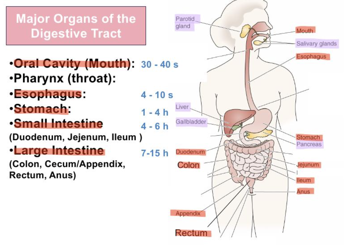
MAJOR ORGANS OF DIGESTIVE SYSTEM
Oral cavity
Pharynx
Oesophagus
Stomach
Small intestine
Large intestine

ORAL CAVITY
30-40s transport
mechanical digestion (teeth), mix with saliva
PHARYNX
propels food towards oesophagus

OESOPHAGUS
4-10s transport
propels food towards stomach

STOMACH
1-4 hours transport
chemical and mechanical digestion

SMALL INTESTINE
Duodenum, jejenum, Ileum
4-6 hours transport
Enzymatic digestion, absorption of water, vitamins, ions
LARGE INTESTINE
Colon, appendix, rectum, anus
7-15 hours
Absorption of water
Compaction of indigestible material before excretion

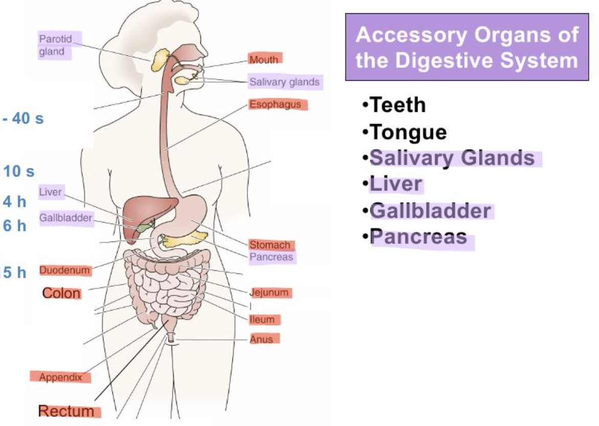
ACCESSORY ORGANS IN DIGESTIVE SYSTEM
Teeth
Tongue
Salivary glands
Liver
Gall-bladder
Pancreas

TEETH
Mechanical digestion
TONGUE
Assists mechanical digestion
Taste

SALIVARY GLANDS
Secrete lubricating fluid with enzymes to break down carbs

LIVER
Secretes bile
Stores nutrients

GALL BLADDER
Storage and concentration of bile

PANCREAS
Exocrine = secrete buffers and digestive enzymes
Endocrine = secrete insulin + glucagon

FOOD TERMINOLOGY
Food → Bolus → Chyme → Faeces or Nutrients

UPPER OESOPHAGEAL SPHINCTER
between pharynx and oesophagus
opens to let bolus enter oesophagus

LOWER OESOPHAGEAL SPHINCTER
between oesophagus and stomach
lets air escape when burping

PYLORIC SPHINCTER
End of stomach
Controls chyme entering the duodenum
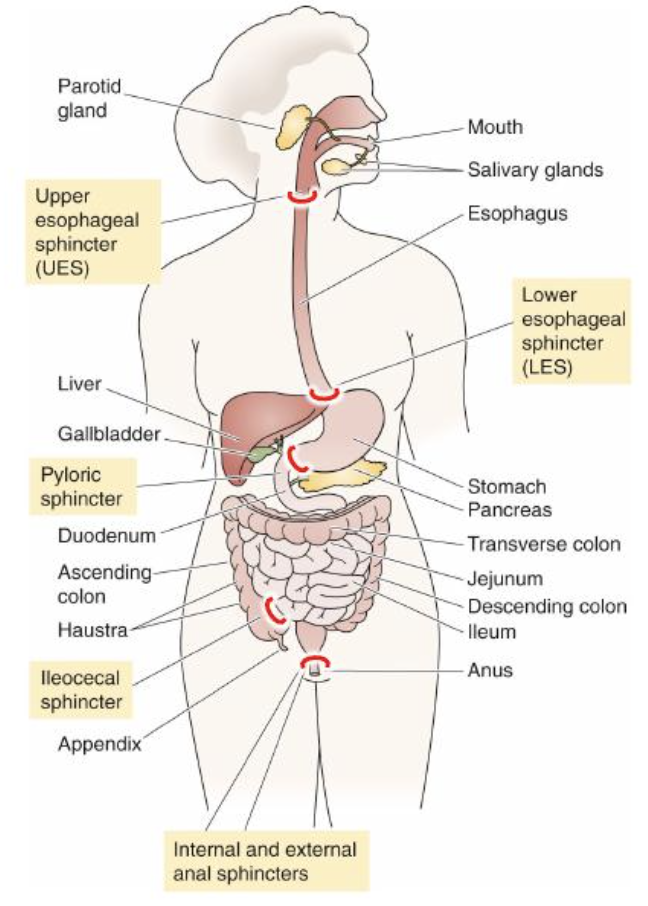
ILEOCECAL SPHINCTER
Between small and large intestine
Controls content entering large intestine

INTERNAL ANAL SPHINCTER
Involuntary
Prevents stool leakage

EXTERNAL ANAL SPHINCTER
Voluntary
Controlled excretion of stool

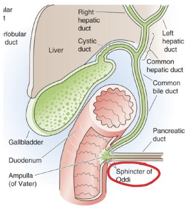
SPHINCTER OF ODDI
Where common bile duct and pancreatic duct join before opening into the duodenum
When closed:
Bile/pancreatic juice can’t enter duodenum
When open:
Bile/pancreatic juice can enter, ensuring the right amount is released when food arrives.
GI TRACT WALL
Mucosa
Epithelial layer with connective tissue, capillaries, neurons, immune cells, and smooth muscle.
Submucosa
Loose connective tissue, larger blood vessels, may have glands.
Muscle Layer
Two smooth layers (inner circular, outer longitudinal) with neurons between
Serosa
Outer connective tissue with squamous epithelial cells.

6 STEPS OF GI TRACT
Ingestion → (Secretion + Digestion + Absorption + Motility) → Defecation
4 PROCESSES OF GI TRACT
Secretion
Moving of material from cells into lumen or ECF
Digestion
Mechanical and chemical breakdown of food into absorbable units
Absorption
Movement of material from GI lumen into ECF (blood)
Motility
Movement of material through the GI tract via muscle contractions
MACROMOLECULES AND THEIR BUILDING BLOCKS
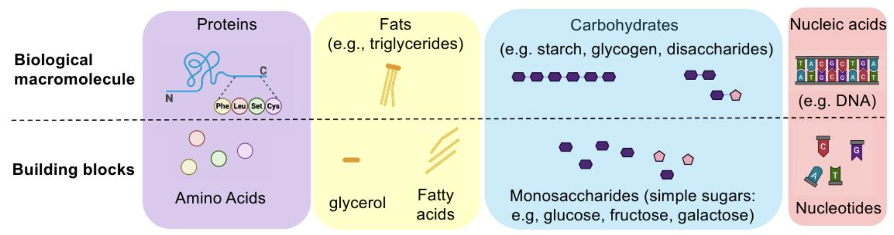
Large macromolecules generally can’t cross GI wall and must be broken down into building blocks.
Macromolecule → Building blocks:
Proteins → Amino acids
Fats → Glycerol + fatty acids
Carbs → Monosaccharides
Nucleic acids → Nucleotides
DIGESTIVE ENZYMES
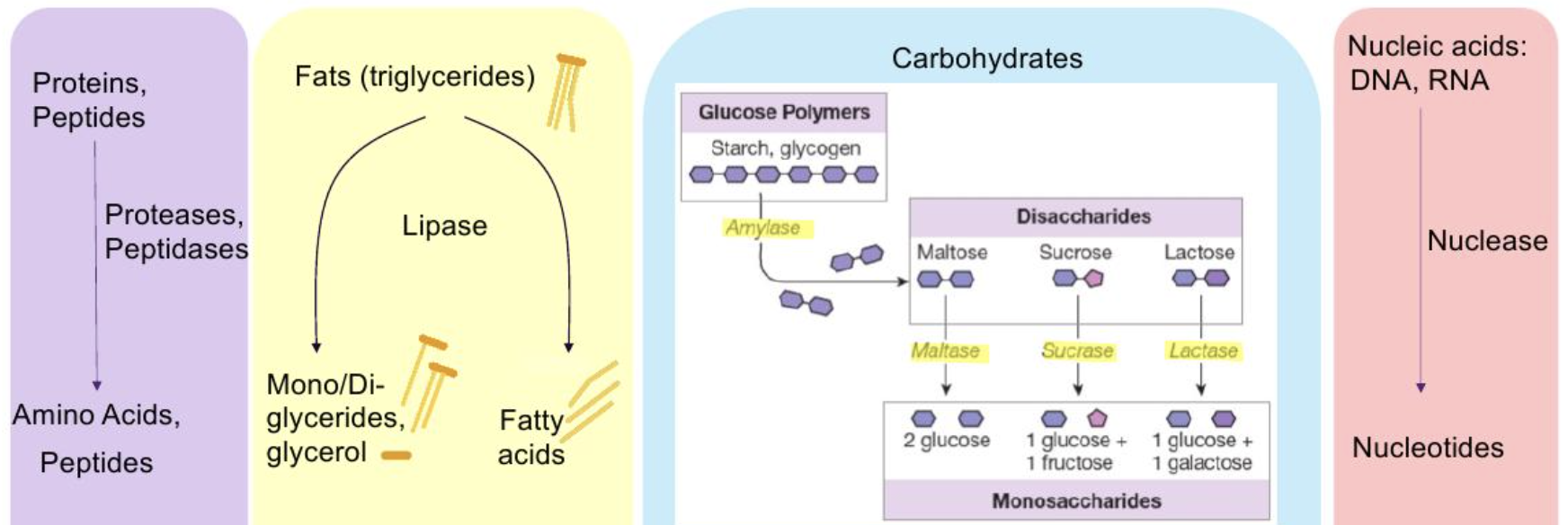
Enzymes are biological catalysts that accelerate reactions without being consumed.
Substrate-specific
Work mostly by hydrolysis (adding water to break a bond, releasing smaller absorbable units)
MACROMOLECULE → ENZYME CLASS → PRODUCT
Proteins/peptides → Proteases, peptidases → Amino acids, peptides
Fats → Lipases → Fatty acids + glycerol
Carbs → Amylase → Disaccharides, monosaccharides
Nucleic acids → Nucleases → Nucleotides
SUBSTRATE → WHERE DIGESTION MAINLY OCCURS
Proteins → Stomach + small intestine
Fats → Oral cavity + stomach + small intestine (mainly)
Carbs → Oral cavity + stomach + small intestine (mainly)
Nucleic acids → Small intestine
SALIVARY GLANDS TYPES
Parotid glands
Submandibular glands
Sublingual glands

DIGESTION IN ORAL CAVITY
Digestion = carbs, Absorption = none.
In the mouth:
Saliva contains salivary amylase mainly from parotid glands
Amylase starts starch breakdown into maltose
Only 5% starch is hydrolysed before swallowing
In the stomach:
Salivary amylase becomes inactive in acidic conditions (pH < 4)
Digestion continues 1hr until food mixes with gastric acid
30% of starches are digested
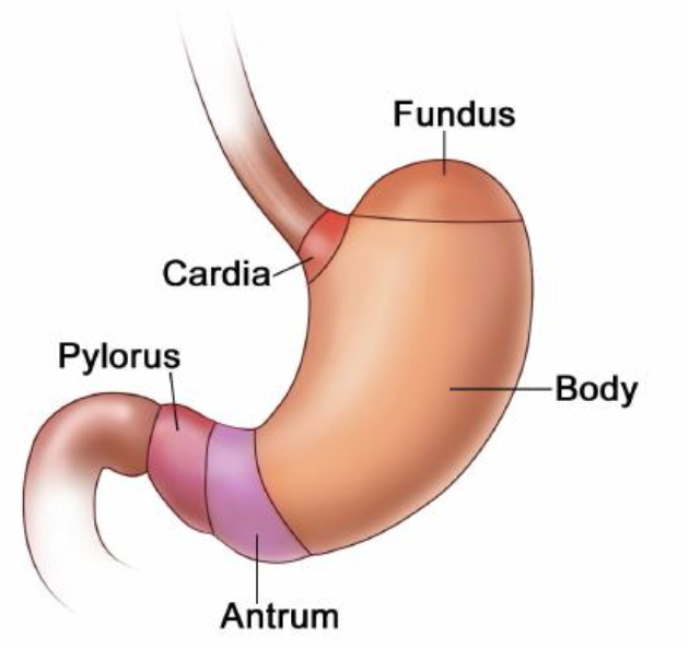
STOMACH SECTIONS
Cardia = nearest oesophagus/heart
Fundus = upper dome
Body = largest section
Antrum + Pylorus = leads into duodenum
STOMACH MAIN FUNCTIONS
Temporarily hold food
Partially digesting food into chyme
Denaturing proteins via HCl
Digesting proteins via pepsin
Digestion = proteins + fats
Absorption = lipid-soluble substances only
GASTRIC SECRETIONS
Secretion → Role
Acid (HCl) → Decreases pH to 1-1.5, kill pathogens/bacteria, denature proteins, activates pepsinogen → pepsin
Mucus → Secretion of water, bicarbonate and mucus, protects wall from acid by buffering
GI Hormones → Gastrin = stimulates acid production + stomach contractions, somatostatin = inhibits acid secretion, suppresses other GI hormones
Digestive enzymes → Pepsin = converts proteins to peptides, Gastric lipase = removes one fatty acid from each triglyceride.
STOMACH CELL TYPES
Mucuous cells → secrete mucus
Parietal cells → secrete HCl
Gastric chief cells → secrete pepsinogen, gastric lipase
Enteroendocrine cells → secrete G cells (gastrin), D cells (somatostatin)
PHASES OF GASTRIC SECRETION
Cephalic phase
Gastric phase
Intestinal phase
CEPHALIC PHASE
Triggered by:
sight, smell, taste or thought of food
Mechanisms:
the hypothalamus and medulla stimulates the vagus nerve, signalling to the stomach to begin secreting gastric juices + gastrin
Outcome:
Prepares stomach for digestion
Accounts for 10-20% of gastric secretion
GASTRIC PHASE
Triggered by:
presence of food in the stomach
Causes stretching and the presence of proteins/amino acids
Mechanisms:
Stretch activates stretch receptors, stimulating gastrin release
pH > 4 = increase in gastrin release
pH < 3 = decrease in gastrin release
More gastrin = more gastric juice = more stomach contractions
Outcome:
60-70% of gastric secretion
NOTE: parasympathetic = increase gastric secretion; sympathetic = decrease gastric secretion.
PANCREAS
Endocrine:
Islets of Langerhans
Secrete insulin and glucose
Regulate blood glucose
Exocrine:
Acinar cells = secrete digestive enzymes
Ductal cells = secrete bicarbonate rich fluid

PANCREATIC JUICE
watery, alkaline solution (pH 8)
neutralises stomach acid entering duodenum
1200-1500mL per day produced
ENZYME → FUNCTION
Proteases → convert proteins to peptides
Carboxypolypeptidase → convert peptides to amino acids
Pancreatic lipase → Convert triglycerides to monoglycerides + fatty acids
Pancreatic amylase → convert starch to maltose
Nucleases → Convert DNA/RNA to nucleotides
GALLBLADDER
small pear-shaped sac on the underside of the liver
Stores and concentrates bile by absorbing water and electrolytes
Honeycomb folds allow it to expand as it fills
BILE RELEASE
Stimulated by hormone CCK →
Gallbladder contracts →
Bile flows into cystic duct →
Common bile duct →
Duodenum
BILE
Produced in the liver
Contains bile salts, which are amphipathic
Emulsification:
Bile salts surround large fat deposits, breaking them into smaller emulsion droplets
Increased surface area:
Lets pancreatic lipase act more efficiently
Micelle formation:
Bile salts package digested fats into micelles for transport to the intestinal epithelial cells where they’re absorbed

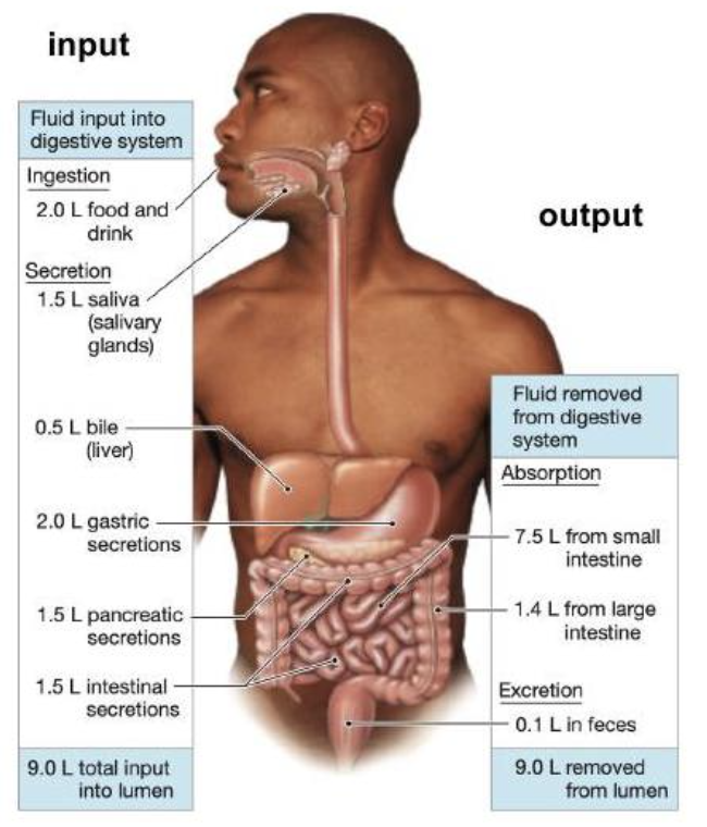
SMALL INTESTINE INPUT INTO LUMEN
9L per day
Food and drink = 2L
Saliva = 1.5L
Bile = 0.5L
Gastric secretions = 2L
Pancreatic secretions = 1.5L
Intestinal secretions = 1.5L

SMALL INTESTINE REMOVAL FROM LUMEN
9L per day
Absorbed in small intestine = 7.5L
Absorbed in large intestine = 1.4L
Lost in faeces = 0.1L
Via homeostasis, output must = input → mass balance
SMALL INTESTINE FUNCTIONS
Chemical digestion
Nearly all nutrient absorption
Chyme mixes with digestive juices slowly, allowing time for absorption
Undigested materials passed on to large intestine
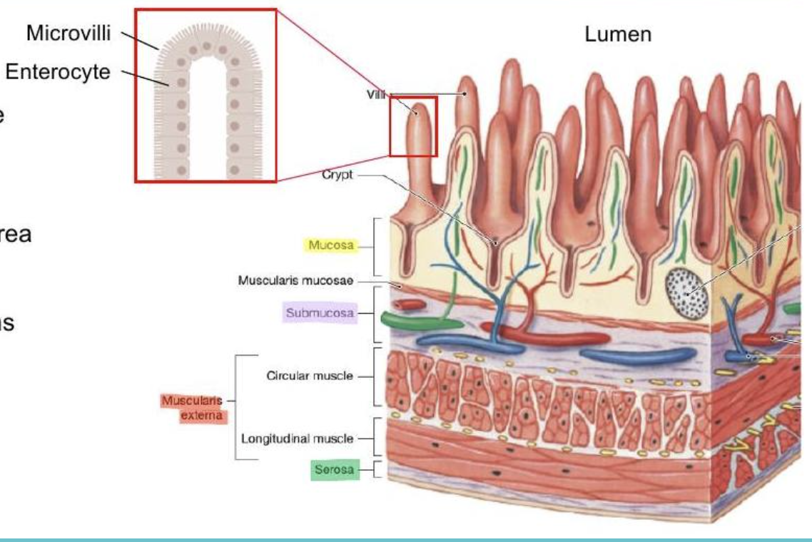
SMALL INTESTINE STRUCTURE
Same four layers as the rest of GI tract
mucosa (SA x3 larger than stomach for greater absorption)
submucosa
muscularis externa
serosa
Villi
finger-like projections
Crypts
Invaginations between villi
Microvilli
Brush border on villi
SMALL INTESTINE SECRETIONS (PROTEASES + PEPTIDASES)
Enterokinase
located at duodenal brush border
activates pancreatic zymogen → trypsinogen → trypsin
Peptidases
located in enterocytes
peptides → amino acids
SMALL INTESTINE SECRETIONS (DISSACHARIDASES)
Maltase
Located at brush border
Maltase → glucose
Sucrase
Located at brush border
Sucrose → glucose + fructose
Lactase
Located at brush border
Lactose → glucose + galactose
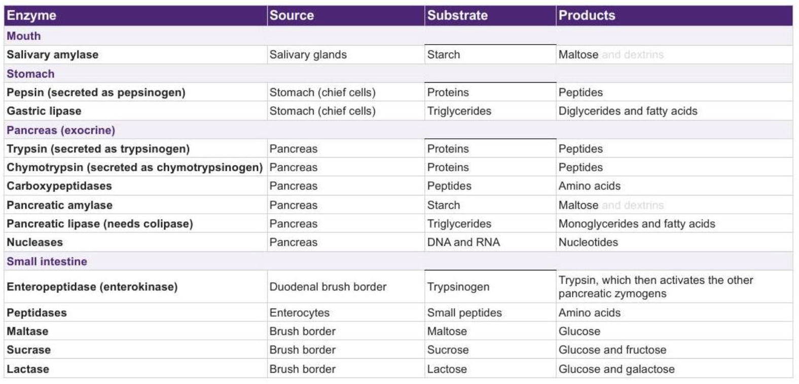
SUMMARY OF ENZYME SECRETIONS
GLP-1 EFFECTS
Effects:
More insulin, less glucagon from pancreas
slower gastric emptying
Less acid secretion
Less appetite and higher satiety
Type 2 diabeties:
Slower emptying flattens glucose peak after meal
extra insulin decreases blood glucose
effective in regulating
GLP-1 CHALLENGES
Challenges:
Half life of 1-2 minutes
useless as an injection
Solutions:
modify with amino acids
can last up to a week
THE INTESTINAL PHASE OF GASTRIC SECRETION
Triggered by:
entry of partially digested chyme entering duodenum
Mechanism:
Hormones released to inhibit further gastric secretion and slow gastric emptying
Secretin = stimulates bicarbonate secretion and flow of bile from liver → gallbladder
CCK = triggers gallbladder + pancreas to contract and release bile + enzymes into duodenum to aid digestion
GIP + GLP-1 = stimulate insulin secretion
LARGE INTESTINE FUNCTIONS
Digestion of remaining food
absorb remaining water, electrolytes (salt) and vitamins
Secretes mucus to ease passage of faeces
Contractions propel faeces towards rectum
Temporarily stores waste until defecation
Defecation is a reflex triggered by rectal dystension
THE COLON AND FAECAL CONSISTENCY
Ascending = fluid/semi-fluid
Transverse = mush
Descending = semi-mush/semi-solid
Sigmoid + rectum = solid
Poor motility = sits longer, increased water absorption → hard faeces → constipation
Excess motility = decreased absorption → faeces don’t solidify → diarrhea

MICROBIOTA
Community of microorganisms on multicellular organisms (ex. bacteria, fungi).
Around 3.9 × 10^13 cells of microorganisms
95% found in GI tract
70-80% in large intestine
FACTORS AFFECTING MICROBIOTA
Diet
antibiotics
lifestyle (stress, sleep, exercise)
Cohabituation (shared living environment is more important than genetics)
Dysbiosis (imbalance - obesity, diabetes, mental health disorder, IBD)
FUNCTIONS OF MICROBIOTA
Digestion (of fibres we cant digest ourselves)
Vitamin synthesis (B and K)
Immune support (train and regulate immune system)
Protection (competes with harmful pathogens for resources)
Signalling molecules (that interact with the host)
Secondary bile acid production
Drug metabolism
PERISTALSIS OF OESOPHAGUS
Contraction behind the bolus
Circular muscles contract behind the bolus, squeezing it forward
Relaxation in front of the bolus
Longitudinal muscles infront of bolus contract to shorten the oesophagus and circular muscles relax to create space for bolus to move forward
Sequential waves
This wave like motion continues down oesophagus in coordinated manner
*gravity helps but not necessary

PERISTALSIS OF STOMACH
Gastric emptying
Antral contractions propel chyme forward and grinds it
Partly open pyloric sphincter lets some chyme through
Stronger contraction = empties more
Gastric mixing
When wave reaches pyloric sphincter it closes tightly
Chyme hitting closed sphincter is tossed back into antrum
Retropulsion = chyme mixes with gastric secretions
Wave starts at upper fundus → pyloric sphincter → antrum.

SEGMENTATION
Short segments of the intestine alternately contract and relax, churning the chyme.
slow, short lived contractions
mainly in small intestine
triggered by filling of stomach and local stretch in small intestine
does not produce much propulsion

MIGRATING MOTOR COMPLEX
Occurs between meals
Peristalsis starts in stomach and travels through small intestine
one cycle = 2 hours
Propels undigested food residue, dead cells, bacteria
‘interdigestive housekeeper’
stimulated by hormone motilin from small intestine
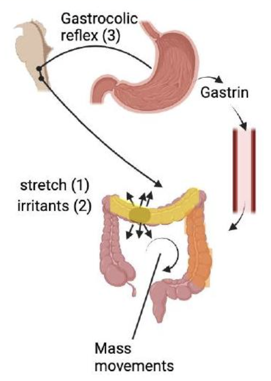
MASS MOVEMENTS
Slow, powerful peristalsis contractions in large intestine that moves waste to rectum.
Occurs 3-5x per day for 10-15 minutes, during or after meals
Triggers:
Stretch of colon wall
Irritants such as chemical or ulcers
Gastrocolic reflex
DEFECATION STEPS
Faeces enters and stretches the sigmoid colon and rectum, triggering the reflex
Stretch receptors activate sensory nerve fibres that signal the spinal cord
Parasympathetic efferent motor fibres are activated and CNS sends signals back to colon and rectum
Sigmoid colon and rectum contract, pushing faeces into anus
Internal sphincters relax in response to parasympathetic activation, letting faeces into anal canal
External sphincters stay closed, holding back faeces
When deciding to defecate, impulses from cerebral cortex relax external sphincter and faeces are expelled.

ENTERIC NERVOUS SYSTEM
The GI tracts own nervous system
100 million neurons
Enteric neurons communicate with each other and work autonomously, but also exchange info with the CNS
Major nerve supply to GI wall and controls motility (including peristalsis)
ENTERIC NERVOUS SYSTEM STRUCTURE
Confined within the GI tract wall from start of oesophagus to internal sphincter and consists of two interconnected nerve plexus:
Submucosal nerve plexus
In the submucosa
Controls local secretions, blood flow and contractions or smooth muscle in mucosal layer
Myenteric nerve plexus
In the muscularis externa
Coordinates motility and peristalsis

ENTERIC NS NEURONS
Intrinsic neurons
Short reflexes
ex. gastrin secretion triggered by stomach stretch
in enteric plexus
allows local reflexes to start, be integrated, and finish inside GI tract
‘little brain’
Extrinsic neurons
Long reflexes
ex. defecation reflex
autonomic neurons that carry signals from CNS to GI tract
respond to stimuli inside or outside of gut

AUTONOMIC CONTROL AND DIGESTIVE SYSTEM
Parasympathetic activation
= enhances digestion
= relaxes sphincters so contents can move through
Sympathetic activation
= inhibits digestion
= contracts circular muscles and keep sphincters closed
LIVER FUNCTIONS
Regulates blood glucose levels
stores glucose as glycogen
synthesises glucose through gluconeogenesis
Vitamin storage
A, D, E, K, B12
Bile production
Nitrogen excretion
Acts as an endocrine organ
releases hormones
Waste management
degrades old/damaged proteins
Iron metabolism
Protein synthesis of various plasma proteins for blood functions
Detoxification
of foreign chemicals
Fat metabolism
Break down of fatty acids and distributes lipoproteins
LIVER ARTERIAL BLOOD SUPPLY
25%
delivered via hepatic artery
O2 rich blood
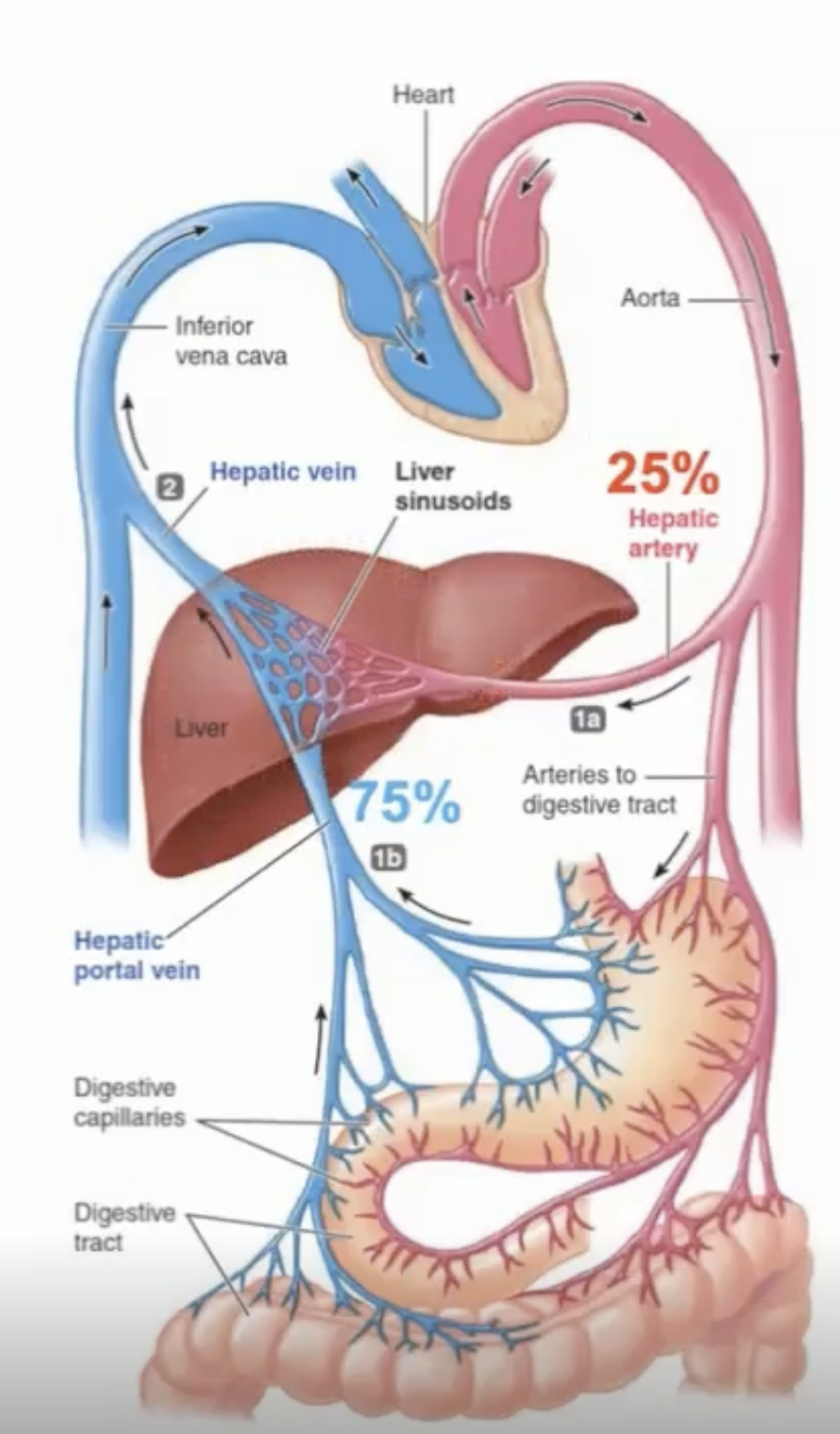
LIVER VENOUS BLOOD SUPPLY
75%
Delivered via hepatic portal vein, carrying blood from digestive tract
02 poor blood
Blood contains
nutrients, drugs, hormones, pathogens, toxins
Blood leaves via hepatic vein

LIVER LOBULES
Liver is organised into hexagonal units called liver lobules
Portal vein + hepatic artery supply blood to portal venuoles + hepatic arterioles which converge at the corners of the lobule
PORTAL TRIAD
Found at each lobule corner, consists of:
a portal vein branch
a hepatic artery branch
a bile duct that drains bile from hepatocytes via bile canaliculi
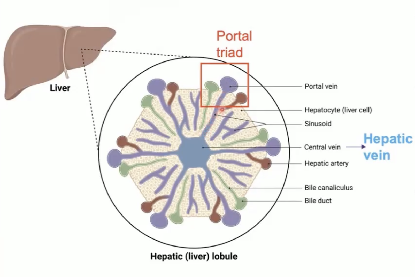
BLOOD FLOW IN LIVER
Portal vein and hepatic artery empties blood into a shared capillary network known as liver sinusoids
Blood from sinusoids is collected by central veins which drain into larger hepatic veins
Hepatic veins then empty filtered blood into inferior vena cava

LIVER LOBULE CELLS
Hepatocytes (80% of all liver cells)
main functional cells of the liver that are responsible for metabolism, detoxification, protein synthesis, and bile production
Liver endothelial cells (LEC)
line the blood vessels (sinusoids) and facilitate exchange of substances between blood and hepatocytes
Stellate cells
Store vitamin A and plays a role in liver fybrosis when activated
Kupffer cells
specialised macrophages that are responsible for phacocytosing pathogens, erythrocytes, dead cells and debris

LIVER ZONATION
Blood flow along lobules radial axis creates gradients of oxygen, nutrients and hormones
Spatial variability results in non-uniform expressions of key liver functions
Ex.
Periportal (zone III)
cholesterol biosynthesis
protein secretion
Pericentral (zone I)
drug detoxification
bile acid production

LIVER PROTEIN SYNTHESIS OF PLASMA PROTEINS
ALBUMIN
60% of secreted/synthesised plasma proteins
maintains osmotic pressure
transports substances in blood
CARRIAGE PROTEINS
Transferrin, ceruloplasmin, transcortin
BLOOD COAGULATION FACTORS
ANTI-CLOTTING PROTEINS
HORMONES
APOLIPOPROTEINS
IMMUNE PROTEINS
LIPOPROTEINS
Essential for transport of lipids in the blood or ECF and are produced by small intestine and liver.
CHYLOMICRONS
located in small intestine
transport dietary fats + cholesterol from gut → lymphatics → blood → muscle
VeryLowDensityLipoprotein
located in liver
Delivers triglycerides synthesised+packaged by liver to peripheral tissue
IntermediateDL
formed from VLDL after TG (triglyceride) offloading
LowDL
formed from IDL
transports cholesterol back to liver
HighDL
reverse cholestryl transport
FAT ABSORPTION + CHYLOMICRON FORMATION
Large fat droplets (from stomach) → emulsified by bile salts → miscelles → pancreatic lipase breaks down fats → fatty acids enter enterocytes → absorbed fats combine with cholesterol + proteins = chylomicron → exported in interstitial fluid → blood → used as energy source.
CHYLOMICRON METABOLISM IN CIRCULATION
At capillary beds of muscles LPL strips out TG → releasing fatty acids + glycerol for tissue uptake → chylomicron returns to liver for further processing,
LIPOPROTEIN CASCADE
Liver releases VLDL → LPL strips triglycerides → IDL → further TG loss → LDL → LDL receptor uptake by liver → cholesterol back to liver
CHOLESTEROL ELIMINATION
In the liver cholesterol is:
re-secreted as VLDL or
converted into bile acids
Bile acids secreted into:
Bile → gut → excretion
to maintain homeostasis
Cholesterol absorbed in intestine:
liver → remainder is excreted as poo
IRON METABOLISM IN LIVER
Sources of iron:
uptake of dietary iron
recycling of haemoglobin via Kupffer cells
Iron transport + metabolism:
Fe2+ leaves cells via ferroportin
in blood, fe2+ binds to ferroportin in plasma
Tf-bound iron can be taken up by cells via TfR1
in the liver iron is stored as ferritin

HEPCIDIN
The liver secretes hepcidin which is a regulator of iron metabolism
an increase in iron levels and storage stimulates hepcidin production
suppressed hepcidin production in iron deficiency
its target is ferroportin
hepcidin binding to ferroportin = degredation = less iron released in blood = decreased iron availability

DETOXIFICATION IN THE LIVER
Xenobiotics = any compound foreign to the organism.
Compounds not taken up by hepatocytes undergo biotransformation:
Phase 1: Modification
Phase 2: Conjugation
Phase 3: Secretion/excretion

PHASE 1 - MODIFICATION
Chemical modification of compounds
Cytochrome P450 enzymes add or expose reactive groups making the molecule more hydrophilic and reactive for phase 2
reactions = oxidation, reduction, hydrolysis
PHASE 2: CONJUGATION
The functional groups introduced in phase 1 are conjugated with polar molecules
this process makes metabolites more hydrophilic, facilitating their excretion
PHASE 3 - EXCRETION/SECRETION
Hepatocytes excrete phase 1 and 2 products into bile or blood
Metabolites actively transported out of hepatocytes using membrane transport proteins
Excretion:
most drugs excreted via kidneys
Larger metabolites excreted into bile and out via faeces
SUPRACHIASMATIC NUCLEUS
The central clock resides in the suprachiasmatic nucleas (SCN) of the hypothalamus
Light is the dominant signal - detected by the retina and relayed to SCN

PERIPHERAL TISSUE CLOCKS
SCN synchronises peripheral clocks present in most cells/organs
peripheral clocks are influenced by feeding
SCN + peripheral clocks + feeding = rhythmic physiology
CIRCADIAN CLOCKS
Humans intrinsic clock is approx 24.5 hours
all known carcadian oscillators are built from negative feedback loops of gene expression

CIRCADIAN RHYTHM - LIVER BLOOD FLOW
Peaks in early phases (2-8 hours)
Trough mid-way through rest phase

CIRCADIAN RHYTHM - BILE SECRETION/EXCRETION INTO GALLBLADDER/DUODENUM
Bile secretion into gallbladder
accumulation rises while fasting
Bile excretion into duodenum
release rises around/after feeding

CIRCADIAN RHYTHM - BASAL GASTRIC ACID SECRETION
Lowest around midday
Rises towards evening/night

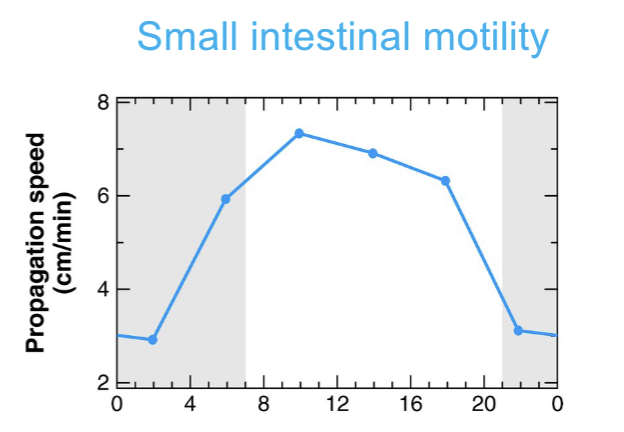
CIRCADIAN RHYTHM - SMALL INTESTINE MOTILITY
peaks around midday
Lowest during night/rest phase