RADS 301
 Call Kai
 Learn
Learn Practice Test
 Spaced Repetition
 Match
Match Flashcards
 Knowt Play
1/126

There's no tags or description
Looks like no tags are added yet.
Name | Mastery | Learn | Test | Matching | Spaced | Call with Kai | Chat |
|---|
No analytics yet
Send a link to your students to track their progress
127 Terms
Which modalities use ionizing radiation?
X-Rays, CT, PET
What are the 3 anatomical planes?
1. Sagittal Plane
2. Frontal Plane
3. Transverse Plane

Highest to lowest radiodensity
Metal, bone, water/soft tissue, fat, air
What does B-mode stand for in ultrasound?
Brightness mode
What does a Doppler ultrasound measure?
Blood flow (mcat moment)
Which of the following statement is true about protons of the body?
a) They are always aligned and stationary.
b) They have intrinsic spin
c) They cannot be manipulated by external magnetic field.
d) The time protons take to recover from displacement to its original alignment with the external magnet field is called T2.
b) They have intrinsic spin
Which of the imaging modalities was the first to be invented?
X-Ray (radiograph / plain film)
Endovascular thrombectomy (EVT) is primarily used to treat which condition?
Acute Ischemic stroke
Which of the following statement is true about Bremsstrahlung radiation?
a) Bremsstrahlung radiation has discrete peaks.
b) Bremsstrahlung radiation is produced when high-energy electrons are deflected by atomic nuclei.
c) Bremsstrahlung radiation is the produced by smashing an electron into an orbital electron.
d) Bremsstrahlung radiation is the result of transition of an outer shell electron to the inner shell.
b) Bremsstrahlung radiation is produced when high-energy electrons are deflected by atomic nuclei.
Which of the following types of radiation are considered ionizing?
1. X-rays
2. Gamma rays
3. Alpha particles
4. Neutrons
5. Microwave
1, 2, 3, 4
Which of the following statements is true about ionizing radiation?
a) The same dose of different types of ionizing radiation causes the same effect on tissue.
b) Ionizing radiation only causes damage to tissue by directly targeting DNA.
c) Generally speaking, the slower the cells divide, the less sensitive they are to ionizing radiation.
d) None of the above
c) Generally speaking, the slower the cells divide, the less sensitive they are to ionizing radiation.
Which of the following is NOT a source of natural background radiation?
a) Radon gas
b) Cosmic rays
c) Radioactive material in the soil
d) Medical X-rays
d) Medical X-rays
Which of the following statements is true about radiation injury?
a) All forms of radiation injury cause immediate tissue damage
b) Radiation injury affects all tissues in the body equally
c) Radiation exposure is safe during pregnancy, as it only affects the mother and not the fetus
d) Exposure to low doses of radiation over time may increase the risk of cancer
d) Exposure to low doses of radiation over time may increase the risk of cancer
Which of the following statements is true about ionizing radiation?
a) The same dose of different types of ionizing radiation causes the same effect on tissue.
b) Ionizing radiation only causes damage to tissue by directly targeting DNA.
c) Generally speaking, the slower the cells divide, the less sensitive they are to ionizing radiation.
c) Generally speaking, the slower the cells divide, the less sensitive they are to ionizing radiation.
Which of the following is NOT a source of natural background radiation?
A) Radon gas
B) Cosmic rays
C) Radioactive material in the soil
D) Medical X-rays
D) Medical X-rays
Which of the following statements is true about radiation injury?
A) All forms of radiation injury cause immediate tissue damage
B) Radiation injury affects all tissues in the body equally
C) Radiation exposure is safe during pregnancy, as it only affects the mother and not the fetus
D) Exposure to low doses of radiation over time may increase the risk of cancer
D) Exposure to low doses of radiation over time may increase the risk of cancer
You may have heard that people get a stent after a heart attack. What is the primary function of a coronary stent?
To open a blockage in the coronary artery
Concerning muscles visualized by ultrasound, which statement is NOT correct?
a) Appears the same, independent of the probe angle
b) Appears different, dependent on probe position
c) Muscles are connected to bone via tendon
d) Muscle tears can be diagnosed easily by ultrasound
a) Appears the same, independent of the probe angle
What Artifact is this from and what is it?
MRI. Acoustic Shadowing.
When a object absorbs or strongly reflects the soundwaves
What is Echo delay
Transducer interprets as “farther” object location
What is Photoelectric effect?
An incoming X-ray photon is completely absorbed, ejecting an electron from the atom.
The atom becomes positively charged; the released photoelectron can cause further ionization. Ionizing radiation can remove electrons from atoms.
Explain photoelectric effect and tissue contrast
Photoelectric absorption increases strongly with atomic number:
- approximately Z³ at a fixed photon energy. Tissue density also affects attenuation.
- Bone stops more X-rays and appears white; air transmits more and appears black.
What is Z³
describes how strongly a material absorbs X-ray photons
Bones have Z = 20, while soft tissues have Z = 7.4
Photons (X-ray and Gamarays)
The rest are therapy purpose
"A photon transfers part of its energy to an electron, then continues in a different direction with less energy. Scattered photons reaching the detector reduce contrast.
https://howradiologyworks.com/scatter-in-x-ray-imaging/"
Who discovered radiology? And how?
Roentgen from Germany. Somehow discovered X-rays while experimenting with cathode rays. A nearby fluorescent screen illuminated, revealing radiation that could penetrate objects and show internal structures.

Which Imaging does not use Ionizing radiation?
Ultrasound (Sound) and MRI (Magnetic fields)
What does it mean when we say attenuated?
X-rays that the body is absorbed. Indicated by the colour white.
What does it mean when we say Radiolucent?
less attenuation = darker, such as air.
What the Strengths of CT
Provides detailed cross-sectional anatomy aka 3D Images
Used for trauma and acute care
Radiographs vs Cts
X-rays: Cheaper, quicker, and low radiation
CT: Rotating x-ray, longer, detailed, higher radiation

Name of CT image?
Coronal
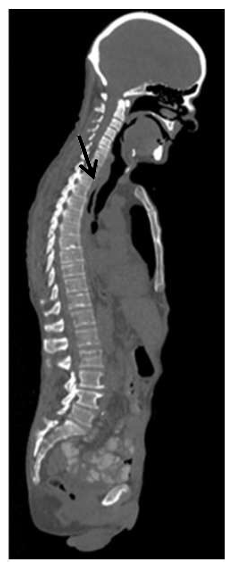
Name of CT image?
Sagittal

Name of CT image?
Axial/Transverse

Answer

On CT, the measure of density is called a
Hounsfield Unit (HU)
What are Contrast studies?
Distinguish structures that look somewhat similar. E.g IV contrast to enhance blood vessels
What is MRI
MRI uses a strong magnet and radiofrequency pulses to generate signals from hydrogen nuclei, abundant in water and fat.
Different sequences emphasize different tissue properties.
Excellent soft-tissue contrast; no ionizing radiation.
Explain the difference between T1 vs T2 MRI
T1:
Fluid is Dark
Fat is Bright
Focuses on Anatomy
T2
Fluid is Bright
Fat is Bright
Focuses on Fluid, edema, pathology
Strengths and Weakness for MRI
Strengths: No exposure to ionizing radiation, good for soft tissues
Weakness: Expensive, long wait time, longer images, metal complication (magnet)
Explain Ultrasound
Uses high frequency sound waves to travel to generate an image
Cheap, no radiation, and real time
ULTRASOUND EXPLAIN THE TWO TYPES OF ECHOES
Hyperechoic: Reflecting many echoes (e.g bone, fat, stones, air)
Hypoechoic: No echoes since ultrasound can pass thru tissue (fluid, black)
What are Acoustic windows
Fluid transmits sound well, allowing deeper structures to be examine
Explain the concept of Ultrasound frequency, depth, and zoom
Higher frequency gives better resolution but less penetration; lower frequency reaches deeper.
e.g using higher frequency on soft tissue, lower frequency on deeper organs
Pulse repetition frequency is separate: fewer pulses per second allow longer listening between pulses.
Read Zoom: Increase resolution interest of area
Write Zoom: Rescans smaller area
Ultrasound applications
Know pregnancy assessment, abdominal organs, heart function, muscles/tendons, and leg-vein clots
What is Echocardiography? What are the two types?
Ultrasound for the heart
TTE: Non invasive. Just chest
TEE: Invasive. In the esophagus
Why use echocardiography?
Echo shows both anatomy and function in real time: chamber size, wall thickness, contraction, valves, and blood flow. No radiation
Describe all the facts of Heart diseases
#1 CAUSE OF DEATH IN WOMEN
1 in 5 Women
What is Coronary calcification? WHats the screening?
Build of calcium and results in atherosclerotic plaque. CT scan.
Two Techniques for Radiographs

What is a supine chest radiograph?
Lying on the back. Position usually set to AP.
AP vs PA effects on imaging?
AP magnifies shadow by 20-25%
Basic Anatomy
Trachea LOL
Explain the Pleural Anatomy
Visceral pleura covers the lung; parietal pleura lines the chest wall.

Right tension pneumothorax
Air accumulates under pressure in the right pleural space. The key mechanism is loss of the normal pressure relationship keeping the lung expanded, followed by increasing pressure in a tension pneumothorax.
Spontaneous pneumothorax
It just happens randomy lol
CT with Contrast
Uses contrast material on blood. This is known as CTPA.
CT strengths and Weaknesses
StrengthsL Bone and Lung detail
Good contrast
Weakness:
Radiation & weight limit
What is a pulmonary embolism?
A pulmonary embolism blocks a pulmonary artery, commonly with a clot
Ray tracing and global illumination
Ray tracing simulates paths of light. Global illumination accounts for indirect lighting and interactions such as shadows and reflections.
Cinematic rendering
photorealistic-looking 3D images from cross-sectional data using lighting models


Main reason Cinematic rendering is better than xray?
Improved diagnosis reveal fractures are that cannot be detected via x-rays
Which complex injuries is cinematic rendering mostly used in?
Pelvic fractures
What is the main weakness of cinematic rendering
Potential for Artifacts: Over-processing could theoretically obscure some subtle details
Cinematic rendering vs Standard Volume rendering
Cinematic uses lighting, and is more computational
Volume uses less detail lighting and is less computational
What is the top grey matter in the brain called? And what’s its main purpose?
Pre-central gyrus. Motor control.
Give me definition of cerebellum
Balance and voluntary moves
Give me definition of Caudate and Lentiform Nuclei
Tuned movements
What is the grey matter in the brain called (bottom)? And what’s its main purpose?
Post-central gyrus. Sensation.


What is the white in the brain called?
Internal Capsule
Name the 4 motor control of the brain
Pre-central gyrus, internal capsule, midbrain, spinal cord
PIMPS without the P
What is Chest CT with contrast called?
Computed tomography Pulmonary angiography. Its non invasive
What is angiography?
The study of vessels
Which of the following type of imaging modality does NOT use ionizing radiation?
A. X-RAY
B. Computed tomography (CT)
C. MRI
D. Fluroscopy
C. MRI
Which of the following imaging modalities is used to screen breast cancer in general population
A. MRI
B. Fluroscopy
C. Ultrasound
D. Mammogram
D. Mammogram
Which of the following is not an advantage of xray?
A. Easily accessible
B. less radiation
C. bone visualization
D. soft tissue visualization
D. soft tissue visualization
Which imaging modality would you use for pulmonary embolism?
A. chest xray
B. PET/CT
C. CT
D. CTPA
D. CTPA
Which of the following imaging modalities is the best non-invasive test for confirming the direct presence of calcification in the coronary arteries?
A. nuclear cardiac imaging
B. catheter cardiac angiography
C. cardiac MRI
D. caridac computed coronary angiography tomography, ie cardiac CT
D. caridac computed coronary angiography tomography, ie cardiac CT
Which of the following statements is/are true about CT?
1. Uses ionizing radiation, Cross-sectional 2D images produced using many x-ray sources
2. Produce very detailed images comparing to X-ray (radiograph)
3. Most commonly used in diagnosis and monitoring of diseases. Variety of applications including different types of cancers, heart diseases, stroke, and etc
All of them are true!
The most common genetic cardiomyopathy with a prevalence of 1 in 500 in the general population is:
a) Coronary artery atherosclerosis
b) Mitral valve disease
c) Hypertrophic cardiomyopathy
d) Myocarditis
d) Myocarditis
What is pneumothorax
air in the pleural cavity
can see a thin white line
entire perimeter of lungs
causes lung to collapse
symptoms include shortness of breath and chest pain can be life threating
The skull protects the brain… = best imaged with
X-ray or CT
Brain tissue is “soft tissue” + water =
best imaged with MRI. CT is faster
Motor function involves the
Frontal Lobe
Sensory function involves the
Parietal Lobe
Neural foramen protects
nerve as it exits
What ultrasound advantages are listed?
No ionizing radiation, generally noninvasive external imaging, real-time images, low cost, and portability.
Why does CT reveal anatomy better than a single radiograph?
CT reconstructs cross-sectional slices, reducing the overlap of structures seen in a projection image.
Which generally attenuates more X-rays: bone or air?
Bone. More attenuation makes it appear whiter; air transmits more X-rays and appears darker.
What does TTE stand for, and where is the probe?
Transthoracic echocardiography; the probe is on the chest wall.

What does TEE stand for, and where is the probe?
Transesophageal echocardiography; the probe is in the esophagus, close to the heart.

Which pleural layer covers the lung surface?
Visceral pleura.
VP
Which pleural layer lines the inside of the chest wall?
Parietal pleura.
VP

Midbrain

Faced Joint