skeletal muscle tissue components
 Call Kai
 Learn
 Practice Test
 Spaced Repetition
 Match
 Flashcards
 Knowt Play
1/83

There's no tags or description
Looks like no tags are added yet.
84 Terms

What is this?
Epimysium
outer layer encircling entire muscle
Dense irregular connective tissues

What is this?
perimysium
Surrounds groups of 10 to 100+ muscle fibres
separates them into bundles (muscle fascicles)
Dense irregular tissue
Endomysium
penetrates interior of each muscle fascicle
Separates muscle fibres from one another
Mostly reticular fibres
What is a tendon?
epimysium, perimysium and endomysium continuous w/ connective tissues
Extends beyond muscle fibres to form tendon attaching muscle to periosteum
Aponeurosis
when connective tissues extends as a broad, flat sheet
What are somatic motor neurons
Neurons that stimulate skeletal muscle to contract
Axon extends from brain/spinal cord to group of skeletal muscle fibres
Diameter and length of mature skeletal muscle
diameter: 10-100 micrometers
Length 10cm or as long as 30cm (12 inch)
What is sacrolemma
plasma membrane of muscle fibre
Multiple nuclei of skeletal muscle fibre located beneath
Has sarcoplasm

T tubules
Thousands of tiny tube shaped invaginations of sarcolemma
form T tubules
tunnel from surface to Center of fiber
Filled with interstitial fluid
Ensures an action potential excites all parts of muscle Fiber at once
Where does muscle action potential travel
travel along sarcolemma and through T tubules
quickly spread throughout muscle fiber
Sacroplasm
cytoplasm of muscle fibre
Substantial amount of glycogen
Contains myoglobin
Myoglobin
red coloured protein
Only found in muscle
Binds oxygen molecules that diffuses into muscle from interstitial fluid
Releases O2 when needed by mitochondria for ATP production
Myofibrils
little threads in sacroplasm
Contractile organelles of skeletal muscle
~2 micrometers in diameter
Extend length of muscle fibre
Make skeleton appear striated

Sarcoplasmic reticulum (SR)
fluid-filled system of membranous sacs
Encircles each myofibril
releases calcium during muscle contraction

What is this
Terminal cistern
Dilated end sacs of the sacroplasmic reticulum
What is a triad
T tubule and two terminal cisterns on either side
What does the sacroplasmic reticulum store and release
stores calcium ions when muscle fibres relaxed
Release Calcium ions from terminal cisterns
—> triggers muscle contraction
Filaments/myofilaments
smaller protein structures in myofibrils
2 thin filaments:1 thick
Thin filaments
8nm in diameter
1-2 micrometers long
Composed of protein actin
thick filaments
16nm in diameter
1-2 micrometers long
Composed of protein myosin

What is this?
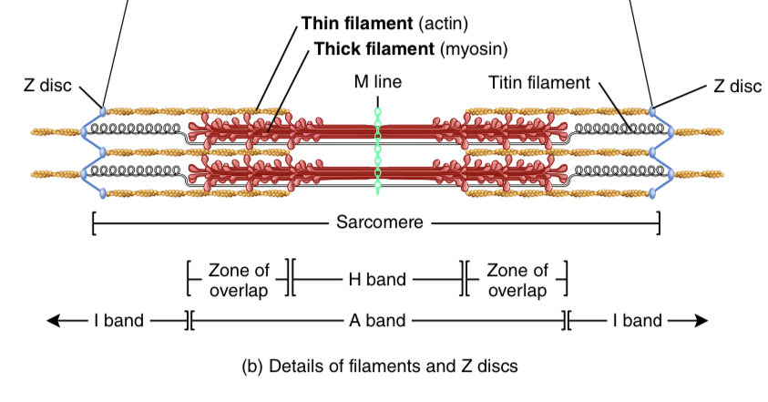
sacromeres
Compartments/basic functional units of myofibril
Z Discs
narrow plate shaped regions o dense protein
Separate each sacromere
A band
darker middle part of sacromere
Extends entire length of thick filaments
Toward each end of A Band is a zone of overlap of thick & thin filaments
I Band
lighter, less dense area containing rest of thin filaments
What create striations of muscle
Alternating I and A bands
H bands
Center of each A band
M line
supporting proteins holding thick filaments tgt at Center of H band

What are the 3 kinds of proteins in myofibril
contractile proteins
generate force during contraction
Myosin + actin
regulatory proteins
switch contraction processes on and off
Tropomyosin + troponin (part of thin filament)
structural proteins
keep thick & thin filaments in proper alignment
Myosin
main component of thick filaments
Functions as motor protein in all 3 types of muscle tissue
300 molecules of myosin form one thick filament
Each molecule shaped like 2 golf clips twisted together
Myosin tails and heads
Motor proteins
pull various cellular structures
Converts chemical energy in ATP to mechanical energy
production of force
What are the 2 binding sites on myosin
actin binding site
ATP binding site
functions as ATPase—> hydrolyzes ATP→ energy for muscle contraction
Actin
main component of thin filament
Molecules form actin filament twisted into helix
Each molecules has a myosin binding site where myosin head attaches
Tropomyosin
strands of Tropomyosin cover myosin binding site of actin in relaxed muscle
Troponin
hold Tropomyosin strands in place
calcium ions bind to troponin
→ troponin changes shape
→ moves Tropomyosin away from myosin binding sites on actin
Structural proteins in muscle
contribute to alignment, stability, elasticity and extensibility of myofibrils
Key proteins: Titin, a-actinin, myomesin, Nebulin & dystrophin
Titin
gigantic
Third most plentiful skeletal muscle protein
stabilizes position of thick filaments
Centralizes A bands
elastic
allows sacromere to return unharmed to resting length after it has been stretched/ muscle contracted
a-actin
Z Discs contains a-actin
Bind to actin molecules of thin filaments and to titan
Myomesin
Form M lime
Holds thick filaments in alignment at M line
Nebulin
long, nonelastic protein wrapped around entire length of each filament
Anchors thin filaments to Z Discs
Regulates length of thin filaments during development
Dystrophin
Acts as molecular shock absorber
help transmit tension generated by sacromeres to tendons
links thin filaments to integral membrane proteins of sacrolemma
Reinforce sacrolemma
When muscle contracted, what happens to bands
H and I narrow/shorten
disappear when muscle maximally contracted
Thin filament ts slide inward and meet at Center of sacromere
briefly list the steps of the contraction cycle (4)
ATP hydrolysis
Attachment of myosin to actin
Power stroke
Detachment of myosin from actin
ATP hydrolysis
myosin ATP binding site hydrolizes ATP to ADP
Energy generated stored in myosin head
Myosin energized
→ myosin head is perpendicular (proper position to bind to actin)
Attachment of myosin to actin
Energized myosin attaches to myosin binding site on actin
referred to as cross bridge
Phosphate group released
Power stroke
position of myosin head goes from 90 to 45 degrees
pulls thin filament toward Center of sacromere
→ generates force
power stroke uses energy from step 1
ADP released after power stroke
Detachment of myosin from actin
at end of power stroke, cross bridge remains until myosin binds to another molecule of ATP
What is excitation contraction coupling
events linking excitation (muscle action potential) to contraction (sliding of filaments)
Occurs at triads of skeletal muscle fibre
What are the two groups of integral membrane proteins linking T tubules and terminal cisterns?
voltage gated Ca²+ channels
located in T tubule membranes
Voltage sensors triggering opening of voltage gated Ca²+ release channels
Prevent Ca²+ from leaving SR
Voltage gated Ca²+ release channels
in terminal internal membrane of SR
What happens when skeletal muscle fibre is excited? (Process of excitation-Contraction Coupling)
1) action potential travels along T tubule
2) Voltage gated Ca²+ channels detect change in voltage
—> go through change in shape
—> causes Ca²+ release channels to open
3) Large amounts of Ca²+ flow into sarcoplasm around thick and thin filaments
4) calcium binds with troponin and changes shape
—> pulls Tropomyosin to move away from myosin binding sites on actin
5) myosin binds to actin to form cross bridges
—> muscle fibre contracts
Ca²+ ATPase pumps
in Terminal cisternal membrane
Use ATP to transport Ca²+ from sacroplasm back to SR
As long as release gates are open, more Ca leaves SR than brought back In by ATPase pumps
calsequestrin
molecules of protein
Bind to Ca²+ allowing more Ca²+ to be stored in SR
What happens to troponin and tropomyosin after muscle contraction
Ca²+ released from troponin
Tropomyosin covers myosin binding sites on actin
Muscle fibre relaxes
Length-Tension relationship
forcefulness of muscle contraction depends on length of sacromeres before contraction
optimum length is 2.0-2.4 micrometers→ allows max contact + tension
longer/shorter sacromeres than optimal →less Zone overlap→ fewer myosin heads make contact w/ thin filaments
Max tension: zone of overlap b/w thick & thin filaments extends from edge of H zone to one end of thick filament

Neuromuscular junction (NMJ)
where muscle action potentials arise
Synapse between somatic motor Neuron and skeletal muscle Fiber
Synapse
region where communication occurs between two neurons
Synaptic cleft
Separates two cells
communicates with neurotransmitter
Part of NMJ
What is at the end of a motor neuron
axon terminal
Divides into cluster of synaptic end bulbs
Synaptic vesicles
in cytosol in each synaptic end bulb
Hundreds of synaptic vesicles
Contain ACETYLCHOLINE (neurotransmitter)
Motor end plate
muscular part of NMJ
30-40 million acetylcholine receptors
Briefly list the steps of nerve impulse eliciting muscle action potential
release of acetylcholine
Activation of ACh receptors
Production of muscle action potential
Termination of ACh activity
Release of acetylcholine
nerve impulses at synaptic end bulb stimulate voltage gated channels on synaptic end plates to open
Ca²+ more concentrated in extracellular fluid
→ travels in thru channels into synaptic end bulbs
Ca²+ stimulates synaptic vesicles undergo exocytosis
→ ACh released into clef
→ travel from motor neuron to motor end plate
Activation of ACh receptors
2 ACh bind to receptor on motor end plate
→ opens ion channel in ACh receptor
when channel open, cations (Na+) flow across membrane
Production of muscle action potential
inflow of Na+ makes inside of muscle Fiber more positive
→ triggers muscle action potential
1 nerve impulse= 1 muscle action potential
Muscle Action potential propagates along sarcolemma/T tubules
SR releases stored Ca²+
Termination of ACh activity
effect of ACh binding is brief bc ACh broken down by AChE
AChE located near motor end plate membrane
Briefly lost the 3 ways muscle fibres produce ATP
Creatine phosphate
Anaerobic glycolysis
Aerobic respiration
Creatine phosphate
excess ATP created when muscle relaxed
used to synthesize creatine phosphate
Creatine steals P from ATP, holds onto it t
Amino acid Energy rich molecule in muscle fibres
Creatine Synthesized in liver kidneys and pancreas then transported to muscle fibers
When ATP needed, creatine kinase breaks down Creatine phosphate
→ P binds with ADP
→ Fast Formation of ATP
Enough energy to contract maximally for 15s

What happens to creatine phosphate when contraction begins
CK transfers phosphate group back to ADP to create ATP
Formation of ATP from creatine phosphate is rapid
→ first source of ATP at beginning of muscle contraction

Anaerobic respiration
creatine phosphate is depleted
→ glucose catabolized to generate ATP
Glycosis (aerobic/anaerobic conditions) breaks down each glucose molecule into 2 ATP + pyruvic acids
Pyruvic acid converted into lactic acid
→ yield 2 lactic acid + 2 ATP
→ liver cells convert lactic acid back to glucose
Aerobic respiration
pyruvic acid enters mitochondria and undergoes aerobic respiration
Yields more ATP than anaerobic but is slower
Each glucose molecule yields 30 or 32 ATP
supplies enough ATP during rest and light/moderate exercises
Supplies nearly all ATP for exercises that are 1hour+
What are 2 sources of oxygen of muscular tissue
1) O2 diffusing into muscle fibers from blood
2) O2 released by myoglobin in muscle fibres
binds to O2 when plentiful, releases when scarce
Motor unit
somatic motor neuron + all muscle fibres it stimulates
All muscle fibres of a neuron contract in unison
What does the total strength of contraction depend on?
size of motor units and number activated at a given time
Twitch contraction
brief contraction of all muscle fibres in a motor unit in response to a single action potential
Lasts 20-200 msec
Myocgram
record of muscle contraction
Latent period:
muscle action potential travels along sacrolemma
Calcium ions released from sacroplasmic reticulum
contraction period:
Ca²+ binds to troponin
Myosin binding sites on actin exposed
Cross bridges form
Tension at peak
relaxation period:
Ca²+ actively transported back into SR
Myosin binding sites covered by Tropomyosin
Myosin heads detach from actin

Refractory period
Period of lost excitability
if muscle fibre receives enough stimulation to contract→ loses excitability
Wave summation
second muscle contraction after refractory period is stronger
Stimuli arriving at diff times can make contractions stronger

Unfused tetanus vs fused
Unfused: skeletal muscle Fiber can only partially relax between stimuli
Fused: skeletal muscle stimulated at higher rate, can’t relax at all
caused by additional Ca²+ while levels of Ca²+ are still elevated from first stimulus

Isotonic contraction
tension in muscle remains almost constant while muscle changes its length
for body movements and moving objects
Eccentric and concentric
Concentric isotonic contraction
great enough to overcome resistance of object to be moved
Muscle shortens, pulls on another structure (tendon)
produce movement + reduce joint angle
Ex: picking up book from table → involves biceps brachii
Eccentric isotonic contraction
length of muscle increases during contraction
Ex: Lowering a book, walking downhill
Isometric contraction
tension generated not enough to exceed resistance of object to be moved
Muscle doesn’t change in length
Ex: holding book with outstretched hand
maintains posture
Energy still used
Briefly types 3 types of skeletal muscle fibres
slow oxidative fibres
Fast oxidative-glycolysis fibres
Fast glycolysis fibres
Slow Oxidative fibres
Half of skeletal muscles
appear dark red bc of many myoglobin & capillaries
Many large mitochondria
ATP by aerobic respiration
slow bc ATPase on myosin hydrolyze ATP slowly
take longer to reach peak tension
resistant to fatigue
posture & running marathon
Fast oxidative-glycolytic fibres
appear dark red bc of many myoglobin & capillaries
Largest fibres
Generate ATP by aerobic respiration
resistance to fatigue
intracellular glycogen level high → generate ATP by anaerobic glycosis
fast bc myosin heads hydrolize ATP fast
contract faster
Walking and sprinting
Fast glycolysis fibres
low myoglobin content, mitochondria, & blood capillaries
white in colour
large amount of glycogen
generate ATP mainly by glycolysis
hydrolyze ATP rapidly → contact strongly and quickly
intense movements of short duration
strength training → inc size strength glycogen content
Why do skeletal muscles lose ability to undergo cell division
hypertrophy (enlargement of cells) rather than hyperplasia (increase numbers in fibres)