BCHE 5190 Dr. Goodwin Exam 3 Review
 Call Kai
 Learn
 Practice Test
 Spaced Repetition
 Match
 Flashcards
 Knowt Play
1/197

There's no tags or description
Looks like no tags are added yet.
198 Terms
what dilemma is dealt with in respiratory electron transport (RET) and oxidative phosphorylation?
there is a cytosolic NADH dilemma
what are the issues of the cytosolic NADH dilemma?
NADH cannot cross the mitochondrial inner membrane
NADH can only access complex I from the matrix-side of the inner membrane
what can eukaryotes do to deal with this cytosolic NADH dilemma?
they have two mechanisms
FAD-dependent glycerol-3-phosphate shuttle
FAD-dependent malate-aspartate shuttle
glycerol-3-phosphate
glycerol-3-phosphate dehydrogenase
G3P
G3P DH
dihydroxyacetone phosphate
DHAP
glycerol-3-phosphate shuttle
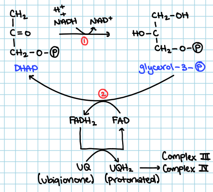
the use of cytosolic G3P DH to convert DHAP to G3P, using NADH and H+ and turning it into NAD+
G3P is converted back to DHAP by inner-membrane bound G3P DH, using FAD and turning it into FADH2. also using UQ to turn FADH2 to FAD and converting UQ to UQH2
are there any H+ translocated at the first step of DHAP conversion to G3P using membrane-bound G3P DH?
there are NO H+ being translocated, BUT 2e- are introduced to the RET
after the conversion of UQ to UQH2, what complexes are used and how many H+ and/or ATP are used?
Complex III 6 H+
Complex IV 1.5 ATP
ubiquinone
ubiquinol
UQ
UQH2
what gives NADH in the cytosol?
glycolysis
what must be available for the two FAD-dependent shuttles of the cytosolic NADH dilemma?
oxygen
draw the G3P shuttle
malate-aspartate shuttle
ims/cytosol: aspartate is converted into oxaloacetate using alpha-ketoglutarate and turning it into Glutamate. NADH is then turned into NAD+, converting oxaloacetate into malate
there are shuttles in the inner membrane which allow the aspartate, alpha-ketoglutarate, and glutamate to travel from the ims/cytosol to the matrix.
matrix: malate from ims/cytosol is converted back to oxaloacetate by NAD+ to NADH and H+. then oxaloacetate is converted to asparatate by Glutamate to alpha-ketoglutamarate
purpose of the malate-aspartate shuttle?
to transport the e- from the cytosol to the matrix so they can be used for oxidative phosphorylation. NADH is impermeable to the inner membrane
what can be expected for the amount of NADH from the cytosol and in the matrix, from the malate-aspartate shuttle?
there is an equal amount in each
what can be expected for the ATP yield from the malate-aspartate shuttle?
no loss of ATP, yielding 2.5 ATP!
the malate-aspartate shuttle more susceptible to what?
equilibrium pressure
what is the P/O ratio of NADH? (P/O = ATP per NADH oxidized)
2.5
which complex is NOT involved in succinate oxidation?
Complex I
The P/O ratio for NADH generated in the cytosol is …
1.5 or 2.5, depending on how NADH into the cytosol processing happens
what element would be a problem for getting NADH in the cytosol via glycolysis?
cyanide
draw the malate-aspartate shuttle

draw the overall reaction of pyruvate carboxylase

biotin
a cofactor required by pyruvate carboxylase and enzymes that catalyzes other similar reactions. it is covalently bound to the enzymes that use it by way of a lysine residue
how many parts does pyruvate carboxylase occur in?
two parts
draw the pyruvate carboxylase mechanism

pyruvate carboxylase
a homotetramer, each subunit composed of four domains
Biotin Carboxylase (BC) Domain (1)
Biotin Carboxy Carrier Domain (1)
Carboxytransferase (CT) Domain (1)
PC Tetramerization Domain (1)
Biotin Carboxyl Carrier Domain (2)
Biotin Carboxylase (BC) Domain (2)
Carboxytransferase (CT) Domain (2)
PC Tetramization Domain (2)

biotin carboxyl carrier domain
transfers between the active site of the BC domain of the subunit 1 and the CT domain of subunit 2

fishing with biotin
transferring intermediates from one active site to another, the biotin carboxy carrier domain parks here and transfers over to biotin carboxyl group

what are the cofactors of RET?
NADH
flavins, FAD/FADH2, FMN/FMNH2
iron-sulfur clusters
ubiquinone (Coenzyme Q)
hemes
copper
NADH cofactor of RET
only appears at the first step of RET, i.e., when the e- transfer to Complex I
HYDRIDE-ONLY DONOR (2e- transfer only H:-)
flavins, FAD/FADHS, FMN/FMNH2 cofactor of RET
serve as adaptors between H:- ONLY DONORS and 1 e- ONLY ACCEPTORS
ACCEPTS H:- for direct and complete reduction
DONATES e- one-at-a-time to one e- only acceptors
FAD/FADHS, FMN/FMNH2 direct and complete reduction
H:- + H+ + FMN → FMNH2
H:- + H+ + FAD → FADH2
where does the direct and complete reduction of FMN and FAD occur?
NADH (complex I)
succinate (succinate DH/complex II)
G3P, membrane-bound G3P DH
fatty acyl-CoAs
draw the oxidation of FADH2, FMNH2
a relatively stable semiquinone radical allows for one-at-a-time e- transfer
flavins frequently donate to Fe/S cluster

iron-sulfur clusters (Fe/S)
one e- only acceptors/donors
DO NOT typically accept or donate e-
although have multiple irons per cluster, only one e- donated at a time
typically bound to proteins via Cys residues, with a special exception to Rieske-type Fe/S cluster
example of multiple irons per cluster
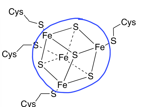
2Fe-2S, 4Fe-4S cluster
4Fe/4S cluster
3 FeIII / 1 FeII → oxidized
2 FeII / 2 FeII → reduced
ubiquinone cofactor of RET
highly hydrophobic, not associated with any one complex or protein
freely mobile within the mitochondrial inner membrane
acts as electron carrier between complexes
capable of one e- and two e- transfers, BUT in RET always one e- transfer
shuttles between one e- only donors and one e- acceptor
Fe/S clusters donate; Fe/S clusters or hemes accept
operates in a “Q cycle”
Q Cycle
as UQ is reduced by Fe/S clusters (e.g. Complex I), H+ must be delivered from somewhere, the matrix
as UQH2 is oxidized, H+ must be off-loaded somewhere, the ims
as UQ/UQH2 redox cycles, there is a net transfer of H+ across the inner membrane (matrix→ims)
draw the Q cycle conceptually

hemes cofactor of RET
prosthetic group for “cytochromes”
Fe-containing prosthetic group → cycles between ferric (FeIII) and ferrous (FeII) oxidation states
ONE e- DONORS/ACCEPTORS
DO NOT DONATE/ACCEPT H+
structure consistent w/ e- transfer properties
Cytochrome C → Heme C
Cyt a3
cytochrome C
heme C
“hexacoordinate”
precludes all binding of other “exogenous” ligands (e.g., O2)
typical arrangement for most hemes in RET
hexacoordinate
all six sites for ligand association are occupied will accomodate e- transfers Fe3+/Fe2+
Cyt a3
binds to its heme differently
“pentacoordinate”
open sites allows for direct binding of O2
open coordination site means that other ligands can also bind (e.g. CN-, N3-, CO)
“heme poisons”
molecules that can lock onto open coordination sites and refuse to let go on the cyt a3 heme
copper cofactor of RET
found only in complex IV
CuA and CuB
CuA
one e- transfer only
CuB
assists cytochrome a3 in O2 binding and reduction

molecule? circled?
nicotinamide adenine dinucleotide (NADH) (reduced)
where the hydride transfer occurs

molecule? circled?
flavin adenine dinucleotide (FAD)
the business end

molecule? circled?
flavin mononucleotide (FMN)
business end

molecule? circled?
ubiquinone (oxidized form)
business end

molecule? circled?
CuA center
business end

molecule? circled?
CuB center
business end

molecule? circled?
Rieske-type [2Fe-2S] cluster

molecule? circled?
[2Fe-2S] cluster
business end
molecule? circled?
[4Fe-4S] cluster
business end
what component does each flavoprotein have in common? ex: Complex I (NADH, coenzyme Q oxidoreductase), Complex II (succinate, coenzyme Q oxidoreductase), fatty acyl-CoA DH, G3P,
Flavins (FMN, FAD) and Fe/S clusters
what cofactor/s are ONLY hydride donors?
NADH
flavins, FAD/FADH2, FMN/FMNH2 (adaptors)
what cofactor/s are ONLY hydride acceptors?
NAD+
FAD/FMN
accepts H:- from H:- only donors to make FADH2/FMNH2
FADH2/FMNH2
transfers one e- (and H+) to a Fe/S cluster to make FADH./FMNH.
FADH. / FMNH.
transfers one e- (and H+) to Fe/S cluster to convert into FAD/FMN
OR
transfers one e- is donated by Fe/S cluster and pairs with FADH./FMNH. and it would pick up a free electron to become FADH2/FMNH2
UQ (CoQ) to UQH. (semiquinone radical/CoQH.)
one e- from a Fe/S cluster and one H+ from the matrix
UQ is fully oxidized
UQH. (CoQH.) to UQH2
one e- from Fe/S cluster and one H+ from the matrix
UQH2 fully reduced
UQH2 to UQH.
one e- accepted by Rieske Fe/S cluster and one H+ to IMS
UQH. to UQ
one e- accepted by heme and one H+ to the IMS
heme a
found in a-type cytochromes
has an aldehyde and a hydrophobic tail
heme b
aka Iron protoporphyrin IX
found in b-type cytochromes
no tail or covalently linked protein like hemes a and b
heme c
found in c-type cytochromes
has two covalently linked proteins by the Cys side chains

which heme is this?
heme a

which heme is this?
heme c

which heme is this?
heme b
why are proteins necessary for RET?
to translocate H+ across the membrane
so cofactors can be held in a close proximity to one another to faciliate e- transfer at sufficient rates
to reduce protential tuning
to prevent premature, incomplete, or uncontrolled reduction of O2
H+ translocation requires which kind of proteins?
transmembrane proteins
why must cofactors be held close together?
to faciliate sufficient transferance of e- at a sufficient rate
what is the typical Å (angstrom) separation between a donor and acceptor?
usually 14Å to produce a e- transfer rate of about ~2,000 per second
for every 1.7Å separation between donor and acceptor, how is the rate affected? what if the distance was 28Å?
the rate DECREASES by a factor of 10
at a distance of 28Å, the rate would be 1/day
how many e- transfers are needed from one end of the RET to the other?
about 30
reduction potential tuning
creates a thermodynamically favorable process step-to-step
“refers to the experimental or structural methods used to deliberately shift the standard reduction potential (E°) of a redox-active center to make it easier or harder to reduce or oxidize”
what tunes the reduction potential of cofactors?
the protein environment
what is an extreme example of a reduction potential tuning?
2Fe-2S (Cys) v.s. Rieske 2Fe-2S (His)
there is a 450 mV difference in potential
the regular clusteres come at the top while the Rieske come “much later”
what is the goal of the reduction of O2?
to make it all the way to 2H2O without the issue of uncontrolled, premature, or incomplete reduction of O2. complex IV makes sure this is possible.
draw the reduction of O2 to H2O2

what is the enzyme that is considered the second line of defense to prevent the incomplete/uncrontrolled reduction of O2?
the protein superoxide dismutase
2O2.- → H2O2 + O2
what is considered the third line of defense to prevent the incomplete/uncrontrolled reduction of O2?
“sacrificisl e- donors” Vitamin C and E
catalase
enzyme that converts H2O2 to H2O and O2
Complex II
aka succinate oxidoreductase, formal biochemical name: succinate:ubiquinone oxidoreductase
~140 kD; 4 subunits
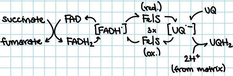
contains: FAD, 3 Fe/S clusters, and 1 heme b
there is no H+ translocation at this complex; however, e- are introduced at RET here
inhibited by thenoyltrifluoroacetone carboxin
how does FAD contribute to Complex II
covalently bound to the enzyme, it is a prosthetic group
is heme b playing a part in e- transfer in Complex II?
no, it is not playing a part in the e- transfer sequence. but it could be a possible defense against O2.- production
draw the catalytic cycle of succinate dehydrogenase/Complex II concerning RET?
what does thenoyltrifluoroacetone carboxin do?
it blocks UQ from binding and reducing
Complex III
biochemical name: ubiquinol:cytochrome C oxidoreductase
500 kD; 2 sets of 11 subunit multimers
one set extends into the mitochondrial matrix
another set extends into the IMS
each 11 multimer set contains a Riekse Fe/S cluster
cytochrome C
there are 4 H+ translocated across the inner membrane for each e- pair transferred
inhibitted by: myxothiazol, antimycin A, stigmatellin
how many Å does Complex III extend into the mitochondrial matrix?
75Å, so it extends more into the matrix than it does the IMS
how many Å of Complex III extends into the IMS?
40Å
what cofactor binds to Complex III in the IMS portion?
cytochrome C
membrane-spanning region of Complex III
each has 11-subunit multimer which contain two UQ/UQH2 binding sites
QP and QN sites
QP
Q “positive,” on the IMS side of the membrane
PIMS