Biology 4U Exam
 Call Kai
 Learn
 Practice Test
 Spaced Repetition
 Match
 Flashcards
 Knowt Play
1/47

There's no tags or description
Looks like no tags are added yet.
48 Terms
OH
Hydroxyl: They make molecules polar which increases the solubility and reactivity of molecules
E,g Glucose, Ethanol
COOH
Carboxyl: Can release H+ ions which can make molecules acidic
E,g - Acetic Acid (vinegar)
NH2
Amino : Can accept H+ ions which makes molecules basic
E,g Amino acids ( leucine, alanie, etc)
SH
Sulfhydryl: They make molecules polar which increases the solubility and reactivity of molecules. They form disulfide bridges
PO4
Phosphate: Can release H+ ions which can make molecules acidic and negatively charged
E,g DNA, ATP
CO
Carbonyl- Ketone: They make molecules polar which increases the solubility and reactivity of molecules
E,g Fructose, Acetone
COH
Carbonyl- Aldehyde: They make molecules polar which increases the solubility and reactivity of molecules
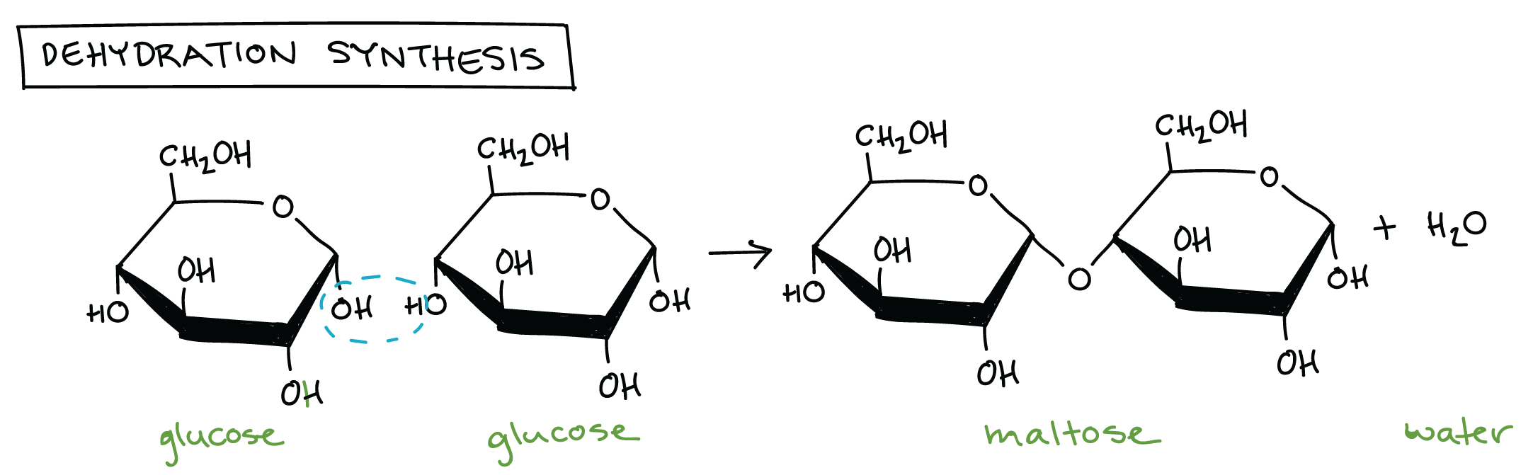
Dehydration Synthesis Reaction
The removal of water to join monomers into polymers. H and OH are removed from monomers
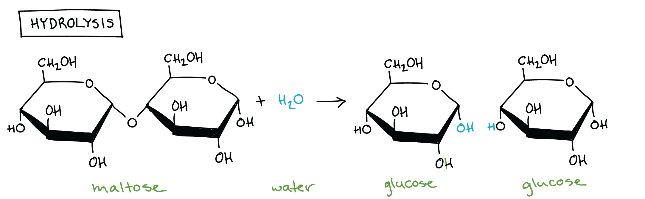
Hydrolysis
The use of water to split polymers into monomers.
Carbohydrates Function and Structure
Organic molecules, that are used for short-term energy storage, energy, cell building
Lipids Function and Structure
Large chains of non-polar hydrocarbons, most are insoluble due to large hydrophobic regions.Main function is long-term energy storage
Fats/Oils: triglycerides = glycerol + 3 fatty acid chains. Unsaturated = liquid, carbon linked with double bonds causing bends. Saturated = solid, carbon linked with singe bonds
Phospholipids: glycerol + 2 fatty acids + phosphate group, hydrophilic head, hydrophobic head e,g cell membrane
Steroids: four carbon rings and a functional group e,g cholesterol
Waxes- carbon or alcohol + large, long chains of fatty acids, waterproof coating
Protein Structure and Function
Very complex structure with there being 3000 types in the human body. e,g hemoglobin, keratin, enzymes, antibodies
Primary Structure: polypeptide chain Secondary Structure: helix or pleated sheet caused by hydrogen bonds Tertiary Structure: complex folding similar to a large knot caused by hydrogen bonding, disulfide bridges Quaternary structure: multiple tertiary structures linked together
Monosaccharides
Monomers also known as simple sugars. They are metabolized during cellular respiration to create energy (ATP). There are triose, pentose and hexose sugars. They are long carbon chains that contain a hydroxyl group (OH) and a carbonyl ( CO, COH). In a dry state they are linear but when they are dissolved in water OH on 5 reacts with COH group on on 1 and a closed ring is formed. If OH is below the plane than that is alpha if above that is beta. e,g glucose, galactose, fructose
Disaccharides
Two monosaccharides joined together through dehydration synthesis. They are used for short-term energy storage and hydrolyzed into simple sugars when energy is needed e,g sucrose, lactose, maltose
Polysaccharides
Long chains of monomers They can be used for structural support and as energy storage.
Storage:
Glycogen- found in animals hydrolyzed in liver and muscles cells during physical exercise. Has more branches than starch and is similar to amylopectin
Starch- mixture of amylose and amylopectin found in plants stored in specialized structures like tubers, taproots,and fruits of grains.
Support:
Chitlin- made up of glucose with nitrogen containing group on the second carbon. This substance is found in fungi walls, exoskeleton of invertebrates
Cellouse- made up of beta glucose monomers . OH groups on parallel chains form hydrogen bonds that create tight bundles (microfibrils) that intertwine to form tough fibres that cannot be digested by humans
Amino Acids
monomers that contain an amino group, a carboxyl group, and an R group which determines its properties. Peptide chains are created through dehydration synthesis
Nucleotides Structure and Function
Monomers of nucleic acids that contain 1-3 phosphate groups, a pentose sugar, and a nitrogenous base. e,g DNA, ATP, RNA
Cell Membrane Structure
Phospholipid- fluid portion (not bonded together) with hydrophilic heads and hydrophobic tails
Proteins- two types, integral- bound to hydrophobic interior and can span the entire bilayer. peripheral- bound to hydrophilic exterior of the membrane (smaller)
Carbohydrates- can attach to proteins and phospholipids to become glycoproteins and glycolipids
Cholesterol - located through the bilayer between phospholipids increases the fluidity of the membrane
Cell Membrane Functions
Phospholipids- acts a barrier separating the inside of the cell from the outside , also regulates movements in and out of the cell
Glycoproteins- allows communication between cells
Proteins- regulates biochemical reactions
Cholesterol- creates attachments between cells
Cell Membrane Proteins
Structural Support Attachment- anchors the cell membrane to inner cytoskeleton, proteins outside the cell and proteins from other cells
Recognition and Communication- Receptors(proteins) have specific binding sites which identify other cells to it. Glycoproteins serve as a chemical identification tags.
Regulates biochemical reactions- receptor proteins (integral) that trigger cellular responses upon the binding of certain molecules (hormones, etc)
Chanel- allows allows materials to pass through cell
Endocytosis
Membrane proteins use cellular energy (ATP) to move molecules and ions against their concentration gradient. This is usually through pumps that have a binding site for ATP
Cells ingest materials trapped by a vesicle that is made from the membrane. Materials are then digested by lysosomes inside the cell
Pinocytosis- liquid engulfed
Phagocytosis-, solid particles engulfed
Receptor- mediated- receptor proteins detect molecules which creates dimples in the membrane
Exocytosis
The of energy to expel waste. Vesicles containing waster fuse with the membrane allowing materials to be released out of the cell
Enzyme Function
They are protein catalysts so they speed biochemical reactions without being consumed in the process. They lower that activation energy which allows reactants to reach the transitional stage. The enzyme’s substrate would be the reactants of a chemical reaction. Enzymes also aid in the breaking a part molecules and joining them together
Enzyme Structure
Enzyme structure is dependent on the shape of its substrate. Enzymes have binding sites that are compatible with with substrates. The specific binding site for the substrate is called the active site. Enzymes will also change shape to be more compatible with substrates which is an induced-fit model.
Competitive and Noncompetitive Inhibitors
C: similar shape to substrate and binds to active site which prevents substrate from binding
N: binds to another site on an enzyme that changes that shape of the enzyme
Glycolysis
cytoplasm, glucose is split, rearranged, oxidized, phosphate is added and removed
Reactants: glucose ATP ,ADP, NADH
Products- NAD+, ATP, ADP +Pi, Pyruvate H2O
Pyruvate Oxidation
matrix, harness energy, oxidation, carbon is released and carboxyl group is removed
Reactants: NAD+, CoA, Pyruvate
Products, CO2, Acetyl-CoA, NADH
Citric Acid Cycle
maxtrix, harness energy- all carbon is released and glucose is oxidized
Reactants- NAD+, Acetyl-CoA, FAD+, ADP, GDP
Products, NADH, CO2, FADH, ATP, GTP
Protein Catabolism
The definition of starvation when an organism breaks down amino acids for energy. The amino group is removed and released as ammonia a waste product. the rest of the structure can be converted into molecules that are used in cellular respiration
e,g leucine —Acetyl-CoA
Alanine— Pyruvate
Lipid Catabolism
Animals build up fat stores when food is unavailable. Triglycerides are broken apart into glycerol + fatty acids. Glycerol can be converted into DHAP or glucose and go through glycolysis. Fatty acids go the matrix and have their acetyl groups removed by enzymes and combined with CoA to create Acetyl-CoA. Which can go through the CAC. Fat’s produce 2x the amount of energy as carbs for equivalent mass
Ethanol Fermentation
NADH gives its hydrogen atoms to Acetaldehyde( pyruvate after CO2 is released) causing ethanol to form. NAD+ is oxidized and can back to glycolysis. Yeast do this which is why they are used in creating alcoholic beverages.
Lactic Acid Fermentation
Occurs in animals and humans during strenuous exercise. Muscle cells need to produce ATP faster than oxygen supplied. So NADH from glycolysis will give its hydrogen atoms to pyruvate in cytoplasm which form lactate. Lactate can cause muscle pain. Lactate can be transported to the l
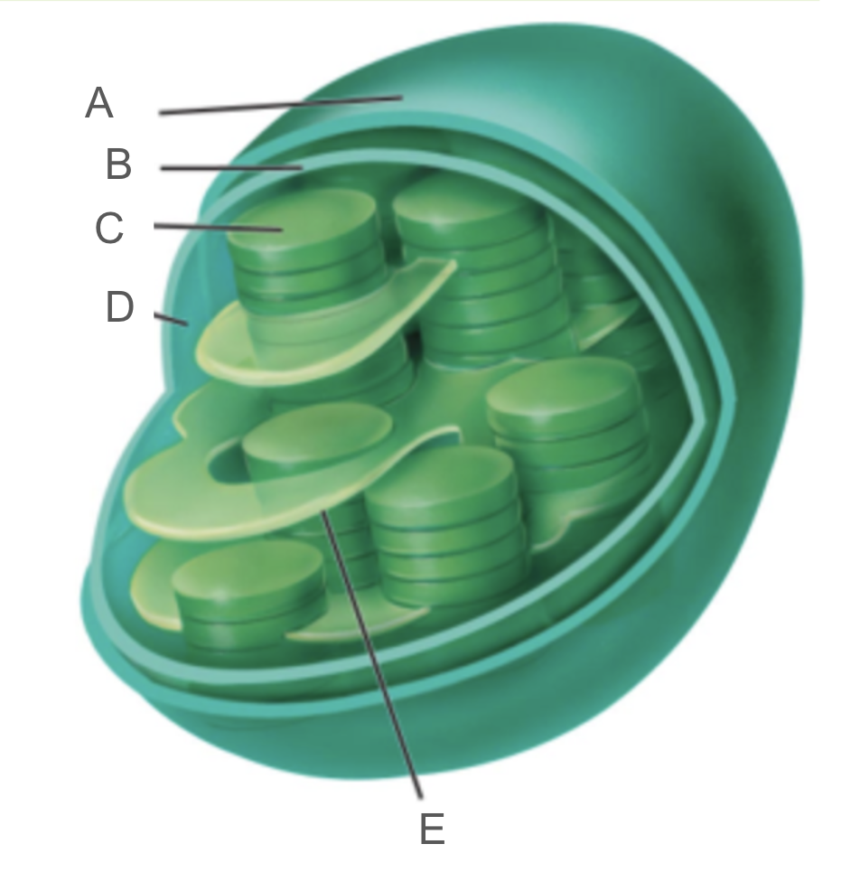
Chloroplast
A: Outer membrane
B: Inner Membrane
C: Thylakoid
D: Stroma
Light Reactions
A photon hits photosystem II causing a molecule inside called P680 to become excited. It then gives its electrons to a primary electron acceptor. This molecule is now positive and extremely electronegative and strips hydrogen atoms (electrons) from water which is why oxygen is a product. One water molecule produces 2 electrons which replace the electrons that were lost from P680. Two electrons that were produced = two photons. The electrons than go through several redox reactions. They go from PQ— cytochrome oxidase—PC and finally they reach photosystem I. The energy is low at this point so a photon strikes the molecule P700 in photosystem I which cause it to become excited. It’s electrons are then transferred to ferredoxin. Electrons combine with protons in the stroma and turn NADPH into NADP+ through NADPH reductase. Through this process an electrochemical gradient develops and ATP is produced through ATP synthesis which is powered by protons going with their concentration gradient
Reactants
12 NADP+, 12 H20, ADP + Pi, 48 photons
Products
12 NADPH, 6 O2, ATP
Calvin Cycle
CO2 is added to RuBP a 5 carbon molecule through the enzyme rubisco. The molecule splits into 3PG. ATP also splits into ADP +Pi which provides energy. NADPH is oxidized turning into NADP+ and gives its electrons to the carbon molecules which create G3P. 1 G3P molecule exits the cell. The other molecules are rearranged into RuBP and and cycle repeats, In order for 1 glucose to be built the cycle must run twice
Reactants
18 ATP, 6 CO2, 12 NADPH
Products
18 ADP + Pi, 1 glucose, 12 NADP+
Photorespiration
Occurs when oxygen binds to rubisco’s active site instead of CO2 which causes 3PG to not be created disrupting the whole calvin cycle. This causes less glucose to be made and usually happens in happens in hot dry weather when stomata are closed. If the stomata are closed to retain water they take in less CO2 especially in the sunlight.
Cam and C4 plants
C: open stomata at night to retain water. This means that they cannot take in CO2 and receive light at the same time. So instead, these plants take in CO2 at night and store them in C4 organic acids. The acids release the CO2 during the day so that CO2 can enter the Calvin Cycle and receive light at the same time
C4: CO2 attaches to PEP which creates oxaloacetate that is converted into malate. Malate leaves the mesophyll cells and enters the bundle sheath cell due to the concentration of CO2 being higher. CO2 will detach which turns malate into pyruvate. Pyruvate is converted into PEP and goes to the mesophyll cells
Photosynthesis and Cellular Respiration
DNA structure
DNA contains a phosphate group, pentose sugar, and a nitrogenous base. DNA is double stranded and runs antiparallel to each other. The 3’ end is the end that ends in sugar and the 5’ end is the end that ends phosphate group. The stands are connected through hydrogen bonds that complementary base pairs create
DNA Replication
DNA replicates semiconservatively meaning that each daughter cell will receive one strand from a parent cell. The strand given is used as a template to create a new strand. The strand first has to be broken apart before it can be used as a template. DNA helicase unwinds the double helix by breaking the hydrogen bonds between complementary base pair. SSBs are added to the exposed strand so that the bonds don’t reform. Replication happens at multiple sites along the DNA strand which causes replication bubbles. RNA primer is added to the strands so that DNA polymerase III can add the complementary nucleotides to the strand . DNA polymerase can only add nucleotides in the 5’ to 3’ direction which creates a leading strand and a lagging strand. The leading is the 3’ to 5’ strand and is a continuous strand. On the lagging strand DNA polymerase III attaches the nucleotides in the opposite direction that DNA helicase is moving. The cause the strand to be built in sections called Okazaki fragments. DNA ligase joins these fragments through phosphodiester bonds. DNA polymerase I removes RNA primer and and replaces it with the correct nucleotides.
PCR
Polymerase Chain Reaction is a lab technique used to replicate specific regions of DNA. It occurs in three steps
Denaturation: temperature is very high (94-96 C). This stage last about 20-40 seconds, the high temperature causes the hydrogen bonds between DNA strands break
Annealing (50-56 C) occurs at a lower temperature so that enzymes can be used without be denatured. DNA primers are added to the stand facing the target sequence in the 5’ to 3’ end so that DNA polymerase III can attach nucleotides to the strand
Elongation( 72 C) DNA polymerase adds nucleotides to the template strand to build the new strand. A specific enzyme needs to be used called Taq polymerase due to the high temperature. The step results in two double stranded DNA molecules. Each step takes about 20-40 seconds which means the PCR only takes 5 minutes in total. The cycle repeats about 30-40 times. After cycle 2, 4 copies of DNA have been creates, after cycle 3, 8 copies of the target sequence has been created with o extra nucleotides
Homeostasis
process that maintains a constant internal environment despite changes to external environment. For the system to work in needs constant feedback
Negative Feedback- mechanism is activated to restore conditions to their original state .
Stimulus— Sensor—Integrator—Effectors— Response
Positive Feedback- encourages and reinforces change and small effect is amplified
Filteration
The high amount of pressure in the glomerulus forces small organic molecules in the blood to move from capillaries to outside the thick knot. Bowman’s capsule surrounds the glomerulus and receives the fluid
Reabsorption
The fluid exits Bowman’s capsule and enters the proximal tubule where most reabsorption occurs . Nutrients diffuse out of the tubules back into the blood. Due to osmosis water will follow do water is reabsorbed as well. At the same time a concentration gradient is being created in the loop of Henle using salt and osmosis. The highest solute concentration is in the inner medulla
Secretion and Concentration
Any waste products like urea, ammonia, nicotine,etc are forced out of the blood into proximal and distal tubules due to active transport because the concentration of waste is higher in the blood than the tubules
Urine leaves distal tubules and enters collecting ducts. The urine goes down the renal pelvis to the ureters and bladder. As it goes down collecting ducts water leaves the ducts in response to concentration gradient concentrating the urine
Regulating Blood Pressure
kidneys release renin into the bloodstream which catalyzes the formation of the hormone angiotensin. It can stimulate ADH causes water to be reabsorbed by distal tubules and collecting ducts, cause arterioles to constrict which increases blood pressure, it can stimulate the proximal tubules to absorb salt causing water to follow due to osmosis
Hormones
Pituitary is the master gland because it secretes hormones that controls most endocrine glands
Protein- made up of amino acids, hydrophilic and target receptors on the outside of cells
Steroid- made up of cholesterol, hydrophobic, targets receptors on the inside
Specific Hormones
Prolactin, anterior pituitary, mammary glands, aids in breast development and milk production
Endorphins, anterior pituitary, pain pathways in the nervous system, inhibits the perception of pain