cell bio 4
 Call Kai
 Learn
 Practice Test
 Spaced Repetition
 Match
 Flashcards
 Knowt Play
1/100

There's no tags or description
Looks like no tags are added yet.
101 Terms
what is the pulse-chase experiment?
Pulse: Add radioactive amino acids to label newly made proteins
Chase: Add nonradioactive amino acids and follow the labeled proteins over time
What pathway did Jamieson and Palade discover?
the secretory pathway: RER → Golgi → secretory vesicles → outside the cell.
Why observe silver grains (trackers) in the mitochondria or ER at later time points?
Silver grains show radioactive proteins
ER labeling indicates proteins entering the secretory pathway. Mitochondria are not part of this pathway
How would you interpret Table II? What conclusions can be drawn?
Compare silver grain distribution over time (observing where the radioactive proteins are located at diff times after made)
Labeling moves from RER→ Golgi → Secretory granules → outside of cell, showing the order of protein transport.
How would you interpret the graph? What is the pathway of secretory proteins?
Radioactive labeling moves from the RER → Golgi → secretory granules
What are yeast secretory mutants?
Yeast cells with mutations that prevent normal protein secretion
How did yeast sec mutants help understand protein transport?
they caused proteins to become stuck at different transport stages, helping scientists identify proteins needed for secretion
How was the mechanism of protein transport uncovered?
Through yeast mutants, pulse-chase experiments, microscopy, and cell-free experiments that identified transport proteins and vesicles
What are microsomes?
Small membrane vesicles formed when the ER breaks apart during cell preparation
What are polysomes?
Multiple ribosomes translating the same mRNA at the same time
What are stripped microsomes?
Microsomes without ribosomes but with their membranes and insertion machinery remaining
What experiment demonstrated that proteins enter the RER lumen co-translationally?
Cell-free translation with microsomes and protease protection
Proteins were protected from proteases when microsomes were present during translation, showing they entered while being made
What is the signal sequence (SS)?
A short amino acid sequence that directs a protein to the rough ER
What is a nascent peptide?
A growing protein chain still attached to a ribosome
What experiment demonstrated that the signal sequence is cleaved co-translationally?
Blobel and Dobberstein compared protein translation with and without microsomes
Microsomes present during translation produced a shorter protein because the signal sequence was removed
How to interpret Blobel and Dobberstein (1975) Gel?
The upper band is the larger precursor protein with its signal sequence
The lower band is the smaller mature protein after cleavage
why is there a strong lower band?
It represents the mature protein after the signal sequence has been removed
Why is there an upper band at later time points in gel?
It represents protein that still has its signal sequence because it was not processed during translation
thicker band = larger protein
thicker band = still has signal sequence in it
What would the gel look like if cleavage happened post-translationally?
The upper band would appear first, followed by a lower band after translation finishes and the signal sequence is removed
What are the key players in ribosome-ER docking?
Signal sequence, SRP, SRP receptor, ribosome, and translocon
What is the function of SRP (Signal Recognition Particle)?
Recognizes the signal sequence, temporarily pauses translation, and directs the ribosome to the ER
What is the function of the signal sequence?
Acts as an address label that directs proteins to the ER
How can you demonstrate that the signal sequence is necessary?
Remove or mutate it
If the protein no longer enters the ER, the sequence is necessary
How can you demonstrate that the signal sequence is sufficient to direct protein to RER?
Attach it to a protein that normally stays in the cytoplasm (doesn’t enter RER)
If that protein enters the ER, the signal sequence contains enough information to direct the protein to the RER.
What happens if you remove or add a signal sequence?
Remove: Protein usually cannot enter the ER
Add: Protein can be directed to the ER
What is the mechanism of SS and SRP recognition and binding?
Ribosome begins making a protein.
Signal sequence emerges and binds SRP.
Translation temporarily pauses.
SRP brings the ribosome to the ER receptor.
Ribosome attaches to the translocon.
SRP leaves, and translation continues into the ER.
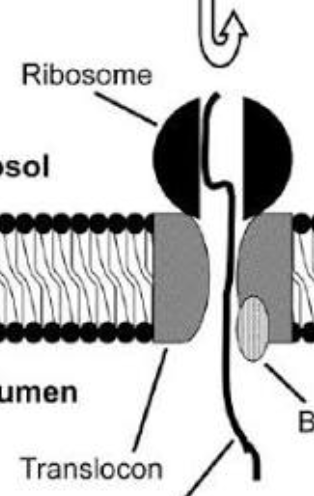
What is a translocon?
a protein channel in the rough ER membrane that allows newly made proteins to enter the ER
Is the translocon open or closed without a translocating protein?
Normally closed to prevent substances from leaking across the ER membrane
How would you test the translocon model?
Block or disrupt the translocon and use protease protection to test whether proteins can still enter microsomes
What is the secretory pathway?
RER → Golgi → vesicles → plasma membrane or outside the cell
What are integral membrane proteins? How are they inserted into the ER?
Proteins embedded in membranes
They enter through the translocon using specific signal-anchor and stop-transfer sequences.
What are the different types of IMP?
Type I, Type II, Type III, Type IV (multipass), and tail-anchored proteins
How is each type inserted into the ER?
Type I: N-terminus enters the ER lumen; stop-transfer sequence anchors the protein.
Type II: Internal signal-anchor; N-terminus faces the cytoplasm.
Type III: Internal signal-anchor in the opposite orientation from Type II.
Type IV: Multiple start-transfer and stop-transfer sequences create multiple membrane crossings.
Tail-anchored: C-terminal membrane anchor is inserted after translation.
Can IMP insertion also be post-translational? What is the mechanism?
Yes.
Tail-anchored proteins use the GET/TRC pathway to reach and insert into the ER membrane after translation.
How do you determine IMP topography during RER biosynthesis?
Determine which regions face the cytoplasm, ER lumen, or membrane using signal sequences, stop-transfer sequences, protease protection, and glycosylation experiments.
What co- and post-translational processes occur in the RER?
Co-translational: Protein entry, signal sequence cleavage, and N-linked glycosylation
Post-translational: Folding, disulfide bond formation, and quality control
What are the steps of O-linked glycosylation? Are they co- or post-translational?
Protein is made first.
Protein travels to the Golgi.
Sugars are added one at a time to serine or threonine amino acids
all steps are post-translational
What is the function of glycosylation?
(attaching carbohydrates (sugars) to proteins/lipids)
Helps proteins fold, stay stable, avoid breakdown, and participate in cell recognition and signaling.
adding sugars helps proteins function properly
How are proteins folded?
Their amino acid sequence determines their shape
Chaperones help them fold correctly, and disulfide bonds stabilize their structure
What are chaperones?
Proteins that help other proteins fold correctly and prevent clumping
What are the functions of BiP, PDI, and Calnexin and Calreticulin?
BiP: Helps protein folding.
PDI: Forms and rearranges disulfide bonds.
Calnexin and Calreticulin: Help glycoproteins fold correctly.
What diseases can result from improper protein folding?
Alzheimer's, Parkinson's, prion diseases, and cystic fibrosis
What is the structure and function of the Golgi?
Flattened membrane sacs called cisternae that modify, sort, and package proteins.
think protein post office
What is the default pathway of protein transport?
ER → Golgi → vesicles → plasma membrane → outside the cell
What is retrograde protein transport?
Backward transport, such as Golgi → ER, to recycle proteins and return misplaced ER proteins
How would you use pulse-chase to observe Golgi compartments?
Label proteins and track them over time as they move through the cis, medial, and trans Golgi.
What are the functions of different Golgi stacks?
Cis: Receives proteins from ER.
Medial: Modifies proteins and carbohydrates.
Trans: Finishes modifications and sorts proteins.
What are the two models for cargo transport through the Golgi?
Vesicular transport: Cargo moves between stationary Golgi cisternae in vesicles.
Cisternal maturation: Cisternae mature from cis to trans while carrying cargo.
What are the pros and cons of vesicular and cisternal?
Vesicular: Explains transport between compartments but does not fully explain movement of large cargo.
Cisternal maturation: Explains large cargo movement but requires Golgi enzymes to recycle backward.
What might be the actual model for transport?
Evidence supports cisternal maturation, but cells can use both mechanisms.
What are recycling and exocytic routes?
Recycling: Returns proteins to earlier compartments or the cell membrane.
Exocytic: Moves proteins toward the plasma membrane or outside the cell
What are transport vesicles?
Small membrane sacs that carry proteins and lipids between organelles
What are coat proteins?
Proteins that help select cargo, bend membranes, and form transport vesicles
How does the cell selectively sort cargo?
Receptors recognize sorting signals on proteins and package them into the correct vesicles
Why are sorting signals important?
They act as address labels that direct proteins to the correct destination
How do vesicles bud and fuse with target membranes?
Cargo is selected.
Coat proteins bend the membrane.
Vesicle buds and separates.
Coat is removed.
Vesicle reaches its target.
SNAREs help membranes fuse.
What are SNAREs?
Proteins that help vesicles attach and fuse with the correct target membrane
v-SNAREs are on vesicles; t-SNAREs are on target membranes.
think guides
What are examples of sorting mechanisms?
KDEL for ER proteins, M6P for lysosomal enzymes, and tyrosine-based or dileucine signals for membrane proteins.
What are the functions of COPI and COPII?
proteins that help transport materials in vesicles between the ER and Golgi
COPII: ER → Golgi.
COPI: Golgi → ER and transport between Golgi compartments
What are important sorting signals?
KDEL, KKXX, mannose-6-phosphate (M6P), tyrosine-based signals, and dileucine signals.
What is the importance of KDEL?
A four-amino-acid sequence that returns soluble ER-resident proteins from the Golgi to the ER.
Why do cell compartments have different pH levels?
Different pH levels control enzyme activity, protein binding, and cargo release
What happens if KDEL is removed from an ER protein?
The protein may escape the ER and be secreted
What happens if KDEL is added to a secretory protein?
It may be returned to the ER instead of being secreted
What are other sorting signals for ER-resident proteins?
KKXX, which helps return ER membrane proteins through COPI vesicles
What is the sorting signal for lysosomal proteins/enzymes?
Mannose-6-phosphate (M6P), which sends the lysosomal enzymes to lysosomes
What are the two models for resident Golgi proteins to sort into correct cisternae?
Vesicular recycling: Enzymes move backward in vesicles to their correct cisternae.
Cisternal maturation: Enzymes recycle backward as cisternae mature forward
What is the endocytic pathway?
process cells use to bring materials from outside the cell into the cell through membrane-bound vesicles
nonspecific endocytosis
receptor-mediated endocytosis
Nonspecific: Takes in extracellular fluid and dissolved materials.
Receptor-mediated: Uses specific receptors to take in selected molecules
think broad selection vs picky selection
What is phagocytosis?
A type of endocytosis where cells engulf large particles, such as bacteria and dead cells
What are clathrin-coated pits?
Membrane areas covered in clathrin that help collect cargo and form vesicles
What are the steps of phagocytosis?
Cell recognizes and attaches to a particle.
Membrane surrounds the particle.
Phagosome forms.
Phagosome fuses with lysosome.
Enzymes digest the material.
What materials enter through phagocytosis? What is their fate?
Bacteria, dead cells, and debris
They are usually broken down in lysosomes
Which bacteria can survive phagocytosis?
Mycobacterium tuberculosis: Prevents normal lysosome fusion.
Salmonella: Survives in a modified vacuole
Listeria: Escapes into the cytoplasm.
What are the major organelles of endocytosis?
Plasma membrane, early endosome, recycling endosome, late endosome, and lysosome
What is the endocytic pathway?
Plasma membrane → early endosome → recycling endosome or late endosome → lysosome
Why is pH important in endosome maturation?
Endosomes become more acidic, causing receptors to release cargo and helping activate enzymes
Why is endosome recycling important? What happens to material not recycled?
Recycling allows the cell to save and reuse important materials, such as membrane proteins and receptors, instead of making new ones
Material not recycled usually goes to lysosomes for breakdown.
What is a lysosome? What is its function?
A membrane-bound organelle containing digestive enzymes that break down proteins, lipids, bacteria, and cellular waste.
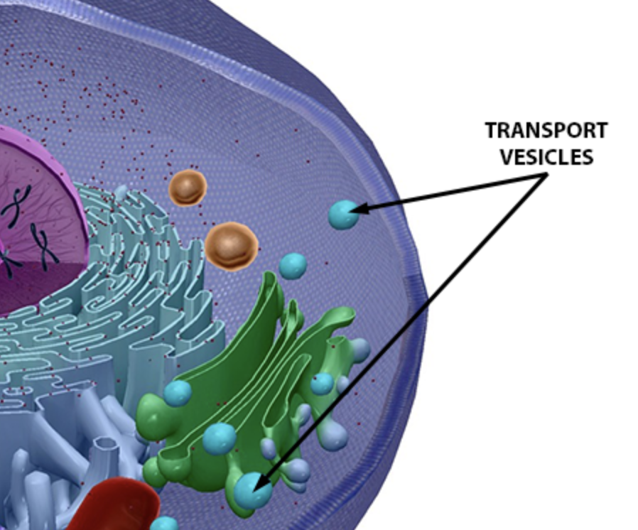
What is the function of transport vesicles/coated pits?
Collect and transport selected materials into, out of and inside the cell
What is the structure of a clathrin-coated vesicle?
A membrane vesicle surrounded by a cage-like clathrin coat containing selected cargo and adaptor proteins.
What other machinery is involved in clathrin-mediated endocytosis (CME)?
(CME = a process where a cell brings materials into the cell using vesicles coated with a protein called clathrin)
Clathrin, adaptin/AP2, dynamin, actin, Rab proteins, and SNAREs
What are the steps of clathrin-coated vesicle formation?
Cargo binds its receptor.
AP2 recognizes the sorting signal.
Clathrin forms a coat.
Membrane bends inward.
Dynamin pinches off the vesicle.
Clathrin coat is removed.
Vesicle delivers cargo to an early endosome.
What signal is recognized by the adaptin complex?
Sorting signals on the receptor's cytoplasmic side, such as tyrosine-based YXXΦ or dileucine signals.
How would you test whether this sequence is crucial for binding?
Mutate or remove the sequence
If adaptin can no longer bind properly, the sequence is important.
What is the function of dynamin?
A GTPase that uses GTP energy to help pinch off vesicles from the plasma membrane
What is the mechanism of vesicle fusion? What are early and late endosomes?
Rab and SNARE proteins help vesicles reach and fuse with the correct compartments.
Early endosomes sort cargo
late endosomes send material toward lysosomes
How can you visualize each step of endocytosis?
Use fluorescently labeled cargo or proteins and follow them with fluorescence microscopy
What is the role of GFP in visualizing proteins involved in endocytosis?
GFP is attached to a protein to make it glow, allowing scientists to track its location and movement.
What receptors enter through CME?
LDL receptors, transferrin receptors, EGF receptors, and some hormone receptors.
What is the role of pH?
Acidic pH helps separate receptors from cargo and directs materials toward recycling or lysosomal breakdown.
What are the steps of LDL transport through CME? What proteins are involved?
LDL binds the LDL receptor.
AP2 recognizes the receptor's sorting signal.
Clathrin forms a coated pit.
Dynamin pinches off the vesicle.
Coat is removed.
Early endosome acidifies and separates LDL from its receptor.
Receptor recycles to the plasma membrane.
LDL moves to the lysosome and is broken down.
What is LDL and why is it important?
Low-density lipoprotein carries cholesterol through the blood to cells for membrane and hormone production.
Why does LDL need to end up in the lysosome?
Lysosomal enzymes break down LDL and release cholesterol for the cell to use.
What happens to the LDL receptor?
It is usually recycled back to the plasma membrane to take in more LDL.
What is the important sorting signal on the LDL receptor?
NPXY, a cytoplasmic sorting signal that helps the receptor interact with adaptor proteins
How would you interpret a graph of LDL uptake and kinetics?
LDL uptake increases with LDL concentration until receptors become saturated
Defective or fewer receptors decrease uptake.
increasing uptake : more LDL
slows down: increasing slowly, receptors are busy
plateaus: receptors are full, uptake is maxed out
Could you use pulse-chase to visualize LDL uptake and kinetics?
Yes. Label LDL, allow cells to take it up, then add unlabeled LDL and track the labeled LDL through endosomes and lysosomes.
How would different mutations affect LDL uptake and kinetics?
LDL receptor mutation: Less LDL binding.
AP2 mutation: Poor cargo recognition.
Clathrin mutation: Poor vesicle formation.
Dynamin mutation: Vesicles cannot separate properly.
Acidification defect: LDL and its receptor may not separate properly.
Why can mutations in LDL uptake or the LDL receptor cause heart disease?
LDL cannot be removed efficiently from the blood, causing high LDL levels and plaque buildup in arteries (atherosclerosis).