H14: Brain CSF
 Call Kai
 Learn
Learn Practice Test
 Spaced Repetition
 Match
Match Flashcards
 Knowt Play
1/31

There's no tags or description
Looks like no tags are added yet.
Name | Mastery | Learn | Test | Matching | Spaced | Call with Kai | Chat |
|---|
No analytics yet
Send a link to your students to track their progress
32 Terms
ventricle
cavities in the brain
lateral ventricles
2
Cavity located in the interior of each cerebral hemisphere; thus there
are 2 lateral ventricles.
The 2 lateral ventricles are the largest of the ventricles
both lateral ventricles are separated by septum pellucidum
Each communicates with the third ventricle by the interventricular
foramen of Monro
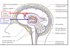
the lateral ventricles are the ________
largest
septum pellucidum
thin membrane that separates lateral ventricles

interventricular foramen of Monro
connects lateral ventricles to third ventricle

third ventricle
1
Narrow, located between the thalamius of each side
Communicates with the fourth ventricle by the cerebral aqueduct of Sylvius (which is
located in the midbrain)
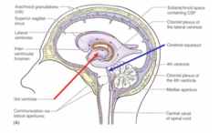
cerebral aqueduct of sylvius
connects 3rd and 4th ventricles

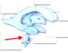
fourth ventricle
1
Cavity located in the pons and medulla
Is continuous with the central canal of the medulla, which in turn, is
continuous with the central canal of the spinal cord
Opens by means of 1 median and 2 lateral aperatures into the
subarachnoid space.
These aperatures are the only means by which
the cerebral spinal fluid formed in the ventricles enters the
subarachnoid space.

fourth ventricle opening
Opens by means of 1 median and 2 lateral aperatures into the
subarachnoid space.
Aperature
an opening or hole
aperature function in fourth ventricle
These aperatures are the only means by which
the cerebral spinal fluid formed in the ventricles enters the
subarachnoid space.
fourth ventricle foramina
2 Luschka
1 Magendie
luschka foramen
2 in the fourth ventricle
lateral

magendie foramen
1 in the fourth venticle
medial
Cerebral Spinal Fluid (CSF)
a clear, colourless, acellular
fluid, similar to blood plasma
choroid plexus
certain sites in the ventricles are lined with this
(EXTRA: modified pia mater together with modified
ependymal cells)
produces CSF

functions of CSF
1.To cushion and support the brain and
spinal cord
2. As a lymphatic system
3. As an environment for neurons
what produces CSF
choroid plexus
CSF Flow SUMMARY
Lateral Ventricle -> Interventricular Foramen of Monroe -> 3rd Ventricle -> Cerebral Aqueduct of Sylvius -> 4th Ventricle -> 2 Foramen of Leuscka and Foramen of Magendie -> subarachnoid space around brain and spinal cord-> Arachnoid Granulations -> Superior Saggital Sinus -> venous system, Away from brain
CSF Flow: lateral ventricles
CSF is formed in the lateral ventricles by the choroid plexus
after it is passed to interventricular foramen of Monro to the 3rd ventricle
CSF Flow: interventricular foramen of Monro
receives CSF from lateral ventricles and brings it to 3rd ventricle
CSF Flow: 3rd ventricle
receives CSF from interventricular foramen and passes it to the cerebral aqueduct of sylvius to the 4th ventricle
CSF Flow: cerebral aqueduct of sylvius
receives CSF from 3rd ventricle and brings it to 4th ventricle
CSF Flow: 4th ventricle
receives CSF from cerebral aqueduct of sylvius and pass through the 3 apentures: foramen of luschka, foramen of magendie, foramen of luschka
CSF Flow: Foramen of luschka and magendie
receives CSF from 4th ventricle and brings it to the subarachnoid space
CSF Flow: subarachnoid space
receives CSF from foramen of luschka, foramen of magendie, foramen of luschka and it travels upward around the brain and downward around the spinal cord
the fluid circulates and eventually enters the arachnoid granulations
CSF Flow: arachnoid granulations
After subarachnoid space, the fluid circulates and eventually enters the arachnoid
granulations which are found as absorptive areas in the
arachnoid mater
after it goes to lateral lacunae
CSF Flow: lateral lacunae
from arachnoid granulations
after it goes to superior sagittal sinus
CSF Flow: superior sagittal sinus
from lateral lacunae
brings it into venous system of the body
CSF Flow: SUMMARY
Is formed in the lateral ventricles by the choroid plexus
2. Passes through the interventricular foramen of Monro
3. Joins the CSF produced in the 3rd ventricle
4. Passes through the narrow cerebral aqueduct of Sylvius
5. Joins with the CSF produced in the 4th ventricle
6. Passes through the aperatures (2 foramen of Lushka, 1 foramen of
Magendie) of the 4th ventricles into the
subarachnoid space
7. It then travels upward around the brain and downward around
the cord
8. The fluid circulates and eventually enters the arachnoid granulations,(top of brain) where CSF is reabsorbed and passes through the lateral lacunae to drain into the superior sagittal sinus and finally into the venous system of the
body
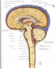
CSF FLOW DIAGRAM

narrowest part of csf pathway
cerebral aqueduct of slyvius
if stenosis occurs here, CSF will back up and could cause hydrocephalus or water on the brain