Nervous system 2401 practical
 Call Kai
 Learn
 Practice Test
 Spaced Repetition
 Match
 Flashcards
 Knowt Play
1/110

There's no tags or description
Looks like no tags are added yet.
111 Terms

Also called (soma) the cell bodies serve as the neurons control center and is responsible for receiving integrating and sending nerve impulses
Cell body

This is a organelle that contains chromatin and nucleoli
Nucleus

Short extensions that branch off the cell body
Dendrites

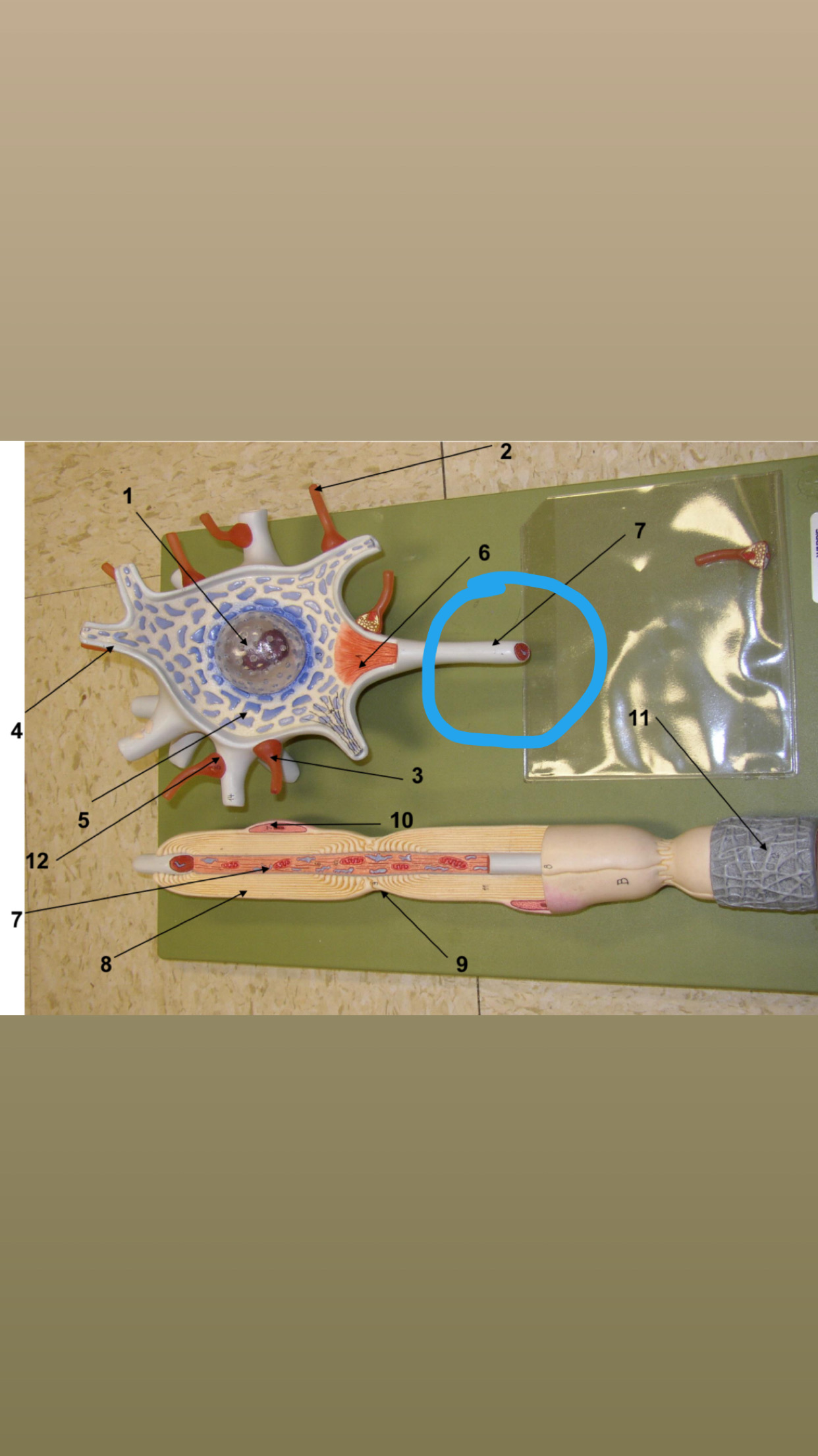
It transmits nerve impulses away from the cell body toward another cell
Axon


Most axons branch extensively at their distal ends telodendria
Telodendria


Also called synaptic terminals/ axon terminals :this is the very en slightly expanded end of an axon
Synaptic knobs


These cells form the myelin sheath around the pns
Neurolemmocytes


The insulating covering around the axon consisting of concentric layers of myelin
Myelin sheath


Are the gaps in the myelin sheath
Nodes ranvier


Triangular region where the axon connects to the cell body
Axon hillock


Areas one neuron communicates with another neuron in a neural pathway
Synapses
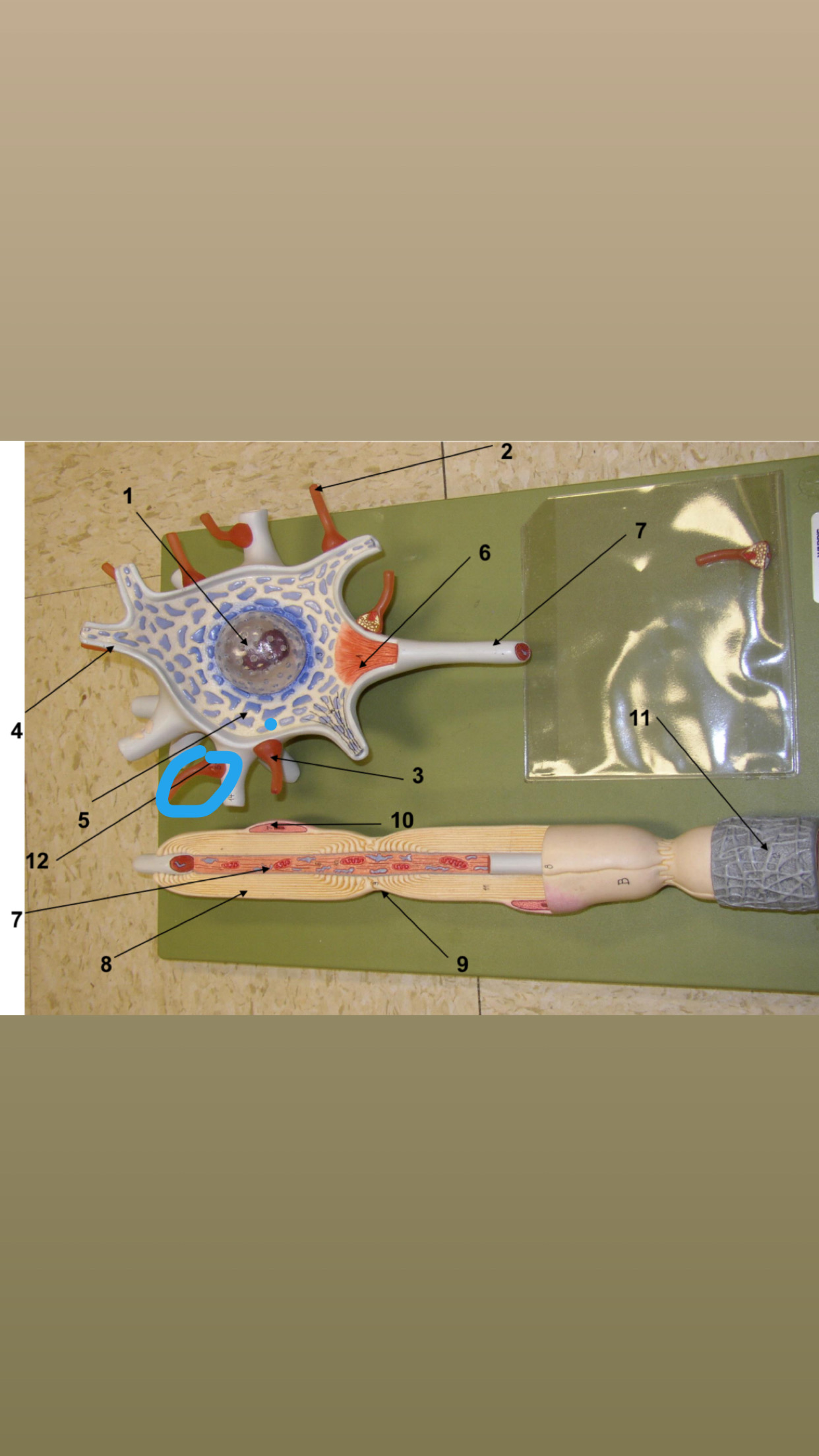

The delicate layer of areolar tissue with capillaries that supply each axon
Endoneurium


This is the large rounded portion of the brain includes the cerebral cortex the nuclei and the white matter
Cerebrum


This part of the brain includes the pineal gland the hypothalamus and the thalamus
Diencephalon

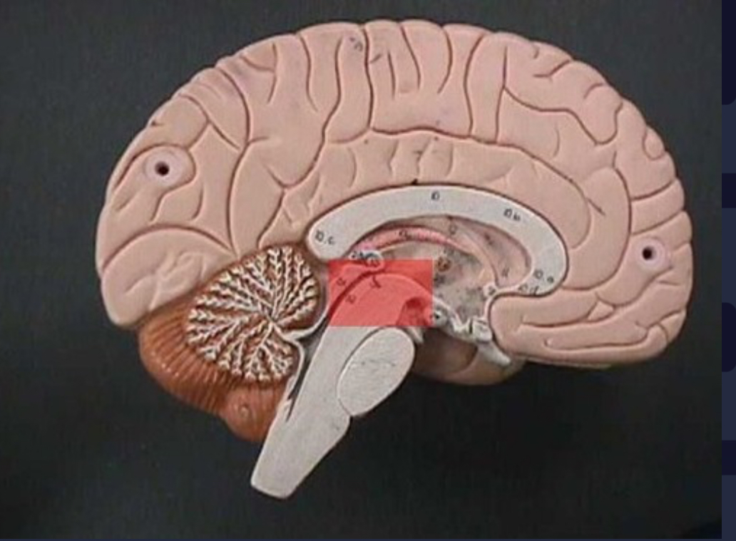
This part of the brain between pins and the Diencephalon
Midbrain


This is part of the brain anterior to the cerebellum and superior to the medulla
Pons


This is the inferior pa of the brain stem
Medulla oblangata


This part lies posterior to the medulla oblongata and pons
Cerebellum


Sensory areas are involved in the perception of sensory information.motor areas control muscular movement
Left cerebral hemisphere


Sensory areas involved in the perception of sensory information. Motor areas control muscular movement
Right cerebral hemisphere


This a deep midsagittal groove that separates the two cerebral hemispheres
Longitudinal fissure


This a deep groove found between the cerebrum and cerebellum
Transverse fissure


This sulcus separates the temporal lobe from the parietal and frontal lobes
Lateral sulcus


Separates the frontal and parietal lobes
Central sulcus

Separates the parietal and occipital lobes and runs in an inferior and anterior direction from the surface of the brain
Parieto-occipital sulcus
This is the fold of cortex just anterior to the central sulcus
Precentral gyrus
This is the fold of cortex just posterior to the central sulcus
Postcentral gyrus
This lobe lies caudal to the central sulcus
Frontal lobe
This lobe lied caudal to the central sulcus
Parietal lobe
This lobe lies laterally,but inferior to the parietal lobe
Temporal lobe
This lobe lies caudal to the parietal lobe
Occipital lobe
This is a small lobe deep to the lateral sulcus. You can only see it by laterally reflecting the temporal lobe
Insula
The layer of gray matter external to white matter. It contains neuronal cell bodies dendrites and short axons
Cortex
Also called cerebral nuclei and basal ganglia) these are paired irregular masses of gray matter external buried deep within the central white matter in the basal region of the cerebral hemisphere
Basal nuclei
This is a bundle of axons linking centers in the left and right cerebral hemispheres
Corpus callosum
This is a thin tract of white matter that connects the hippocampus
Fornix
This group of axons in a tract passing to and from the cortex between the cerebral nuclei
Internal capsule
This is a cone shaped gland coated in the posterior portion of the roof of the Diencephalon.
Pineal gland
This is a large oval structure located on either side of the third ventricle
Thalamus
This is the floor of the Diencephalon
Hypothalamus
This is a thin stalk that extends from the hypothalamus to attach to the pituitary gland
Infundibulum
This is an endocrine organ that sits in the sella turcica of the sphenoid bone.
Pituitary gland
These are paired masses nuclei in the hypothalamus that process sensations related to smelling and that controls swallowing
Mammillary bodies
These are 4 bumps in midbrain. It is made up of the 2 superior colliculi and 2 inferior colliculi.
Corpora quadrigemina
These help visually track moving objects and control reflexes such as turning the eyes and head in response to a visual stimulus
Superior colliculi
These control reflexive turning of the head and eyes in the direction of a sound
Inferior colliculi
These are motor tracts located on the anterolateral surfaces of the mesencephalon
Cerebral peduncles
These are transverse fibers that connect the one to the cerebellum
Middle cerebellar peduncles
These are two longitudinal ridges that flank the ventral midline of the medulla
Pyramids
This is a narrow band of cortex along the midline of the cerebellum
Vermis
This is the white matter of the cerebellum. It is called this because its distribution pattern resembles the branches of a tree
Arbor vitae
(VEN-trih-kuhls) (These are found in the cerebral hemispheres. They are C-shaped.)
lateral ventricles
(This is an opening between the lateral ventricles.)
interventricular foramen
(This ventricle is located between the cerebral hemispheres in the diencephalon, around the thalamus.)
third ventricle
(also called cerebral aqueduct) (This a slender canal that connects the third ventricle with the fourth ventricle.)
mesencephalic aqueduct
(This ventricle is located between the pons and cerebellum.)
fourth ventricle
(This is an opening in the roof of the fourth ventricle that allows the CSF to flow into the subarachnoid space.)
median aperature
(These are paired openings in the roof of the fourth ventricle that allow the CSF to flow into the subarachnoid space.)
lateral aperatures
(This is a layer of ependymal cells and the capillaries that lie within the pia mater. This is where cerebrospinal fluid is made.)
choroid plexuses
(This is the tough, fibrous, outermost layer of the dura mater that forms the periosteum of the cranial bones.)
periosteal layer of dura mater
(This is the inner layer of the dura mater and is usually fused to the periosteal layer except at the dural venous sinus-large veins that drain blood from the brain.)
meningeal layer of dura mater
(This is the middle layer of the meninges and is made of a delicate web of collagen and elastic fibers.)
arachnoid mater
(This is the innermost layer of the meninges.)
pia mater
(This is a horizontally oriented fold of dura mater that separates the occipital and temporal lobes of the cerebrum from the cerebellum.)
tentorium cerebelli
(This is a large, sickle-shaped vertical fold of dura mater, located in the midsagittal plane. It projects into the longitudinal fissure between the left and right cerebral hemispheres; it attaches to the crista galli of the ethmoid bone)
falx cerebri
(thick, opaque outer layer)
dura mater
(delicate, transparent middle layer that is attached to the undersurface of the dura mater)
arachnoid mater
(if present)
spinal cord
corpus callosum
corpora quadrigemina
pineal body
(These contain somatic motor nuclei, which provide nerve impulses for contraction of skeletal muscles.)
anterior horns
(These contain autonomic motor nuclei that regulate the activity of smooth muscle, cardiac muscle, and glands.)
lateral horns
(These contain somatic and autonomic sensory nuclei.)
posterior horns
(These contain bundles of axons.)
anterior funiculi (column)
(These contain bundles of axons.)
lateral funiculi (column)
(This is a thin strip of gray matter that surrounds the central canal of the spinal cord. It is filled with CSF and is continuous with the ventricle system of the brain.
gray commissure
(This is a bundle of nerve fibers (axons) that cross the midline of the spinal cord.)
white commissure
(This is a fissure in the anterior, middle part of the spinal cord.)
anterior median fissure
(This is a deep groove in the posterior, middle part of the spinal cord.)
posterior median sulcus
(The dorsal roots contain sensory axons only.
The cell bodies of these sensory neurons are located in a posterior root ganglion.)
dorsal roots
(also called posterior root ganglia) (singular = ganglion) (The cell
bodies of the sensory neurons in the dorsal roots are located here in the dorsal root ganglia. horn of the spinal cord.)
Each ganglion is attached to the dorsal root. They carry sensory signals to the dorsal gray
dorsal root ganglia
(also called anterior roots) (The ventral roots contain motor axons only that lead to muscle or glands. These motor axons arise from cell bodies in the anterior and lateral horns of the spinal cord.)
ventral roots
(also called posterior rami) (singular = ramus) (The dorsal ramus is the
smaller of the two main branches. It innervates the deep muscles of the back and the skin of the back.)
dorsal rami
(also called anterior rami) (The ventral ramus is the larger of the two main branches. It splits into multiple other branches, which innervate the anterior and lateral portions of the trunk, the upper limbs, and the lower limbs. Many of the anterior rami go on to form nerve plexuses.)
ventral rami
(These are additional rami which contain axons associated with the autonomic nervous system.)
rami communicantes
(also called sympathetic trunk ganglia or paravertebral ganglia)
(The cell bodies of ganglionic autonomic neurons are house here)
autonomic ganglia
(This is the space "below" the arachnoid.)
subarachnoid space
(This is a space "above" the dura mater. It is filled with adipose tissue and blood vessels.)
epidural space
central canal
gray horns
white funiculi (columns)
(These are individual axons)
nerve fibers
(These are groups of nerve fibers (axons) bound into bundles.)
nerve fascicles
(This is a layer of dense irregular tissue that surrounds each nerve fascicle.)
perineurium
(This is a layer of dense irregular connective tissue that binds the fascicles together into a nerve. It surrounds the entire nerve.)
epineurium
cervical nerves