neuroanatomy and imaging
 Call Kai
 Learn
 Practice Test
 Spaced Repetition
 Match
 Flashcards
 Knowt Play
1/34

There's no tags or description
Looks like no tags are added yet.
35 Terms
What is the basic anatomy of the brain split into
3 divisions
Forebrain
Midbrain
Hindbrain
Hindbrain
Oldest part of brain
Controls functions needed for life -autonomic functions
Includes cerebellum and most of brainstem
Why is this part of the brain important
Important for optometric practice as some px have “chiari malformations “ where the brainstem is forced through the magnum foramen -hole at the base of the skull
-these px often have issues with headaches ,diplopia, nystagmus and photophobia
Midbrain
Top of the brainstem
Responsible for saccades and managing visual attention -superior colliculus and eye movements , upper eyelid and pupil reactions (oculomotor nucleus)
Damage to part of midbrain
Can cause diplopia , nystagmus , vertical gaze palsy and tracking difficulties as well as impaired pupil reflexes
What does forebrain include
Includes cerebral cortex and limbic system

What does damage to this part cause
Affects how the brain interprets information
Px may experienced visual field loss, blurred vision, diplopia and poor spatial awareness
Cerebral cortex
Divided into two hemispheres each with 4 lobes
-folded surface provide large surface area for neurons , the more folded the brain the cleave re the animal
-bumps are “gyri” and the grooves are “sulci”
these two provide landmarks
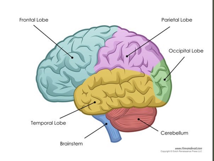
Cerebral cortex image

Frontal lobe and its role
Largest of the 4 lobes
In charge of higher executive functions ,planning,problems ,solving ,reasoning , judgement
-responsible for personality and emotions and impulse control
includes brocas area which is main region for producing speech
What is the frontal lobe the most common area for
Brain damage , changes such as loss of speech, remembering words, executive dysfunction ,socially inappropriate behaviour
-also involved in neurodevelopment conditions ,ASD, ADHD
Parietal lobe
is the somatosensory part of the brain -interpretation of sensory information
Includes touch,pressure, temperature, and pain
What is parietal lobe responsible for
Creating a map of the world called spatial attention and orientation
Main part of the brain involved in reading and writing
-is also where visual perception and attention occurs
Sensory motor cortex
Area found at the join between the frontal and parietal lobes

Temporal lobe
Holds primary auditory cortex
Is in charge of oculo vestibular responses -balance and related eye movements
-home to wernikes area -responsible for the comprehension of speech and links to Broca’s area in the frontal lobe
Responsibility of temporal lobe
Memory, object and facial recognition
-px with temporal lobe defects can suffer with prosopagnosia (faces) or anomia (objects) as well as aphasia (language)
Occipital lobe
Completely devoted to visual processing
Links to parts of the temporal and parietal lobe to complete the pathway
Vision is thought to take up 30-50% of the brain
Dorsal and ventral pathways
Two anatomically distinct and functionally specialised pathways for processing different aspects of vision
-identified in 1982 by ungerleider and mishkin and build on by milner and goodale 1992 to form “perception and action” model of visual processing
Dorsal pathway
Beings in occipital lobe and travels to parietal
“Where” or “perception” pathway
Involved in locating objects in spacen
Ventral pathway
Beins in occipital lobe and travels to temporal lobe
“What” or “action” pathway
Involved in processing shapes ,textures ,patterns and colours
influence of dorsal and ventral “perception and action” pathways
remain highly influential
however there’s newer evidence suggesting huge numbers of of inter stream interactions which has changed the idea that we have functionally independent pathways
-complex visual functions likely to involve collaboration between the two specialised visual streams as well as other areas of the brain
What is the limbic system known as
Survival centre
Responsible for emotion and behavioural expression
What is the limbic system made up of
Thalamus - relay centre for sensory information
Hypothalamus - regulates hormones as well as controls body temperature, appetite and flight or fight response
Hippocampus - Houses emotions, learning and short term memory
Amygdala- also holds emotions, long term memory and fear
Pituitary gland - essential for hormones and reproduction
(Parts of limbic system run very cos to the optic nerve tract and chiasm)
Magnetic resonance imaging MRI
based on principles of nuclear magnetic resonance NMR
-uses very powerful magnet to align the hydrogen atoms in our body
-a brief radio signal then temporarily knocks the atoms out of alignment
-when they return to their aligned positions they emit a small amount of energy which can be measured
-different tissues and structures have different amounts of hydrogen atoms and so give different amounts of energy allowing us to distinguish them
Pros and cons of MRI
Good for imaging soft tissues of the body
Poor temporal resolution - delay in signal so wouldn’t pick up recent changes e.g haemorrhages
Contradicted for px with magnetic iron based foreign bodies
Imaging techniques - computerised tomography CT
Most similar to X ray but with greater spatial resolution
-multiple narrow beams of radiation sent through the body at different angles
-then received by a computer which uses an algorithm to create 2D or 3D image
What is CT scan good for
Imaging soft tissue , bones and blood vessels
-tissues or diff densities appear in different shades of grey , white areas habe maximum density
-similar to OCT to read , whiter areas let less light through
(Possible risk of cancer due to radiation exposure?)
MRI more detailed but CT more faster, better for trauma
Imaging techniques - positron emission tomography PET
Uses radioactive tracers as a radiation source introduced into the px body
usually ingested but also IV if needed
This radioactive substance can be detected in the blood by special CT scan (MRI also used occasionally)
PET scan
The more active parts of the brain use more blood and therefore show up on the scan
-the radioactive substance decays quickly (half life) and is not harmful
Mainly used for brain scans , blood vessels and tumours

Imaging technique MEG magnetoencephalography
Detects weak magnetic fields from above the surface of the head produced by electrical activity within the brain
When is MEG used
In a special room shielded from earths magnetic field and with coils kept at very low temps
Virtually silent, non invasive and quick
-well suited to clinical applications as it can be readily used in children and other populations who may not tolerate demanding procedures
-not as precise as other imaging techniques
Electroencephalography EEG
Electrophysiological technique for the recording of electrical activity arising from the human brain
How does EEG work
Microelectrodes placed on the scalp record normal electrical activity from the brain
Signal is amplified using software
Trace can be then monitored to check for abnormal activity
-excellent temporal sensitivity, can tell you when in the brain
When is EEG used
Primarily in epilepsy and brain disorders , but also in visual function research
Summary table of different imaging techniques
