BIOL 1062 - Test 1
 Call Kai
 Learn
Learn Practice Test
 Spaced Repetition
 Match
Match Flashcards
 Knowt Play
1/54

Earn XP
Description and Tags
A set of vocabulary flashcards based on the BIOL 1062 Test 1 outline, including test structure rules, human organization, and digestive system concepts.
Name | Mastery | Learn | Test | Matching | Spaced | Call with Kai | Chat |
|---|
No analytics yet
Send a link to your students to track their progress
55 Terms
List the Biological Organization of humans from least to most complex
atoms → molecules → cells → tissues → organs → organ systems → organism.

What is Homeostasis?
The state of steady internal physical and chemical conditions maintained by living systems.

What does the Anterior/Ventral Cavity consist of?
Thoracic Cavity
divided by the diaphragm
Abdominal Cavity
Pelvic Cavity

What organs can be found in the Anterior/Ventral Cavity?
Thoracic Cavity:
Lungs
Heart
Abdominal Cavity:
Digestive Organs (stomach, intestines)
Pelvic Cavity:
Urinary Bladder
Reproductive Organs
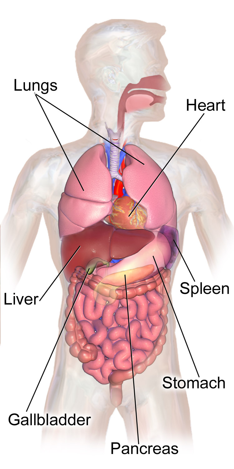
What does the Dorsal/Posterior Cavity consist of? What organs are apart of it?
Cranial Cavity
Brain
Spinal Cavity
Spinal Cord

What are the 4 types of membranes in the body?
Mucous Membrane
Serous Membrane
Synovial Membrane
Meninges
Where can you find Mucous Membranes? What is their structure and function?
Line the Digestive, Respiratory, and Urinary organs
Made of an epithelium covering loose connective tissue
Contain Goblet Cells that secrete Mucous
Protective function
Mucous moves due to Cilia

Where can you find Serous Membranes? What is their structure and function?
Line the Thoracic and Abdominal Cavities; covers the organs
Made of epithelium and loose connective tissue
Secretes Serous Fluid
Keeps membranes lubricated
Supports and compartmentalizes; prevents organs from dangling
What are the three types of Serous Membranes that are different based on their location?
Pleura: Lines the Thoracic Cavity and Lungs
Pleura (Plural) = lungs (plural)
Peritoneum: Lines the abdominal cavity
Pericardium: Lines the Cardiac Cavity

Where can you find Synovial Membranes? What is their structure and function?
Lines: freely movable joints
Knee joints, elbow joints, etc.
Secretes Synovial Fluid
Like WD-40 for joints
Made of loose connective tissue

Where can you find the Meninges? What is their structure and function?
Found in the Dorsal Cavity
Protects the Brain and Spinal Cord
Secretes Cerebrospinal Fluid (CSF)


Label the following diagram of Anatomical Directional Terms


Label the following diagram of Anatomical Directional Terms


Use the correct Anatomical Directional Term to answer the following:
Chin is ___________ to the nose
Ears are ___________ to the nose
Nose is ____________ to the ears
Knee Joint is __________ to the hip joint
Shoulder is ___________ to the wrist
Wrist is ____________ to the fingers
Chin is inferior to the nose
Ears are lateral to the nose
Nose is medial to the ears
Knee joint is distal to the hip joint
Shoulder is proximal to the wrist
Wrist is proximal to the fingers

What are the four types of tissues found within the human body?
CMEN
Connective
Muscle
Epithelial
Nervous

What is the function and structure of Epithelial Tissue?
Function:
Protects underlying structures
Secretion, Absorption, Filtration, Excretion (SAFE)
Structure:
Depends on (2):
Number of Cell Layers:
Simple: one layer
Stratified: multi-layered
Pseudostratified: appears multi-layered
Cell Shape:
Squamous (Squished): flat
Cuboidal: cube-like shape
Columnar: column-like shape

Describe Simple Squamous tissue
structure
function
where is it found in the body
One Layer (Simple) of Flat Cells (Squamous)
Ideal for Diffusion
Found in:
Alveoli of lungs
Blood vessels

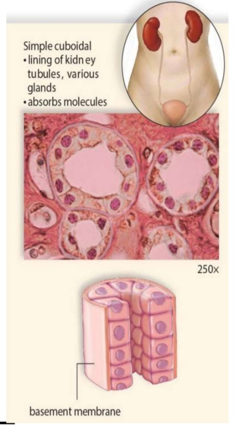
Describe Simple Cuboidal Tissue
structure
function
where is it found in the body
One layer (Simple) of cube-like cells (Cuboidal)
Ideal for Absorption and Secretion
Found in:
Lining of Kidney Tubules
Urinary Organs
Describe Simple Columnar Tissue:
structure
function
where is it found in the body
One layer (Simple) of column/rectangular cells (Columnar)
Ideal for Absorption and Secretion
Found in:
Lining of Small Intestine
Uterine Tubes

Describe Pseudostratified/Ciliated Columnar tissue
structure
function
where is it found in the body
Appears multilayered but only single layered (pseudo=fake)
Each cell touches the basement membrane (bottom surface)
Ideal for Sweeping Action due to Cilia
sweeps away unwanted material
Found in:
Lining of Trachea
Small Intestine

Describe Stratified Squamous tissue
structure
function
where is it found in the body
Multi-layered (Stratified) flat cells (Squamous)
Ideal for Protection (areas exposed to wear/tear, constant use)
Found in:
Mouth
Esophagus
Lining of nose
Vagina

Describe Connective Tissue
structure
function
where is it found in the body
Located beneath Epithelial Cells
All Epithelial Cells rest on Connective Tissue
Binds organs together and fills space
Found everywhere in the body

Describe Muscular Tissue
structure
function
where is it found in the body
Composed of cells called Muscle Fibers
Used for movement (within/of the body)
Found within the body (3)
Named on:
Appearance
How many nuclei
How they are controlled

Describe Smooth Muscle
Appearance
How many nuclei
How they are controlled
Where might they be found in the body?
Smooth appearance
Lacks Striations (Stripes)
ex: zebra has lots of striations
Cells are spindle-shaped with one nucleus
Involuntary Control
ex: bolus moving down esophagus
Found in:
Walls of hollow internal organs
Passageways throughout the body

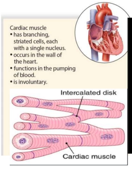
Describe Cardiac Muscle
Appearance
How many nuclei
How they are controlled
Where might they be found in the body?
Cells are separate/individual but are bound at each end at intercalated disks (branched part)
Has branching, striated cells, each with a single nucleus
Involuntary Control
Electrical Impulses flow when the heart muscle contracts
Found only in the wall of the heart
Describe Skeletal Muscle
Appearance
How many nuclei
How they are controlled
Where might they be found in the body?
Striated due to alternating light and dark bands
Cylindrical, long and multi-nucleated due to fusion of several cells into a fiber
Voluntary Control
Usually attached by tendons to bones of skeleton

Describe Nervous Tissue
structure
function
where might it be found in the body?
Composed of two types of cells:
Neurons:
Conduct electric nerve signals/impulses
Neuroglia
Support and nourish neurons
Found in:
Brain
Spinal Cord
Nerves

What is the arrangement of layers in the gastrointestinal organs? (Innermost to outermost)
Mucousa (innermost facing the lumen: hollow cavity)
Submucousa
Muscularis
Serosa (outermost)


Label the following Digestive System Diagram

What are the functions of the Digestive System
Ingestion: taking in food (eating)
Digestion: breaking down food (to smaller molecules)
Mechanical Digestion (physical breakdown)
Chemical Digestion (chemical breakdown; enzymes)
Absorption: absorb nutrients from digestive organs into bloodstream
Defecation: remove waste products
What are the names of the food as it travels through the digestive system (tract)?
Food
Into the mouth
Bolus
after being chewed and “condensed into a blob"
Chyme
Bolus + Gastric Juice in the stomach
Feces
After passing through intestines

Where are the macromolecules digested throughout the digestive system? What enzymes are involved?
Protein:
Stomach: Gastric Juice
Cleaving of HCl activates Pepsinogen to Pepsin
Breaks polypeptides into smaller peptides
Small Intestine: Trypsin
Carbohydrate:
Mouth/Salivary /Tongue: Salivary Amylase
Pancreas: Pancreatic Amylase
Lipids:
Mouth: Lingual Lipase
Stomach: Gastic Lipase
Breaks triglycerides into diglycerides and fatty acids
Liver + Gallbladder: produces Bile
Bile Salt is an emulsifier: breaks down lipids
Pancreatic Lipase
Nucleic Acids:
Small Intestine: Nucleases
What is the difference between primary and accessory digestive organs?
Primary Digestive Organ:
organs which food passes through
Accessory Digestive Organ:
Organs that food does not pass through
What are the six Primary Digestive Organs?
Mouth
Pharynx
Esophagus
Stomach
Small Intestine
Large Intestine

What are the five Accessory Digestive Organs?
Tongue
Salivary Glands
Liver
Gallbladder
Pancreas

What digestion/enzymatic functions occur at the mouth?
Three functions:
Ingestion:
Receives food
Digestion: Two types of digestion:
1. Mechanical Digestion:
Breaking down to smaller pieces
2. Chemical Digestion:
Salivary Amylase: digests carbohydrates
Lingual Lipase: digests lipids
Absorption:
Oral Mucosa absorbs simple and small molecules (glucose, alcohol) into blood stream

What digestion/enzymatic functions occur at the pharynx?
Passage carrying food between mouth and esophagus
Shared passage for food and air
“crossroad between digestive and respiratory system”
Epiglottis covers the glottis, preventing the bolus from entering the trachea/lungs
No digestion or absorption occurs

What digestion/enzymatic functions occur at the esophagus?
Passageway of smooth muscle carrying bolus between pharynx and stomach
Peristalsis (rhythmic contractions): moving the bolus down the esophagus with periodic contraction and relaxation of smooth muscle
No digestion or absorption occurs

What digestion/enzymatic functions occur at the stomach?
Mechanical Digestion:
churning of food:
contractions: uneven surface (Rugae) + movement
Chemical Digestion:
Proteins:
Gastric juice:
Cleaving of HCl activates Pepsinogen into Pepsin
Polypeptide + Pepsin → Smaller (Oligo) peptide
Lipids:
Gastric Lipase
Breaks triglycerides into diglycerides and fatty acids

What digestion/enzymatic functions occur at the Small Intestine?
Primary site of absorption of water and nutrients
Villi and microvilli increase surface area of small intestine for nutrient absorption
Blood capillaries absorb:
Carbohydrates, proteins, nucleic acid
Small Lymphatic Capillary:
Fatty acids: too big and hydrophobic to absorb on their own
packaged with proteins and absorbed via lacteals
Most Chemical Digestion takes place in the Duodenum
Nucleic acid digestion
Main absorption happens in the Jejunum and Ileum

What digestion/enzymatic functions occur at the Large Intestine?
Defecation takes place in the Large Intestine
No Digestion occurs in the Large Intestine

Describe the structures and mechanism involved in swallowing
When food is swallowed:
Uvula closes off the nasopharynx
Prevents Bolus from entering nasal cavity
Epiglottis covers the glottis
Forces the Bolus to pass down the esophagus
Person does not breathe when swallowing

What is the role of Villi and Microvilli in the Small Intestine?
They increase the surface area of the small intestine for nutrient absorption

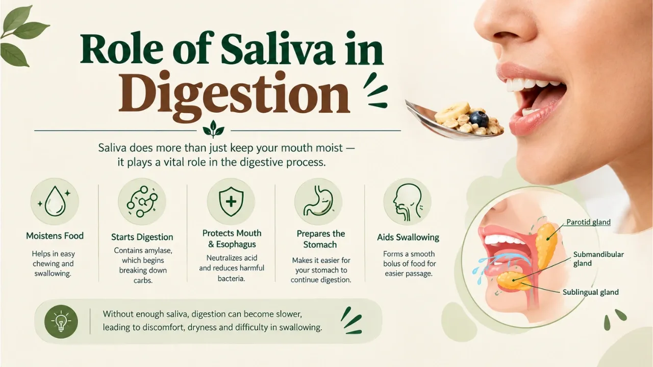
What is the function and chemical contributions of the Salivary Glands?
Secretes Saliva
H2O + Proteins → Salivary Amylase + IGA
Primary Digestion of Carbohydrates:
Salivary Amylase + Polysaccharide →Oligosaccharide → Disaccharide
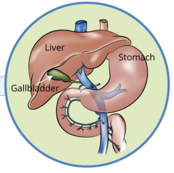
What is the function and chemical contributions of the Liver?
Two Functions:
Detoxification
Hepatic Portal Circulation
Producing Bile:
Contains Two Parts:
Bilirubin:
Byproduct of destroying + recycling RBC
Bile Salts
Hydrophobic and Hydrophilic Side


What is the function and chemical contributions of the Gallbladder?
Stores Bile
Release Bile to Duodenum
Reabsorbs excess water

What is the function and chemical contributions of the Pancreas?
Endocrine Function:
Produce and release hormones directly into the bloodstream to control blood sugar
Exocrine Function:
Produce and secrete digestive enzymes and Sodium Bicarbonate (NaHCO3)
NaHCO3 neutralizes stomach acid entering the Duodenum
Creates Pancreatic Juice
What enzymes (4) can be found in Pancreatic Juice? What macromolecule do they work on?
Pancreatic Lipase (Lipids)
Triglycerides → glycerol + 3 fatty acids
Final step of Lipid Digestion
Pancreatic Amylase (Carbohydrates)
Polysaccharide, Oligosaccharide → Disaccharide
Trypsin (Proteins)
Cleaving Trypsinogen → Trypsin
Small Polypeptides → Smaller Peptides
Nuclease (Nucleic Acid)
RNA/DNA → Nucleotides
Describe the Hepatic Portal Circulation
Blood from the digestive organs passes into the liver via the hepatic portal vein
Liver detoxifies blood before it is circulated via the hepatic vein into the inferior vena cava (heart)

Describe the nervous input of Defecation
Accumulation of feces in the rectum causes it to stretch
Stretching initiates nerve signals for rectal muscle to contract and anal sphincter to relax
In order to defecate, both must be relaxed
If not a good time to defecate, brain sends signals to stop relaxing and keeps contracting

What is the role of the Enteric Nervous System?
Regulates gut function
How does the mucus layer differ in the small intestine and the large intestine?
Small Intestine (A):
One layer
No bacteria
Large Intestine (B):
Two layers
Outer mucus layer:
Contains good bacteria
Inner mucus later:
No bacteria

What is the role of Enteroendocrine Cells?
Regulates digestion and hormones (metabolism)

What is the role of Stem Cells?
Used for cell renewal
Unspecialized cells that can change into many different specialized cells

What is the role of Goblet Cells?
Releases a slippery fluid called Mucin
Mainly consists of a carbohydrate chain
Creates Mucus when mixed with water and glycoproteins
