Red Meat Pathology 1
 Call Kai
 Learn
 Practice Test
 Spaced Repetition
 Match
 Flashcards
 Knowt Play
1/26

There's no tags or description
Looks like no tags are added yet.
27 Terms
What are the 6 most common lung conditions you will see in sheep upon PMI?
1) Enzootic pneumonia
2) Fibrinous pneumonia
3) Ovine pulmonary adenocarcinoma
4) Muellarius infection
5) Dictyocaulus infection
6) Abscesses

Describe the classic gross features of an enzootic pneumonia (suppurative pneumonia)…
Cranioventral distibution; consolidated to those fields
Usually bilateral
Causes range from:
Mycoplasmas
Pasteurella multocida
Partial rejection (just the lungs)
Describe the classic gross features of a fibrinous pneumonia…
Cranioventral distribution
Pleura covered by fibrinous exudate
Can be bilateral or unilateral
Most common cause:
Mannheimia haemolytica
Partial rejection
Describe the classic gross features of an OPA…
Large, consolidated pink areas of the lungs
Usually in the center, firm and raised
Will also see abundant, frothy fluid in the trachea
Partial rejection

Describe the classic gross features of a muellarius infection…
A type of “verminous pneumonia”
Multifocal “lead shot” lesions found on the dorsal aspects of the lungs
More chronic cases can have larger spots along the dorsal aspect of the lungs that seem to coalesce
Can see worms and eggs in alveoli upon histology
Partial rejection
Really quickly review the life cycle of Muellerius capillaris…
Adults produce eggs in the lungs of sheep… eggs hatch in lungs
Arrested development
Once L1, are shed in feces
Snail eats them; L1→ L3 in snail, temperature dependent development
Faster in warmer temps
May overwinter in snail
Once L3, leave snail and onto grass
L3 ingested by sheep, migrate from GI to lungs via mesenteric lymph nodes, thoracic duct, heart, and pulmonary arteries
Describe the classic gross features of a dictyocaulus infection…
Consolidation of the CAUDAL part of the lungs
Adults will live in bronchi, can be seen
Partial rejection

Really quickly review the life cycle of Dictyocaulus filaria…
Lamb eats L3 in pasture
L3 penetrate SI mucosa; L3 → L4 in mucosa
L4 travel to lungs via blood and lymph; penetrate capillaries and enter alveoli
L4 migrate to bronchioles
L4 → Adult in bronchi
Eggs hatch in lung and migrate up trachea
Coughed up
Swallowed
Released in feces
L1 → L2 → L3 in grass
Pilobilus fungi help to distribute to clean grass, which sheep then eats
Dictyocaulus filaria is the sheep one… D. viviparus is the cow one
Describe the classic gross features of an abscess…
Hard to miss
Often happen due to secondary opportunistic bacteria
Trueperella pyogenes
Staphylococcus aureus
Partial rejection if only lung abscess
What are 3 less common lung lesions you could see?
1) Hydatidosis
2) Caseous Lymphadenitis → TB
3) Maedi visna viral infection

Describe the classic gross features of hydatidosis…
Intermediate stage of Echinococcus granulosus
Definitive host is dog; intermediate is sheep
Will see cysts usually in the lungs; may find them in the liver
Focal lesion
Fluid filled with a thick capsule
IS ZOONOTIC; check farmer and family
Partial rejection
Describe the classic gross features of caseous lymphadenitis…
Caused by inhalation of bacteria or bloodstream/lymphatic spread from an existing infection elsewhere in the body
Usually caused by knicking when shearing with dirty equipment
Corynebacterium pseudotuberculosis specifically
Need to check the rest of the carcass in this case for other infected lymph nodes
FULL REJECTION
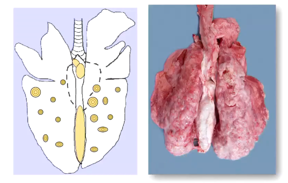
Describe the classic gross features of the Maedi visna virus…
INTERSTITIAL PNEUMONIA
Lung thick and rubbery, unable to deflate normally
No exudate whatsoever
Partial rejection
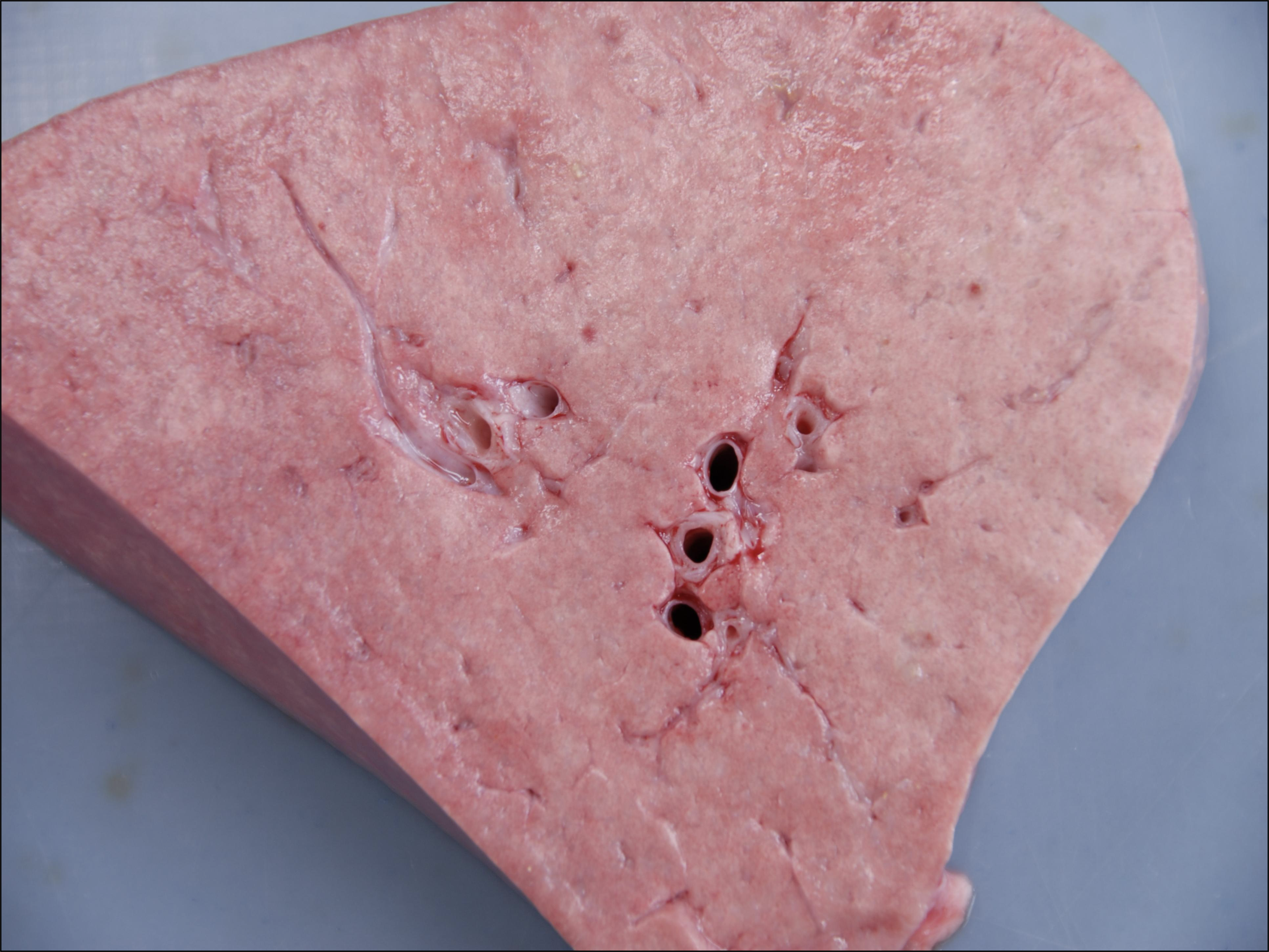
What is a common lesion you may see in the sheep lungs / heart that ISN’T pathological, but instead is caused perimortem by improper electrical stunning technique?
Blood splashing
Caused by the sudden blood pressure increase due to electrical stunning
Ecchymotic or petechial hemorrhages on the lungs, heart, and skeletal muscle
If happening to lots of sheep, need to check stunning procedure; amperage may be too high (must be at least 1.0A to ensure proper stun)

HEART, MUSCLE, and BONES
What are 2 major cardiac lesions you could see upon PM in a sheep?
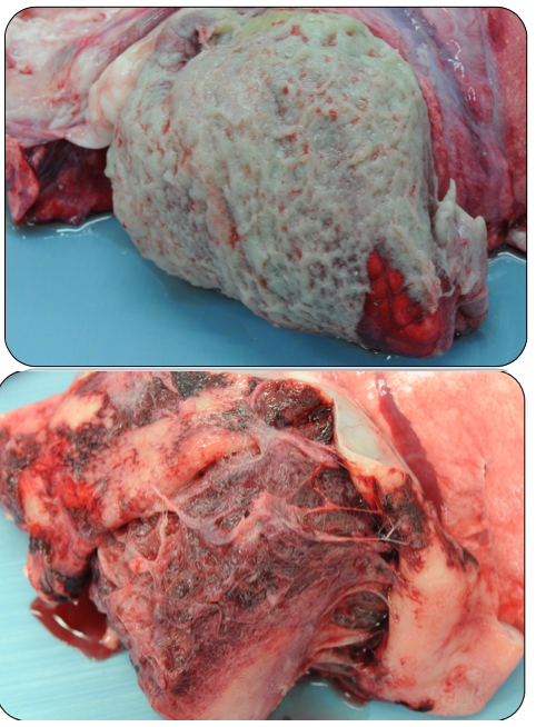
1) Pericarditis
Characterized by:
Surrounding exudate around the pericardium
fibrous / constrictive mostly, signs of chronic pericarditis; acute (fibrinous) much less common to see in abattoir (see images)
2) Cysticercosis
Cysticercus ovis COMES FROM dog tapeworm, Taenia ovis
Sheep are intermediate hosts… ingest infected dog feces, larvae migrate into muscle and heart and encyst there (C. ovis)
C. ovis is essentially the intermediate stage of Taenia ovis
Found in SKELETAL MUSCLE, CARDIAC MUSCLE, and the DIAPHRAGM
So if you’re doing a PM and see cysts in the heart, need to check entire carcass… sheep may have generalized cysticercosis, and will need to be totally rejected
What is osteomyelitis, and how does it present in sheep in a PMI?
Is INFECTION of the bone… can happen anywhere in sheep (legs, vertebrae) but most commonly is seen in the jaw
Common pathogens:
Trueperella pyogenes,
Fusobacterium necrophorum,
Staphylococcus aureus,
Streptococcus spp,
Pseudomonas aeruginosa
If infection is localized to the mandible alone, then can remove the jaw and use the rest of the carcass
Unfortunately, because of this, sheep don’t eat very well in the first place… if body is also emaciated in addition to this, total rejection of the carcass
LIVER
Which 2 diseases account for 90% of the liver damage detected in sheep in the abattoir?
Fasciolosis
Cysticercosis
Describe fasciolosis… how does it present on the liver in the migratory phase? Mature phase? Chronic phase?
Fasciola hepatica can infect both lambs and adults
Causes death in acute infections
Emaciation in chronic infections
Migratory phase
Damage looks traumatic / inflammatory
Multiple hemorrhagic tunnels
Burrowing metacercaria = hemorrhagic tracts
Fibrin or fibrous, hard tags on the surface of the liver that look like mineralization
LESIONS CONCENTRATED TO LEFT LOBE
Mature phase
Adult reaches bile duct; bile duct is swollen and visible on the liver, full of flukes that will be seen grossly
Chronic phase
Will see more clinical signs:
Anemia, hypoproteinemia
Loss of metabolic process (emaciated)
Atrophy of left lobe, and hypertrophy of right lobe
Liver looks distorted and mottled
Partial rejection

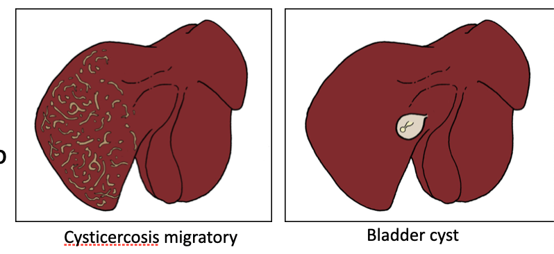
Describe how cysticercosis affects the liver… which parasite is the one that targets the liver directly in its intermediate stage? What does the migratory phase look like?
Caused by the intermediate stage of Taenia hydatigena
Migratory stage
Cysticerci migrate from liver to peritonium… gives you characteristic, serpentine-shaped tracts along the edges of the left lobe
Filled with fibrous material after healing; are thickened and pale, making them stand out
Will later develop a bladder cyst when it gets to the peritoneum… thin walled cyst, where you can see the scolex
May have just one or multiple… if multiple, whole carcass is rejected
**** can arrest in development in the liver; tiny little cysts
Partial rejection if only a few cysts
What is one parasitic condition in sheep that can affect the BRAIN?
Coenurosis / Coenurus cerebralis
AKA Gid
AKA parasitic induced cerebral cysts
Is caused by the LARVAL form of the dog tapeworm, Taenia multiceps
Enters the CNS hematogenously from SI tract (doesn’t target it, just gets there by mistake)
Coenurus cerebralis = immature stage of Taenia multiceps
Clinical signs: neurologic
Blindness, nystagmus, deviation of the head and eyes, ataxia, and collapse

What are some control measures you could take to minimize the incidence of Taenia spp. infections in sheep?
don`t feed farm dogs with uncooked meat /raw diet
remove carcasses of dead sheep as soon as possible
dispose of sheep carcasses as ABP category 2
regular dosing of dogs with anthelmintic (praziquantel)
at least every three months
farmer to put notes in public footpaths running through sheep fields explaining the disease risks and encouraging dog walkers to worm their dogs regularly
KIDNEYS and GI
What are the 3 main kidney conditions you may see on PMI?
1) Polycystic kidney
Congenital defect; either one or both effected
Seen only in young sheep
2) Renal neoplasia
Only in older animals
If cancer is localized to just the kidneys, then partial rejection
3) Embolic nephritis
COULD BE CAUSED BY TB
Check other areas of the body to confirm ^
** For these, LOCAL REJECTION ALWAYS unless you suscpect uremia or septicemia

What is Johne’s disease?
Disease of the ILEUM
Caused by Mycobacterium avium (subspecies paratuberculosis)
Ileum will appear thickened and yellow, looks like a brain
Need to check mesenteric lymph nodes; can spread through lymph and blood to other parts of the GI tract
Rejection of gut

What disease of the SI might you find as an incidental finding in a sheep over 5 years of age?
Small Intestinal Adenocarcinoma
Starts in the ileum, may spread to other organs (spreads via contact)
Occassional weight loss and ascites, but otherwise no clinical signs
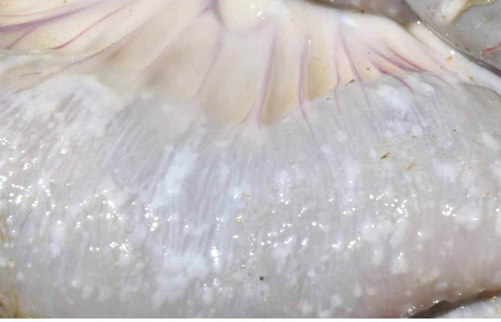
What is one big reason an entire sheep carcass may be rejected?
EMACIATION
Lack of fat on body
Lack of omental fat around the GI tract
What is one pathological reason an entire sheep carcass may be rejected just by looking at the color of its skin alone?
PALLOR due to ANEMIA, because this indicates…
Parasites
Haemonchus contortus
Fasciola hepatica
Tick-Borne haemoparasites (Babesia, Theileria)
Nutritional Element Imbalances
Iron Deficiency
Copper Deficiency
Anaemia of Inflammatory/Chronic Disease
Haemorrhagic Conditions (Bleeding abomasal ulcers, organ rupture, trauma)
What is one AESTHETIC reason an entire sheep carcass may be rejected?
Carotenoid pigmentation
Accumulation in the adipose tissue
Rejected on aesthetics
But safe for consumption
Causes
Ingestion of feeds rich in carotenoids, such as fresh green pasture, lucerne (alfalfa), and other green plants
Breed predisposition
Nonsense mutation in the beta-carotene oxygenase 2 (BCO2) gene (Våge and Boman 2010.)
Should be differentiated from jaundice (icterus)