PHYSIOLOGY 101
 Call Kai
 Learn
Learn Practice Test
 Spaced Repetition
 Match
Match Flashcards
 Knowt Play
1/323

There's no tags or description
Looks like no tags are added yet.
Name | Mastery | Learn | Test | Matching | Spaced | Call with Kai | Chat |
|---|
No analytics yet
Send a link to your students to track their progress
324 Terms
Central Nervous System
Includes the spinal cord and brain (which includes the cerebrum, cerebellum, and brain stem)
Central Nervous System Functions
To process and coordinate information such as sensory information, motor commands, and more complex brain functions such as memory and learning.
Peripheral Nervous System
Comprised of spinal nerves and cranial nerves.
Spinal Nerves
Extend into the periphery of the body originating from the spinal column
Cranial Nerves
Extend from the brain to peripheral structures in the head such as the eye and nose.
Peripheral Nervous System Function
To transmit information which can be done via carrying sensory information from the body to central nervous system or carrying commands from the central nervous system to the body.
Spinal Cord
Found within protective membranes (meninges) and encased in a bony vertebral column. In the centre there is a very small central canal which is filled with cerebrospinal fluid. It is divided into 32 segments which are also the origin of the spinal nerve, and starts just below the brain extending to the sacral region. It is also made up of nervous tissue.
Cerebrospinal Fluid (CSF)
Important for protecting the spinal cord from injury.
Spinal Regions
There are 4 regions. The cervical region which has cervical spinal nerves. The thoracic region which has thoracic spinal nerves. The lumbar region which has the lumbar spinal nerves. The sacral region which has the sacral spinal nerves.
Spinal Cord Function
Transmit sensory information to the brain and transmit motor commands from the brain. It is also involved in integrating information and involved in the autonomic control of spinal reflexes.
Spinal Segments
Each segment in the spinal cord has a pair of dorsal root ganglia which are located close to the spine.
Ganglia
A swelling on a peripheral nerve fibre that contains a collection of neurons.
Dorsal Root Ganglia
A collection of cell bodies of sensory neurons located toward the back of the spinal cord (with dorsal meaning back).
Dorsal Roots
Located between the spinal cord and dorsal root ganglia. Formed by the axons of sensory neurons and carry sensory information to the spinal cord.
Ventral Roots
Formed by axons of motor neurons and located at the front of the spinal cord (with ventral meaning front).
Spinal Information
Sensory information enters the spinal cord through the back and motor commands leave the spinal cord from the front.
Gray Matter
Region of the nervous system that has a high density of neuronal cell bodies providing the characteristic colour.
Nuclei
A group of neuronal cell bodies that have the same function; sensory nuclei and motor nuclei which are both located in different regions of grey matter.
White Matter
Region of the nervous system that has a high density of axons. It surrounds grey matter and carries information into and out of the central nervous system along nerve tracts.
Grey Matter Functions
Integrates information and initiates commands that will enter the peripheral nervous system. It contains nuclei and neuroglia which help support the neurones located in the grey matter. Theres also unmyelinated axons.
Ascending Tracts
Carry information to the brain
Descending Tracts
Carry information away from the brain
Tract
Collection of axons in central nervous system that have a common origin and destination. Are little grooves in tissue in the spinal cord.
Nerve
Bundle of axons usually in the peripheral nervous system (optic nerve is the only nerve in the central nervous system).
Spinal Nerve
Every spinal segment is attached to a pair of spinal nerves, which extend into the periphery forming peripheral nerves that innovate body tissues and organs. It is a structure encasing lots of neurones, both sensory and motor neurones, with mixed nerves containing both. Therefore, spinal nerves are a convergence of dorsal and ventral roots (axons).
Connective Tissue Surrounding Spinal Nerves
Epineurium is the outer layer. Perineurium is the middle layer. Endoneurium is the inner layer. It is a protective mechanism for nerves.
Spinal Neurone and Nerve Peripheral Distribution
Sensory neurones and motor neurones can extend to different regions of our periphery to do a function or gain information about a function. Spinal nerve distribution is important clinically as it informs where an injury occurs within the nervous system.
Dermatome
Bilateral region of skin surface monitored by a single pair of spinal nerves. Clinically, damage to a spinal nerve or ganglia results in loss of sensation in corresponding skin region.
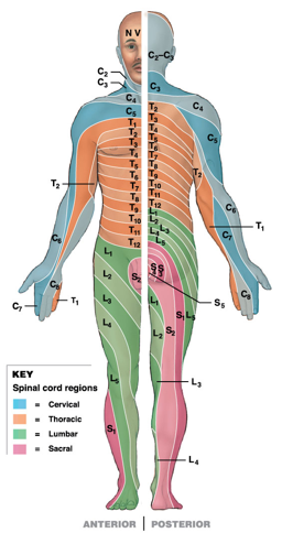
Dermatomes
Peripheral Neuropathies
Regional losses of a sensory or motor function due to trauma or compression and the dermatome provides clues to the region of spinal cord that is injured.
Nerve Plexus
Complex interwoven network of nerves that makes vital connections in the nervous system
Spinal Reflexes
Quick autonomic responses controlled only by the spinal cord and triggered by specific stimuli. The stimuli information is integrated by the spinal cord rather than the information being transmitted to the brain. Can be complex or simple depending on number of neurones in circuit.
Circuit or pathways
Enables actions within the nervous system. Neurones communicate with other functionally related neurones in circuits or neuronal arrangements. Requires two cells with cell bodies, axons, axon terminals, and a synapse.
Synapse
Region of interaction between the axon terminal and the cell body of two different neurones. In a circuit it can be with one neuron and axon terminals synapsing more than one neurone, or multiple neurons where the axon terminals synapse with one neuron.
Neurones in circuit
Interconnected sensory neurons, motor neurons, and interneurons.
Interneurons
Form a neuronal pool to interpret information, as well as interpret the planning and coordination of sensory information and motor commands.
Spinal Reflex Steps
A typical spinal reflex has 5 steps. Arrival of stimulus and activation of receptor, activation of a sensory neuron, information processing in the CNS, activation of a motor neuron, and response by a peripheral effector.
Somatic Reflexes
Associated with involuntary control of the muscular system, such as the stretch reflex (knee jerk reflex)
Visceral Reflexes
Associated with changes in internal systems and can be referred to as autonomic reflexes
Monosynaptic Reflex
A sensory neuron synapses directly with a motor neuron creating one synapse. They are very fast and simple. In the grey matter of the spinal cord the axon terminal of the sensory neuron synapses directly with the cell body of the motor neuron. So, the information travels along the sensory neuron, and once activated it will be integrated and processed in the spinal cord, and then the information for a command to occur will transfer along the motor neuron to the effector.
Stretch Reflex
Tendon hammer activated the receptor (muscle spindle), stretching the muscle spindle which activates the sensory neuron. Stimulation and activation of the motor neuron which then results in rapid contraction of the effector.
Muscle Spindle
Located within skeletal muscle and made up of neuronal fibers or nerve (intrafusal) fibers. Surrounding these fibers are large extrafusal fibers which perform muscle contraction. Tendon hammer stretches the intrafusal fiber, which stimulates the sensory neuron. Information is integrated by the spinal cord which then which then stimulates the motor neuron that activated the extrafusal fibre producing a reflex response.
Muscle Spindle Reflex
When you add a load to your arm, it drops slightly before coming back to normal position. The load is causing the muscle spindle to stretch which activated a sensory neuron. Information as an action potential is the command traveling along the sensory neuron through the root ganglion into the spinal cord through dorsal roots. Sensory neuron then synapses with the motor neuron, which causes the skeletal muscle to contract and arm to return back to normal.
Polysynaptic Reflex
Complicated reflex. A sensory neuron synapses with a pool of interneurons before synapsing with a motor neuron creating multiple synapses, meaning that more than one muscle group can be activated at the same time. This allows information to travel from one spinal segment to another, and although most is integrated at the spinal cord, some is relayed and process in the brain, meaning several reflexes may take place to produce a coordinated, controlled response.
Interneurons
Can be both excitatory post synaptic potentials or inhibitory post synaptic potentials, meaning some muscles can be stimulated while others can be inhibited.
Golgi Tendon Reflex (Polysynaptic Reflex)
Protects muscles from excessively heavy loads by causing the muscle to relax and drop the load. The organ is the sensory receptor with nerve endings interwoven with collagen fibres at the interface of a tendon and skeletal muscle. Stimulating the receptor activated sensory neurons which stimulates inhibitory neurons in the spinal cord that then synapse with the motor neurons inhibiting their activity. Skeletal muscles therefore cannot create enough tension or force to hold the load.
Inhibitory Postsynaptic Potential
When the membrane potential becomes more negative to -90mV, therefore being further away from the threshold value required to stimulate a neuron, which then inhibits it, meaning information can no longer flow through a motor neuron, which enables the neuron to relax.
Withdrawal/Flexor Reflex
Polysynaptic reflex. Moves affected parts of the body away from a stimulus, as one muscle contracts the opposing muscle relaxes (reciprocal inhibition). So, agonist muscle may be contracting, and antagonist muscle may be relaxing helping the contraction take place. So sensory information will activate a pool of interneurons with one stimulatory creating excitatory postsynaptic potential in a motor neuron, whilst an inhibitory interneuron is also stimulated which created inhibitory postsynaptic potential in membrane of motor neuron.
Crossed Extensor Reflex
Flexor reflex and crossed extensor reflex working together at the same time. Collateral polysynaptic reflex. Occurs when you step on something sharp, and the limb goes through the reflex and through flexion to lift up, whilst the other limb straightens and goes through extension to support ourselves.
Ipsilateral Reflex Arcs
The sensory stimulus and motor response occur on the same side
Contralateral Reflex Arcs
The sensory stimulus and motor response occur on opposite sides
Major Regions of the Brain
Cerebrum, cerebellum, diencephalon, brainstem, limbic system, and cranial nerves
Brainstem
Midbrain, the pons, and medulla oblongata
Dinecephalon
Thalamus, hypothalamus, pineal gland, and pituitary gland
Medulla Oblongata
Component of the brainstem involved in transmission of information between the spine and brain. Autonomic (acting subconsciously) nuclei controlling visceral (wet bits within physiology) activities including the cardiovascular and respiratory rhythmicity centre.
Cardiovascular Centre
Adjusts heart rate, heart contractions, and blood flow
Respiratory Rhythmicity Centre
Regulates the breathing rate
The Pons
Important component that links the cerebellum with the midbrain, diencephalon, cerebrum and spinal cord. It is the location of nuclei and tracts involved in processing information that is sent to and from the cerebellum.
Pneumotaxic Respiratory Centre
Inhibits inspiration
Apenustic Respiratory Centre
Stimulates inspiration
Midbrain
Processes and regulates hearing (auditory) and visual information and also controls alertness. There are different sensory nuclei present including, superior colliculi, inferior colliculi, and substantia nigra.
Superior Colliculi
Grouping of sensory nuclei located within the membrane, that processes visual information and controls the reflex movements of eyes, head and neck in response to visual stimuli tracking (ability of our eyes to watch something moving left to right).
Inferior Colliculi
Sensory nuclei that receives hearing information from the medulla oblongata and pons. Also controls the reflex movements of the head, neck, and torso in response to auditory stimuli such as loud noise.
Substantia Nigra
Neuronal activity involved in the subconscious control of muscle tone and learned movements in the cerebrum is inhibited. Parkinson’s disease is associated with the loss of neuronal activity in this sensory nuclei.
Cerebellum
Important component of brain with cauliflower appearance. Coordinates movement at the subconscious level, and coordinated information to maintain body balance and equilibrium through monitoring proprioceptive information (monitoring where we are in space). Fine tunes learned movement such as walking (gait), riding a bicycle, and playing an instrument. Alcohol affects function causing inability to walk in straight line.
Thalamus (Diencephalon)
Filters ascending sensory information to the cerebral cortex. Relays information from cerebellum to the cerebral cortex. Crude recognition of pain, temperature, and pressure.
Cerebral Cortex
Collection of neurons that form the outermost sheet of the brain. Characterised as grey matter at the outermost part of the brain.
Hypothalamus (Diencephalon)
Major regulation of homeostasis, control’s autonomic function, subconscious control of skeletal muscle, coordinates the nervous system and endocrine systems, regulates body temperature, circadian rhythms, emotions and behaviour.
Circadian Rhythm
The regulation of the 24 hour sleep wake cycle
Pineal Gland
Small gland that secretes the hormone melatonin which regulates circadian rhythms (sleep wake cycle)
Limbic System
Functional grouping that establishes emotional states, and links conscious functions of cerebral cortex with autonomic functions of brain stem. Facilitates memory storage and retrieval.
Cerebrum
Largest part of the brain that controls all conscious thoughts and intellectual functions. Processes somatic sensory and motor information.
Cerebrum Parts
4 lobes, the frontal lobe, parietal lobe, temporal lobe, and occipital lobe.
Occipital Lobe
Associated with vision
Temporal Lobe
Associated with hearing
Frontal Lobe
Associated with sensory information
Parietal Lobe
Associated with motor information
Functional Principles of Cerebrum
Each cerebral hemisphere receives sensory information from, and sends motor commands to, the opposite side of the body. The two hemispheres have different functions, although their structures are alike. Correspondence between a specific function and specific region of cerebral cortex is not precise.
Cranial Nerves
12 pairs connected to the brain that extends up from the brain stem to peripheral effectors such as the eyes and nose. Includes sensory nerves, special sensory nerves, motor nerves, and mixed nerves.
Sensory Nerves
Carry somatic sensory information to the brain, including tough, pressure, vibration, temperature, and pain.
Special Sensory Nerves
Carry sensations such as smell, sight, hearing, and balance.
Motor Nerves
Axons of somatic motor neurons that extend from the cranium to allow a response to occur.
Mixed Nerves
Similar to spinal nerves, contain a mixture of motor and sensory fibres/nerves
Receptors
Specialised cells that monitor specific conditions in the body or external environment. Detects a stimulus and converts to an action potential which travels to the CNS via sensory neurons. Classified by the type of stimulus that excites them. Each receptor has a characteristic sensitivity which is called receptor sensitivity, such as a touch receptor vs taste (chemical) receptor.
Sensory Pathway
Stimulus needs to depolarise a sensory receptor for the initiation of an action potential. This stimulus will produce a graded change in the membrane potential of a receptor cell. If this change is strong enough, the receptor will generate an action potential. The information will then be transferred along the axon of the sensory neuron to the CNS where the information will be processed.
Action potential
A signal, piece of information, command.
Depolarization of Sensory Receptor
A stimulus produces a graded change in the membrane potential of a receptor cell.
Action Potential Generation
If the stimulus depolarizes the receptor cell to threshold, action potentials develop in the initial segment.
Propagation
Axons of sensory neurons carry information about the type of stimulus (touch, pressure, temperature) as action potentials to the CNS.
CNS Processing
Information processing occurs at every relay synapse. Sensory information may be distributed to multiple nuclei and centres in the spinal cord and brain.
Modality
A particular way in which something exists. A stimulus can be one of many forms, and this is called (the term).
Receptor Sensitivity
The classification of receptors by the type of stimulus that excites them. For example, a touch receptor will detect stimulation created by touch. They can be stimulated by pain, sound waves, and any one of these forms is called a modality. Once a sensory receptor has been activated the information is transferred along a sensory neuron to particular sensory processing areas in the brain or the primary somatosensory cortex or just sensory cortex.
Nociceptor
Pain and itch
Thermoreceptor
Temperature change (increase and decrease)
Chemoreceptor
Chemical concentrations such as concentration of carbon dioxide.
Mechanoreceptor
Tactile receptors, baroreceptors, and proprioceptors
Tactile receptor
Generally located just under our skin and enable detection of change in touch, pressure, and vibrations.
Baroreceptors
Detects change in organ pressure such as a change in blood pressure detected from within smooth muscle of blood vessels.
Proprioceptors
Muscle contraction and position of joints, so where we are in space, our body balance, and our equilibrium.
Simple sensory receptors structure (e.g. pain)
Have free nerve endings (which have little receptor specificity but may respond to tissue damage by pain sensations), unmyelinated axon, and a cell body. This means the stimulation will depolarise in the membrane in the free nerve endings and potentially create an action with sufficient stimulus and depolarisation, potentially allowing the information or command to travel along the neuron as an action potential.