AE 1.8 - Great Vessels
 Call Kai
 Learn
 Practice Test
 Spaced Repetition
 Match
 Flashcards
 Knowt Play
1/37

There's no tags or description
Looks like no tags are added yet.
38 Terms
Aortic wall (layers & functions)
intima
thin, inner lining
smooth layer for blood flow
media
thick, middle muscle layer
for constriction & dilation
pulsatile flow
adventitia
outer layer
anchor, connect, & support aorta

Major segments of the thoracic aorta
aortic root
ascending aorta
aortic arch
descending aorta

Branches of the aortic arch
innominate artery
right subclavian
right common carotid
left common carotid
left subclavian

Basic function of the aorta as a conduit & reservoir
conduit
high flow of oxygen rich blood to whole body
blood from LV —> systemic circuit of arteries
reservoir
acts as elastic buffer, propels blood forward
expands during systole —> store large part of SV
recoils during diastole —> to maintain continuous blood flow

Standard echocardiographic windows used to evaluate each portion of the aorta
PLAX & PSAX
ascending aorta
descending aorta
Apicals (4, 2, long axis)
descending thoracic aorta
Subcostal
abdominal aorta
Suprasternal
ascending, arch, descending aorta
Right sternal border
ascending aorta (more distal)

Advantages & limitations of TTE for assessing the aorta
advantages
quick
portable
non invasive
can see aortic root well
limitations
sensitivity not sufficient distal to aortic root
descending thoracic aorta imaged less easily and accurately
misses IMH (inter mural hemorrhage) and PAU (ulcer)
Proper aortic measurement technique
Timing
Aortic root & distal: end-diastole (AV closed, one beat before it opens)
Aortic annulus/LVOT: mid-systole (AV open at peak systole)
Measurement conventions & orientation
leading edge to leading edge
made perpendicular to long axis of the aorta
should be interpreted when indexed to an individual’s height & weight
Common locations where specific aortic measurements are obtained
Aortic root (sinus) – largest diameter from the right coronary sinus of Valsalva to the non coronary cusp
Sinotubular junction – measured where the curvature of the sinus transitions to the straight walls of the ascending aorta
Mid-ascending aorta – measure at the level of the right pulmonary artery
Distal ascending aorta – between right PA and the right innominate artery, usually found in RSB window
Aortic arch – between the origin of the right innominate artery and the left subclavian artery
Descending aorta – measure between the left subclavian artery and the diaphragm (proximal, mid and distal descending)
Abdominal aorta – measure between the diaphragm and the iliac bifurcation
Definition of an aneurysm
dilation of aorta ≥ 50% of normal diameter
with intact vessel layers

Common locations of thoracic aneurysms
Ascending aorta
Descending aorta
Aortic arch
Thoracoabdominal

Risk factors & associated conditions (Thoracic Aortic Aneurysms)
Risk factors
Familial/Genetic (20%)
Marfan Syndrome
Bicuspid aortic valve — related aortopathy
Ehlers-Danlos syndrome type IV
Loeys-Dietz syndrome
Acquired
Hypertension
Family History
Atherosclerosis
Trauma
Tobacco use
Advanced age
Male
Types of aneurysms
True
contain all 3 layers
Saccular aneurysm
Fusiform aneurysm
most aortic aneurysms
False
Pseudoaneurysm
rare
contained ruptures of the aorta
most of the aortic wall has been breached
luminal blood is held in only by a thin rim of the remaining wall or adventitia
result of penetrating or blunt trauma, penetrating atherosclerotic ulcers
Dissecting aneurysms
Sinus of valsalva
congenital anomaly where there is weakening of one of the sinuses resulting in dilation
most common is the right sinus

Role of echocardiography in evaluating aneurysms
Identify the presence of aneurysm
Measure max diameter of aneurysm
Define extent of the aneurysm
Determine involvement of the aortic valve, arch vessels
Assess for aortic dissection
Detect mural thrombus (difficult in TTE)
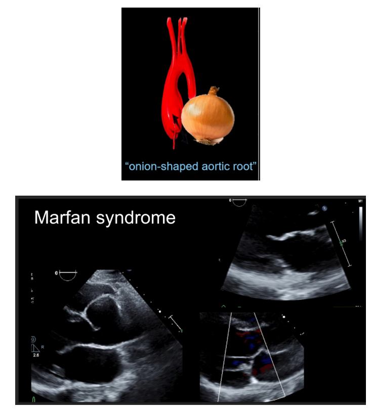
Marfan Syndrome
clinical features
Early glaucoma
Early cataracts
Detached retina
Dislocated lens
Nearsightedness
Spontaneous pneumothorax
Dural ectasia (swelling of the spine)
echocardiographic features
Proximal aorta dilation (onion-shaped)
Sinotubular junction diameter effacement
Pulmonary artery dilation
Myxomatous mitral valve
Aortic Insufficiency
Mitral regurgitation
Aortic dissection
Rupture of Sinus of Valsalva aneurysm
Turner Syndrome
clinical features
Hypertension
Hearing loss
Vision problems
Kidney problems
Autoimmune disorders (celiac disease, Hashimoto’s thyroiditis, diabetes)
Skeletal (Scoliosis, kyphosis, brittle bones)
Learning disabilities
Infertility
echocardiographic features
Associated with bicuspid aortic valve (BAV)
Coarctation of the aorta
Mitral valve prolapse or regurgitation
Cardiac dextroposition
Ventricular septal defect
Thoracic aortic aneurysms and dissections
Most of these patients will also demonstrate abnormal aortic arch anatomy
Because of their stature, aorta size index must be used
Ehlers-Danlos Syndrome (vascular type)
clinical features
Overly flexible joints
Joint dislocations
Early-onset arthritis
Fragile skin may develop scarring
Walls of blood vessels, intestines or uterus can rupture
echocardiographic features
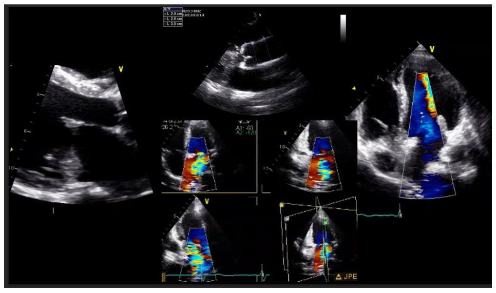
Aortic root dilation
Mitral valve and aortic valve prolapse
Aneurysm of the sinus of Valsalva
Dilatation of the proximal innominate artery
Membranous ventricular septal aneurysm
Loeys-Dietz Syndrome
clinical features
Scoliosis
Premature fusion of the skull bones
Easily bruise
Spontaneous pneumothorax
Hernias
Immune system-related problems (food allergies, asthma, eczema, inflammatory bowel disease)
echocardiographic features
Aortic root aneurysms and dissections
Dissections can occur at dimensions smaller than in other inherited disorders
Marked aorta tortuosity (winding)
Pulmonary artery dilation
Bicuspid Aortic Valve (related aortopathy)
clinical and echocardiographic features
Aortic dilation may occur in 20% to 30% of patients with BAV
Aortic dilation typically involves the aortic root and ascending aorta
Aneurysm may occur even in absence of significant valve dysfunction

Basic pathophysiology (Aortic Dissection)
Stanford classification (Type A vs. Type B)
Major risk factors (Aortic Dissection)
Typical clinical presentation (Aortic Dissection)
Role & limitations of echocardiography (Aortic Dissection)
Characteristics of the true & false lumen
Common complications of Aortic Dissection
General treatment differences between Type A & Type B aortic dissections
Typical location (Coarctation of the Aorta)
Common associated congenital abnormalities (Coarctation of the Aorta)
Echocardiographic findings (Coarctation of the Aorta)
Characteristic Doppler findings (Coarctation of the Aorta)
Normal anatomy & function (Pulmonary Artery)
Normal branching pattern (Pulmonary Artery)
Standard echocardiographic windows used to visualize (pulmonary artery & branch pulmonary arteries)
Normal measurement technique (Pulmonary Artery)
Pulmonary hypertension
basic echocardiographic findings & clinical significance
Pulmonary artery aneurysm
basic echocardiographic findings & clinical significance
Pulmonary artery stenosis
basic echocardiographic findings & clinical significance
Pulmonary embolism
basic echocardiographic findings & clinical significance