Practical 2 Histology
 Call Kai
 Learn
Learn Practice Test
 Spaced Repetition
 Match
Match Flashcards
 Knowt Play
1/18

There's no tags or description
Looks like no tags are added yet.
Name | Mastery | Learn | Test | Matching | Spaced | Call with Kai | Chat |
|---|
No analytics yet
Send a link to your students to track their progress
19 Terms
Smooth Muscle

Connective Tissue

Ciliated Cell

Goblet Cell

Trachea
Lined by resp epithelium, cartilage, smooth muscle, glands, immune tissue, adventitia

Bronchus
Irregular patches of cartilage, limited smooth muscle, sero/mucous glands, resp epithelium


Bronchiole
No cartilage, smooth muscle present, few to no glands, decreasing numbers of goblet cells, resp epithelium in simple ciliated columnar

Alveolar Duct

Respiratory Bronchiole

Terminal Bronchiole

Alveoli
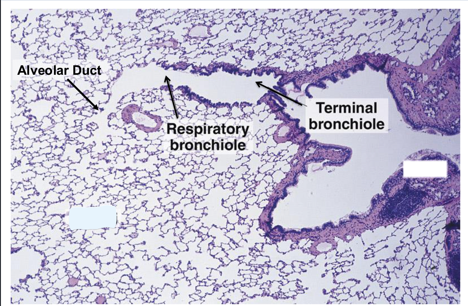
Alveolus

Endocardium
Inner most layer(simple squamous) and underlying CT. Conducting Cells can be found in CT

Myocardium
Cardiac muscle cell layer. Largest layer. Sparse CT between cells

Epicardium
Outermost layer, simple squamous cell layer and underlying CT

Cardiac Myocyte
Branching networks of fibers, single centrally located nucleus. Striation due to presence of sarcomeres. High vascularity

Purkinje Fibers
large diameter, pale staining cytoplasm


Cardiac Skeleton
4 rings of dense fibrous CT

Intercalated Discs
Tight junctions, desmosomes, and gap junctions
