FINISH . ANATOMY CH 14 PERIPHERAL NERVOUS SYSTEM
 Call Kai
 Learn
 Practice Test
 Spaced Repetition
 Match
 Flashcards
 Knowt Play
1/57

There's no tags or description
Looks like no tags are added yet.
58 Terms
sensory receptors
pick up stimuli from inside or outside the body
afferent
nerves
bundles of peripheral axons
cranial
spina
most are mixed— sensory and motor nerves
ganglia
clusters of peripheral neuron cell bodies
motor endings
axon boutons (axon terminals/terminal boutons) of motor neurons
innervates effectors— muscle fibers and glands
efferents
peripheral sensory receptors
picks up sensory stimuli and transmits electrical signals into CNS
two categories
free nerve endings of sensory neurons
monitors general sensory information
complete receptor cells
specialized epithelial cells or small neurons
picks up SPECIAL sensory information
exteroreceptors
sensory receptors by location
picks up info from outside the body
located at or near body surfaces
receptors for touch, pressure, pain, and temperature
receptors of special sense organs
interoreceptors
sensory receptors by location
recieves info from inside the body— viscera
licated in digestive tube, bladder, lungs
monitor variety of stimuli
changes in chemical concentration— gases H+, salts
stretching of tissue
temperature
taste stimuli
proprioceptors
sensory receptor by location
also an encapsulated nerve ending/mechanoreceptor
monitors degree of stretch in locomotory organs
located in
skeletal muscles
tendons
joints
ligaments
sends info to CNS
muscle spindles
type of proprioceptor
measures changing length of a muscle
embedded in perimysium between muscle fascicles
three types of fibers
INTRAFUSAL MUSCLE FIBERS— modified skeletal muscle fibers
ANNULOSPIRAL ENDINGS— located around intrafusal fibers, stimulated by rate and degree of stretch
FLOWER SPRAY ENDINGS— monitors spindle ends (contractile parts) responds only to degree of stretch
golgi tendon organs
type of proprioceptor
located near muscle-tendon junction
monitors tension in tendons
joint kinesthetic receptors
type of proprioceptor
sensory nerve endings in joint capsules
4 types
lamellar corpuscles
bulbous corpuscles
free nerve endings
receptors resembling tendon organs
mechanoreceptors
sensory receptor by stimulus
responds to mechanical forces
touch, pressure, stretch, vibration, itch
BARORECEPTORS— monitors blood pressure
thermoreceptors
sensory receptor by stimulus
detects temperature
chemoreceptors
sensory receptor by stimulus
detects chemicals
taste
chemicals H+, NA+
smell
photoreceptors
sensory receptor by stimulus
responds to light
nociceptors
sensory receptor by stimulus
detects pain
free nerve endings
sensory receptor by structure
also proprioceptors— joint kinesthetic receptors
responds to pain and temperature
itch receptors in dermis
epithelial tactile complexes
modified free nerve ending
slowly adapting mechanoreceptors for light touch
hair follicle receptors
modified free nerve ending
wraps around hair follicle
rapidly adapting mechanoreceptors for light touch
bending of hairs
encapsulated nerve endings
sensory receptor by structure
enclosed in specialized CT capsules
amplifies stimulus or filters out irrelevant stimuli
all mechanoreceptors
tactile (messiner’s) corpuscles
lamellar (pacinian) corpuscles
bulbous corpuscles (ruffini endings)
proprioceptors
tactile (messiner’s) corpuscles
encapsulated nerve ending/mechanoreceptor
spiraling neuron ending surrounded by unipolar schwann cells
located in dermal papillae
rapidly adapting
receptors for fine, discriminative touch
occurs in sensitive, hairless areas of skin
lamellar (pacinian) corpuscles
encapsulated nerve ending/mechanoreceptor
also a proprioceptor— joint kinesthetic receptor
single unipolar neuron ending surrounded by layers of schwann cells
located in dermis
rapidly adapting
senses deep pressure and vibration
bulbous corpuscles (ruffini endings)
encapsulated nerve ending/mechanoreceptor
also a proprioceptor— joint kinesthetic receptor
located in dermis
unipolar neuron ending enclosed in thin, flattened capsule
slowly adapting receptors for pressure and touch
monitors continuous pressure
cranial nerves
nerves that attach to the brain and pass through specific foramina of the skull
CN I and II attach to forebrain; all others attach to brain stem
primarily serves head and neck structures
CN X is the only cranial nerve that extends into abdomen
olfactory CN I

optic CN II

oculomotor CN III

trochlear CN IV

trigeminal CN V

abducens CN VI

facial CN VII
pathway
enters temporal bone through internal acoustic meatus
branches
temporal, zygomatic, buccal, mandibular, cervical

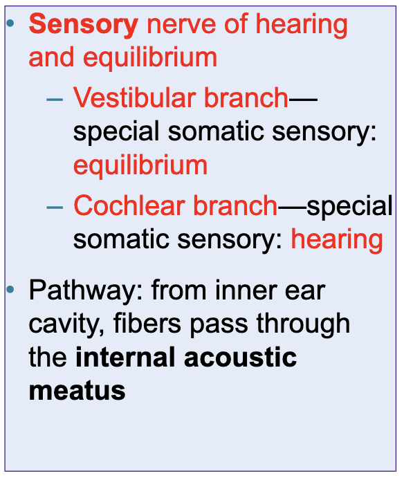
vestibulocochlear CN VIII
glossopharyngeal CN IX
pathway
through jugular foramen to pharynx

vagus CN X

accessory CN XI

hypoglossal CN XII

spinal nerves
30 pairs of nerves that connect to spinal cord
named for where they come out of spinal cord
8 cervical pairs
C1 is superior to first verebra, but C8 is inferior to seventh vertebra
every spinal nerve below cervical region exits inferior to the vertebra of the same number
12 thoracic pairs
5 lumbar pairs
5 sacral pairs
1 coccygeal pair
dorsal root
connects spinal nerves to spinal cord
contains sensory fibers
cell bodies located in the ____ ___ ganglion
ventral root
connects spinal nerves to spinal cord
contains motor fibers
neuron cell bodies arising from anterior gray column
dorsal and ventral rami
spinal nerves branching
both contain sensory and motor fibers
rami communicantes
connects spinal nerves to sympathetic chain ganglia
dorsal rami
innervates deep back muscles
follows a neat segmented pattern in line with their emergence point from vertebral column
ventral rami
innervates anterior thoracic and abdominal wall
only in thoracic region
arranged in simple segmented pattern
runs anteriorly, one deep to each rib— INTERCOSTAL NERVES
each gives off lateral and anterior CUTANEOUS branches to the skin
hilton’s law
states that a joint is innervated by any nerve that innervates the muscles that move it or skin over it
nerve plexus
network of nerves
formed by ventral rami of all spinal nerves
except T2-T12
occur in 4 regions
CERVICAL
BRACHIAL
LUMBAR
SACRAL
fibers from ventral rami converge, diverge, and redistribute
each muscle in a limb receives its nerve supply from more than one spinal nerve

cervical plexus
nerve plexus
deep to sternocleidomastoid, extends into posterior triangle of the neck
formed by ventral rami of first four cervical nerves (C1-C4)
most are sensory cutaneous nerves of the neck, back of head, and superior part of the shoulder
some innervate muscles of the anterior back
phrenic nerve
nerve of cervical plexus
most important nerve of plexus
formed from C3-C5
provides somatic motor and sensory innervation to the diaphragm

brachial plexus
nerve plexus that innervates upper limb
lies partially in neck and axilla
formed by ventral rami of cervical nerves C5-C8 and most of the ventral ramus of T1
may recieve small contributions from C4 or T2
cords give rise to main nerves of the upper limb
roots/rami, trunks, divisions, cords (really tired drink coffee)

lateral and medial cords
brachial plexus cords composed of nerves
musculotaneous
median (both cords)
ulnar
posterior cord
brachial plexus cord composed of nerves
axillary
radial

lumbar plexus
nerve plexus that innervates lower limb
L1-L4
lies within psoas major muscle in the posterior abdominal wall
smaller branches innervate the posterior abdominal wall and psoas muscle
two main branches
femoral nerve
obturator nerve
sacral plexus
nerve plexus that innervates lower limb
arises from L4-S4
caudal to the lumbar plexus
some fibers from the lumbar plexus contribute to this plexus via LUMBOSACRAL TRUNK
the two plexuses are often considered together
nerves branch from SCIATIC NERVE
TIBIAL NERVE
medial and lateral plantar nerves on sole of foot
COMMON FIBULAR NERVE
supplies all of the muscles in the posterior region of lower limb
supplies cutaneous innervation on the posterior leg/sole of foot
superior and inferior gluteual nerves
sacral plexus nerves
innervates gluteal muscles
pudendal nerve
sacral plexus nerve
innervates muscles of the perineum
stimulate erection, voluntary inhibition of defecation and urination
dermatome
region of skin innervated by a single spinal nerve
innervated by all cutaneous branches of a single spinal nerve except C1
named by spinal nerve level
for lower limb— lumbar nerves innervate anterior surface; sacral nerves innervate posterior surface

shingles (herpes zoster)
disorder of the PNS
viral infection
stems from childhood chicken pox
often brought on by stress
mostly experienced by people over age 50
migane headache
disorder of the PNS
relates to sensory innervation of the cerebral arteries
arteries dilate, compress, and irritate sensory nerve endings
peripheral neuropathy
disorder of the PNS
pathological condition of peripheral nerves
symptoms of sensory nerve involvement
parasthesia, pain, burning, loss of sensation
symptoms of motor nerve involvement
muscle weakness and paralysis
causes
trauma, repetitive use
systemic disorders
HIV, diabetes, vitamin B deficiency