The Autonomic Nervous System and Visceral Reflexes
 Call Kai
 Learn
 Practice Test
 Spaced Repetition
 Match
 Flashcards
 Knowt Play
1/69

There's no tags or description
Looks like no tags are added yet.
70 Terms
Autonomic nervous system, ANS (visceral motor system): function and primary targets
A motor nervous system that controls glands, cardiac muscle, and smooth muscle.
Primary target organs of the ANS: Viscera of thoracic and abdominal cavities. Some structures of the body wall—cutaneous blood vessels, sweat glands, and arrector muscles
Function: Carries out actions involuntarily, without our conscious
intent or awareness
• Visceral effectors do not depend on ANS to function, but only to
adjust their activity to the body’s needs
• Severing ANS nerves to organs results in exaggerated responses
(denervation hypersensitivity).
Viceral reflexes
Unconscious, automatic, stereotyped responses to stimulation involving visceral receptors and effectors; controlled by ANS
Visceral reflex arc:
1. Receptors—nerve endings that detect stretch, tissue damage, blood chemicals, body temperature, and other internal stimuli
2. Afferent neurons—lead to CNS.
3. Integrating center—interneurons in the CNS.
4. Efferent neurons—carry motor signals away from the CNS.
5. Effectors—carry out end response.

Name the two subsystems of the ANS
sympathetic and parasympathetic
Sympathetic division
• Prepares body for physical activity: exercise, trauma, arousal,
competition, anger, or fear; fight-or-flight reaction
• Increases heart rate, BP, airflow, blood glucose levels, etc.; reduces
blood flow to the skin and digestive tract
Parasympathetic division
• Calms many body functions reducing energy expenditure and
assists in bodily maintenance; resting and digesting state
• Digestion and waste elimination
Autonomic tone
Normal background rate of activity that represents the balance of the two systems according to the body’s needs.
• Neither division has universally excitatory nor inhibitory
effects.
Examples of parasympathetic tone:
• Maintains smooth muscle tone in intestines
• Holds resting heart rate down to about 70 to 80 beats per minute
Example of sympathetic tone:
• Keeps most blood vessels partially constricted and maintains blood
pressure
How is ANS involved in both PNS and CNS
ANS has components in both the central and peripheral
nervous systems
• Control nuclei in the hypothalamus and other brainstem
regions
• Motor neurons in the spinal cord and peripheral ganglia
• Nerve fibers that travel through the cranial and spinal
nerves.
ANS motor pathway (vs somatic motor pathway)
• In somatic pathway, a motor neuron from brainstem or
spinal cord issues a myelinated axon that reaches all the
way to skeletal muscle
• In autonomic pathway, a signal must travel across two
neurons to get to the target organ, and cross a synapse
where these two neurons meet in an autonomic ganglion
• First neuron in pathway: cell body in the brainstem or spinal cord;
its axon (preganglionic fiber) extends to autonomic ganglion
• At ganglion, first neuron synapses with second neuron; ACh is the
neurotransmitter
• Second neuron in pathway: cell body in autonomic ganglion; its
axon (postganglionic fiber) extends to target cells;
neurotransmitter is either ACh or NE.
The ANS nerve exits through th lateral horn on the spinal cord.

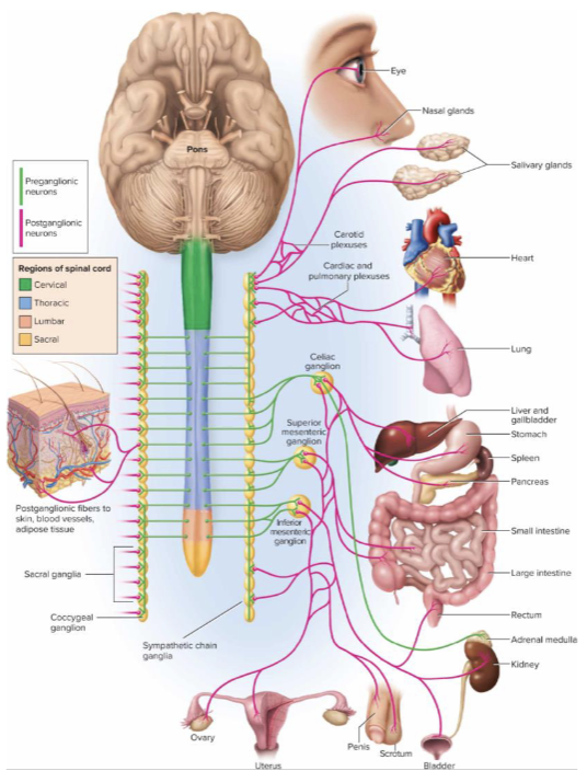
Anatomy of the sympathetic division:
• Its pathways arise from thoracic and lumbar regions of the
spinal cord (so also called thoracolumbar division)
• Relatively short preganglionic and long postganglionic
fibers
Thoracolumbar NS division
AKA sympathetic NS Its pathways arise from thoracic and lumbar regions of the spinal cord (so also called thoracolumbar division)
Sympathetic NS division general pathway
• Cell bodies of preganglionic neurons in lateral horns and nearby
regions of spinal cord gray matter; fibers exit spinal cord by way of
spinal nerves T1 to L2
• Lead to nearby sympathetic chain of ganglia (paravertebral
ganglia)—a series of longitudinal ganglia adjacent to both sides of
the vertebral column from cervical to coccygeal levels
• Usually 3 cervical, 11 thoracic, 4 lumbar, 4 sacral ganglia, and 1
coccygeal ganglion
• Sympathetic nerve fibers are distributed to every level of the body.
Image of The Sympathetic Chain Ganglia

Schematic of the Sympathetic Nervous System
Communicating rami
Each paravertebral ganglion is connected to a spinal
nerve by two branches: communicating rami
Preganglionic fibers and White communicating ramus
Preganglionic fibers are small myelinated fibers that travel from
spinal nerve to the ganglion by way of the white communicating
ramus (myelinated)
Postganglionic fibers and the gray communicating ramus (unmyelinated))
Postganglionic fibers leave the ganglion by way of the gray
communicating ramus (unmyelinated), and by other routes.
• Gray ramus returns to the spinal nerve
• Postganglionic fibers extend the rest of the way to the target organ
Draw out the general neuropatways through the sumpathetic chain ganglion
From the lateral horn of gray matter of the spinal cord trhough the spinal nerve through the white ramus of the communicatng rami then there are 4 options from there: (1) it synapse iwth a postganglionic neruon and that axon then travels through the white ramus into the spinal nerves, (2) it travels up or down the sympathetic nerve/ other sympathetic ganglions, (3) travels throiugh the splanchnic nerve.

Abdominal aortic plexus: anatomy (three collateral ganglia)
Collateral ganglia contribute to a network called the abdominal aortic plexus
• Wraps around abdominal aorta
• Three major collateral ganglia in this plexus
• Celiac, superior mesenteric, and inferior mesenteric ganglia
• Located at points where arteries with the same names branch off aorta
• Postganglionic fibers accompany these arteries and their branches to the target organs
Solar plexus
Collective name for the celiac and superior mesenteric ganglia
• Nerves radiate from ganglia like rays of the sun

Neuronal divergence in the sympathetic division
Neuronal divergence predominates in the sympathetic
division
• Most sympathetic preganglionic neurons synapse with 10
to 20 postganglionic neurons
• One preganglionic neuron can excite multiple
postganglionic fibers leading to different target organs
• Has relatively widespread effects.
Adrenal (suprarenal) glands: adrenal cortex, and adrenal medulla.
Paired glands located on superior poles of the kidneys; each is two glands with different
functions:
• Adrenal cortex—outer layer; secretes steroid hormones
• Adrenal medulla—inner core; essentially a sympathetic
ganglion. Consists of modified postganglionic neurons without dendrites,
axons.
Adrenal medullaL what nerve stimulate it and what hormones does it secrete
• Stimulated by preganglionic sympathetic neurons
• Secretes a mixture of hormones into bloodstream: the
catecholamines—85% epinephrine (adrenaline) and 15%
norepinephrine (noradrenaline).
Sympathoadrenal system
Sympathoadrenal system—collective name for adrenal medulla
and sympathetic nervous system; closely related in development
and function.
general Anatomy of the parasympathetic division: ganglionic lengths
• Its pathways arise from the brain and sacral regions of the
spinal cord (so also called craniosacral division)
• Relatively long preganglionic and short postganglionic
fibers
General pathway fro the parasympathetic division
• Cell bodies of preganglionic neurons in midbrain, pons, and medulla, or sacral spinal cord segments S2 to S4
• Preganglionic fibers travel through cranial or sacral nerves, end in terminal ganglia in or near target organs (intramural ganglion—embedded within organ wall)
• Synapse with postganglionic neurons in ganglia
• Postganglionic fibers cover rest of the (short) distance to target organ.
Neural divergence of the parasympathetic NS
Parasympathetic division is relatively selective in
stimulation of target organ—very little neural divergence
Parasympathetic fibers leave the brainstem in the
following cranial nerves:
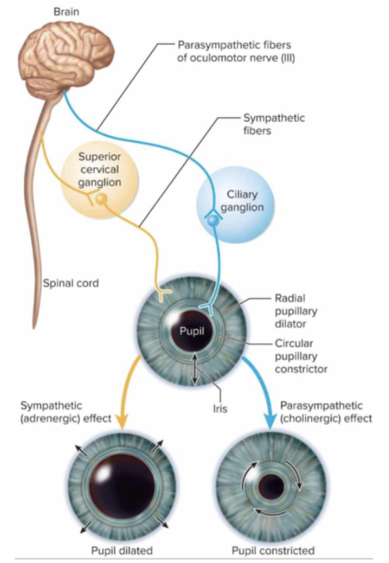
• Oculomotor nerve (CN 3)—thicken lens and narrow pupil of eye
• Facial nerve (CN 7)—regulate tear, nasal, and salivary glands
• Glossopharyngeal nerve (CN 9)—regulate parotid salivary gland
• Vagus nerve (CN 10)—innervate viscera as far as proximal half of
colon; cardiac, pulmonary, and esophageal plexuses in chest
give off anterior and posterior vagal trunks.
What area of the spine are parasympathetic fibers in the spinal cord
Remaining parasympathetic fibers arise from S2 to S4 of
spinal cord
Spinal nerve pathway fo parasympathetic division
• Fibers travel in anterior rami of spinal nerves, then form pelvic
splanchnic nerves that lead to inferior hypogastric plexus
• Some fibers synapse at this plexus; most pass through, travel
through pelvic nerves to terminal ganglia.
What are the target organs of the
Target organs: distal half of colon, the rectum, urinary bladder, and
reproductive organs.

Enteric plexus (Enteric NS)
the nervous network of the digestive tract
• Composed of 500 million neurons found in the walls of the
digestive tract (5× more neurons than spinal cord)
• Unlike rest of ANS, it does not arise from the brainstem or
spinal cord
• Like the rest of ANS, it innervates smooth muscle, glands
• Has its own ganglia and reflex arcs
• Regulates motility of esophagus, stomach, and intestines
and secretion of digestive enzymes and acid
• Normal digestive function also requires regulation by
sympathetic and parasympathetic systems.
Megacolon
massive dilation of bowel accompanied by
abdominal distension and chronic constipation
Hirschsprung disease
hereditary defect causing absence of enteric plexus
• No innervation in sigmoid colon and rectum
• Constricts permanently and will not allow passage of feces
• Feces becomes impacted above constriction
• Megacolon—massive dilation of bowel accompanied by abdominal distension and chronic constipation
• May be colonic gangrene, perforation of bowel, and infection of peritoneum (peritonitis)
• Usually evident in newborns who fail to have their first bowel movement.
Divisions of ANS often have opposite effects on an organ
Two reasons this is possible:
• Sympathetic and parasympathetic fibers secrete different
neurotransmitters (norepinephrine and acetylcholine)
• The receptors on target cells vary. Target cells respond to the same neurotransmitter differently depending on the type of receptor they have for it. There are two different classes of receptors for acetylcholine and two classes of receptors for norepinephrine.
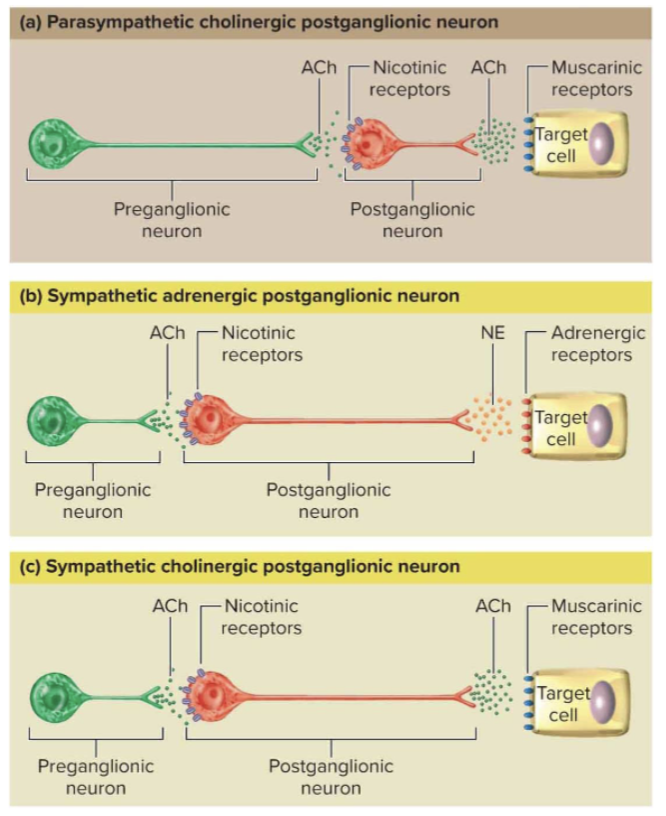
Acetylcholine (ACh) in the context of ANS
is secreted by all preganglionic neurons in both divisions and by postganglionic parasympathetic neurons
Cholinergic neruons
Neurons that secrete ACh are called cholinergic neurons
Chlinergic receptor
Any receptor that binds ACh is called a cholinergic
receptor.
Muscarinic receptors: definition and example.
Found on cardiac muscle, smooth muscle, and gland cells with cholinergic innervation
• Act through second-messenger systems
• Receptor subtypes exist, often providing contrasting effects on
organs
• Example: ACh excites intestinal smooth muscle, but inhibits
cardiac muscle
Nicotinic receptors
Found on postganglionic neuron cell bodies in autonomic ganglia; on cells of adrenal medulla; and neuromuscular junction of skeletal muscle fibers
• Binding of ACh to receptor is always excitatory
• Opens ligand-gated ion channels, produce excitatory postsynaptic potential.
Norepinephrine (NE)
is secreted by nearly all sympathetic postganglionic neurons
Adrenergic fibers
Nerve fibers that secrete NE are called adrenergic fibers
Adrenergic receptors
Receptors for NE are called adrenergic receptors
Alpha-adrenergic receptors
• Usually excitatory
• Two subclasses use different second messengers (α1 and α2)
Beta-adrenergic receptors
• Usually inhibitory
• Two subclasses with different effects, but both act through cAMP as a second messenger (β1 and β2).
Autonomic effects on glandular secretion
are often an indirect result of their effect on blood vessels
• Increased blood flow, increased secretion
• Decreased blood flow, decreased secretion
How long do affects last on sympathetic evrsus parasympathetic? and Why?
Sympathetic effects tend to last longer than
parasympathetic effects
• NE released by sympathetic fibers can be reabsorbed by axon
terminal, diffuse to adjacent tissues, or pass into bloodstream
• ACh from parasympathetic fibers broken down quickly at synapse
What are some other neuotransmitters that ANS fibers release?
Some ANS fibers release other neurotransmitters in
addition to ACh or NE
• Examples: enkephalin, substance P, neuropeptide Y, somatostatin,
neurotensin, gonadotropin-releasing hormone, nitric oxide (NO).
Neurotransmitters and Receptors of the Autonomic
Nervous System.
Dual innervation
most viscera receive nerve fibers from both parasympathetic and sympathetic divisions.
Even when both division innervate a single organ,
innervation is not always equal or exert equal influence.
Dual innervation not always necessary for ANS to produce
opposite effects in on an organ
Dual innervation Antagonistic effects
oppose each other
• Can be exerted through dual innervation of same effector cells
(example: heart rate)
• Can be exerted because each division innervates different cells
(example: muscles of iris)
Dual innervation Cooperative effects:
two divisions act on different effectors to produce a unified overall effect
• Example: saliva production—parasympathetic stimulates serous
cell secretion and sympathetic stimulates mucous cell secretion
Dual Innervation pathway of the Iris
Control without dual innervation. What effectors receive only sympathetic fibers
Adrenal medulla, arrector muscles, sweat glands, many blood
vessels
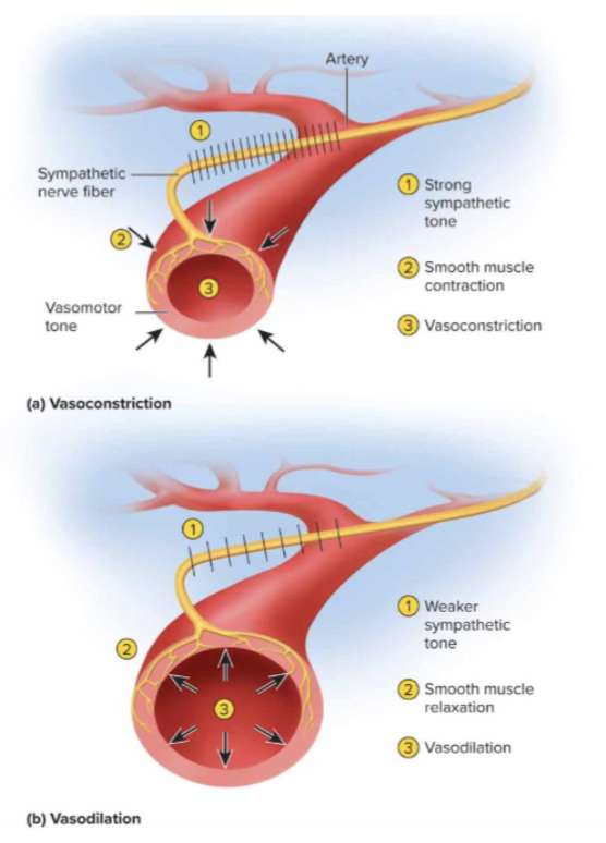
Vasomotor tone
vessels in a continual state of partial constriction
Control without dual innervation: regulation of blood pressure and routes of
blood flow. Vasodialation and vasoconstriction.
• Baseline sympathetic tone maintains vasomotor tone—vessels in a continual state of partial constriction
• Increase in firing frequency—vasoconstriction; decrease in firing frequency—vasodilation
• Can also shift blood flow from one organ to another as needed
• During stress, blood vessels to muscles and heart dilate while blood
vessels to skin constrict.
The ANS is regulated by several levels of the CNS: name the structures of the CNS that ANS is regularted by.
Cerebral cortex and limbic system, hypothalamus, midbrain, pons, and medulla oblongata, spinal cord.
Cerebral cortex and limbic system in flunce on ANS
emotions influence the ANS because of the connections between our limbic system and the hypothalamus
Hypothalamus influence on ANS:
contains nuclei for primitive functions—hunger, thirst, sex
Midbrain, pons, and medulla oblongata affects on ANS:
contain nuclei for cardiac and vasomotor control, salivation, swallowing, sweating, bladder control, and pupillary changes
Spinal cord affect on ANS:
reflexes for defecation and micturition
• We control these functions because of our control over skeletal muscle
sphincters; if the spinal cord is damaged, the smooth muscle of bowel and
bladder is controlled by autonomic reflexes built into the spinal cord.
Neuropharmacology
study of effects of drugs on the nervous system
Sympathomimetics
enhance sympathetic activity
• Stimulate receptors or increase norepinephrine release
• Cold medicines that dilate the bronchioles or constrict nasal blood
vessels
Sympatholytics
suppress sympathetic activity
• Block receptors or inhibit norepinephrine release
• Beta-blockers reduce high BP interfering with effects of epinephrine
and norepinephrine on heart and blood vessels
Parasympathomimetics and parasympatholytics
enhance parasympathetic effects, while parasympatholytics suppress them
Prozac
a selective serotonin reuptake inhibitor (SSRI)—blocks
reuptake of serotonin to prolong its mood-elevating effect
MAO inhibitors
block enzyme monoamine oxidase (MAO) to slow the breakdown of monoamine neurotransmitters; aid in treating depression
Caffeine
competes with adenosine (the presence of which causes sleepiness) by binding to its receptors.