Lecture #23: Digestive system (II): Alimentary canal
 Call Kai
 Learn
Learn Practice Test
 Spaced Repetition
 Match
Match Flashcards
 Knowt Play
1/144

There's no tags or description
Looks like no tags are added yet.
Name | Mastery | Learn | Test | Matching | Spaced | Call with Kai | Chat |
|---|
No analytics yet
Send a link to your students to track their progress
145 Terms
1. Ingestion
2. Motility
3. Secretion
4. Hormone release
5. Chemical digestion
6. Absorption
7. Elimination
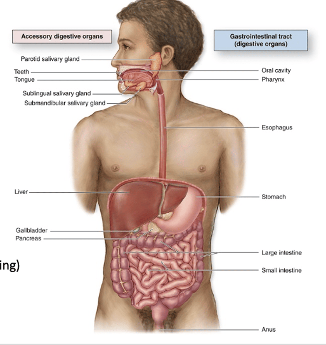
What are the functions of the digestive tract? (7)
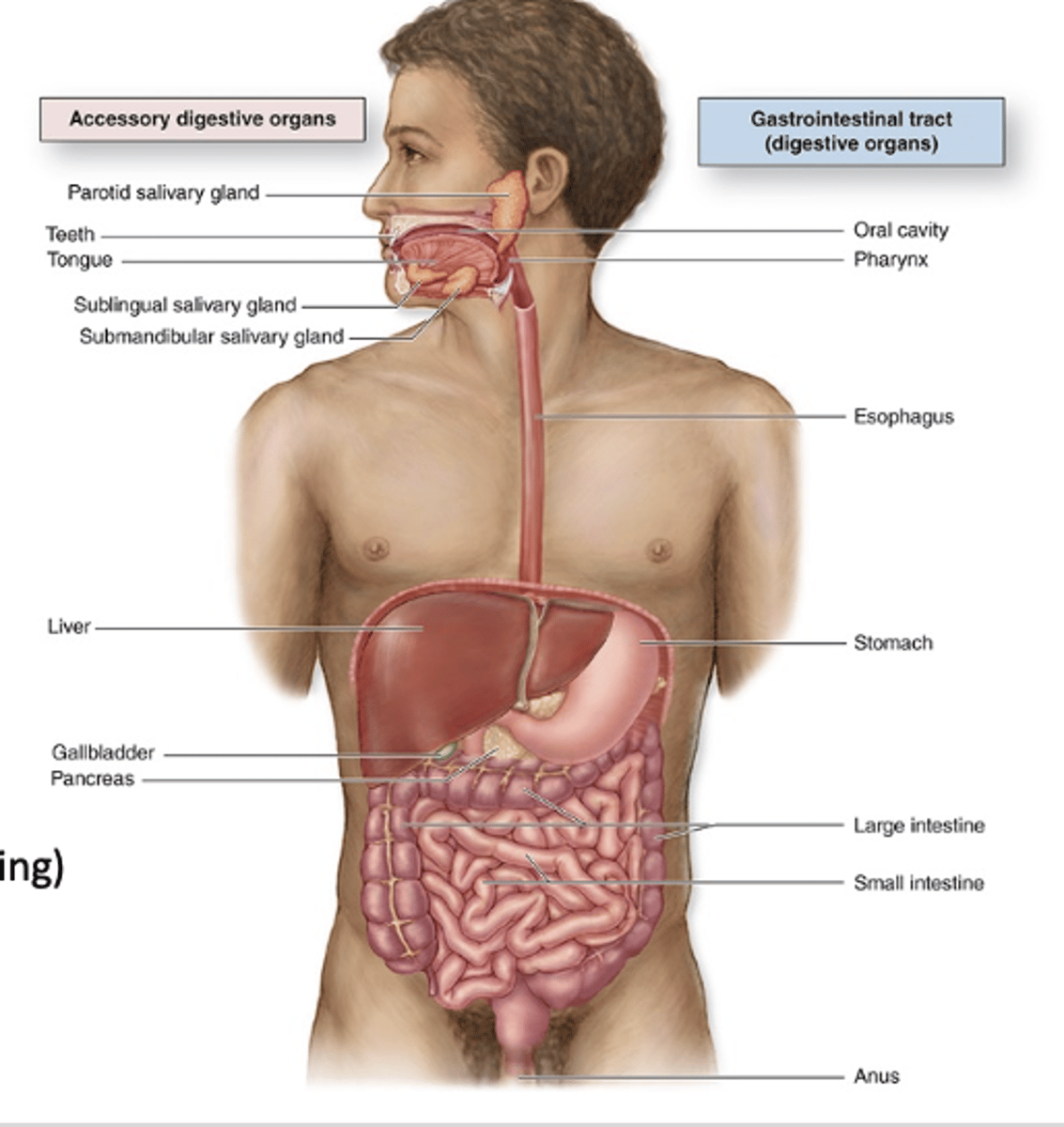
Mechanical digestion such as mastication, mixing, and churning.
Chemical digestion (enzymes)
What do accessory digestive organs do?
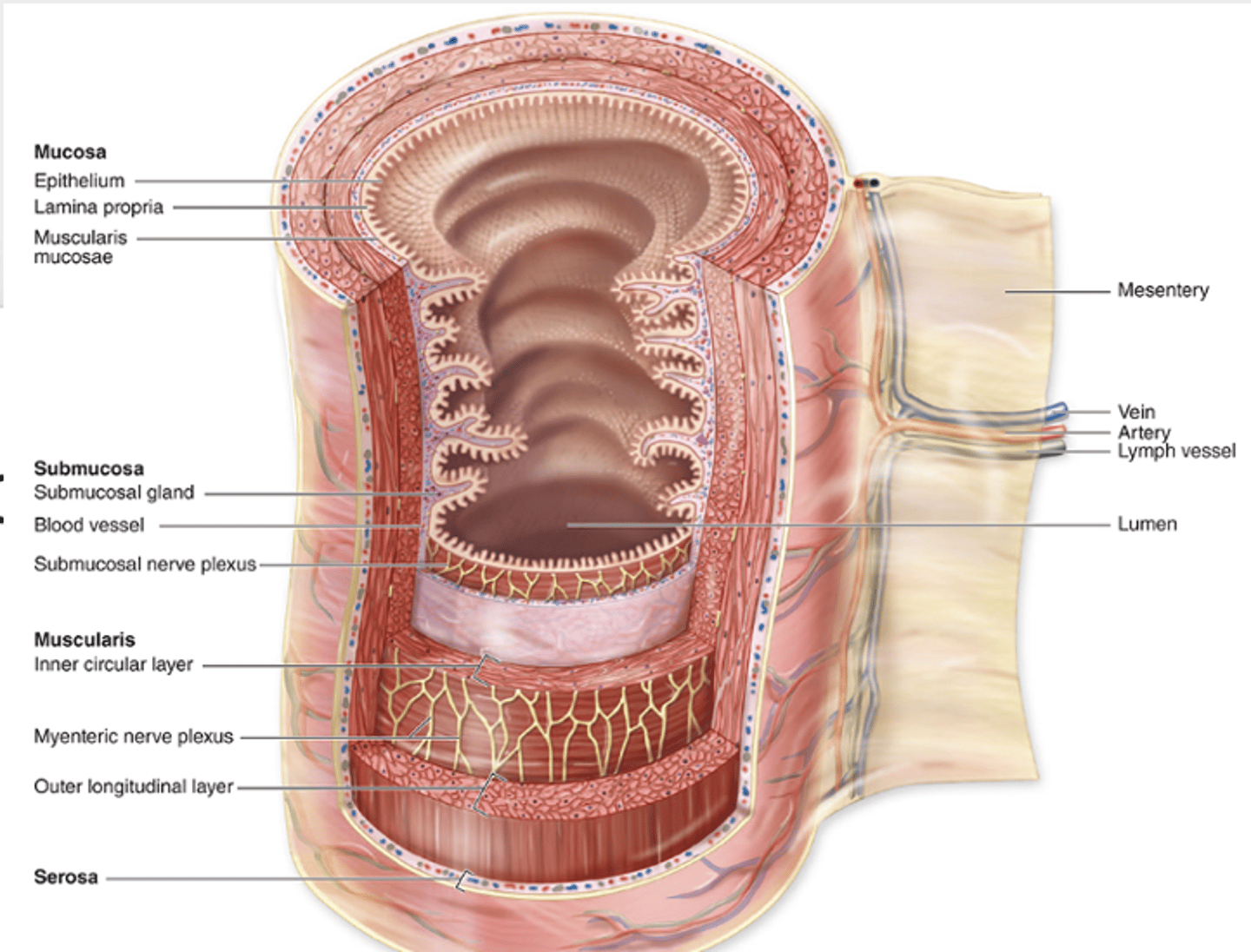
1. Epithelial lining
2. lamina propria (loose CT)
3. muscularis mucosae.
What are the three components of the mucosa in the digestive tract?
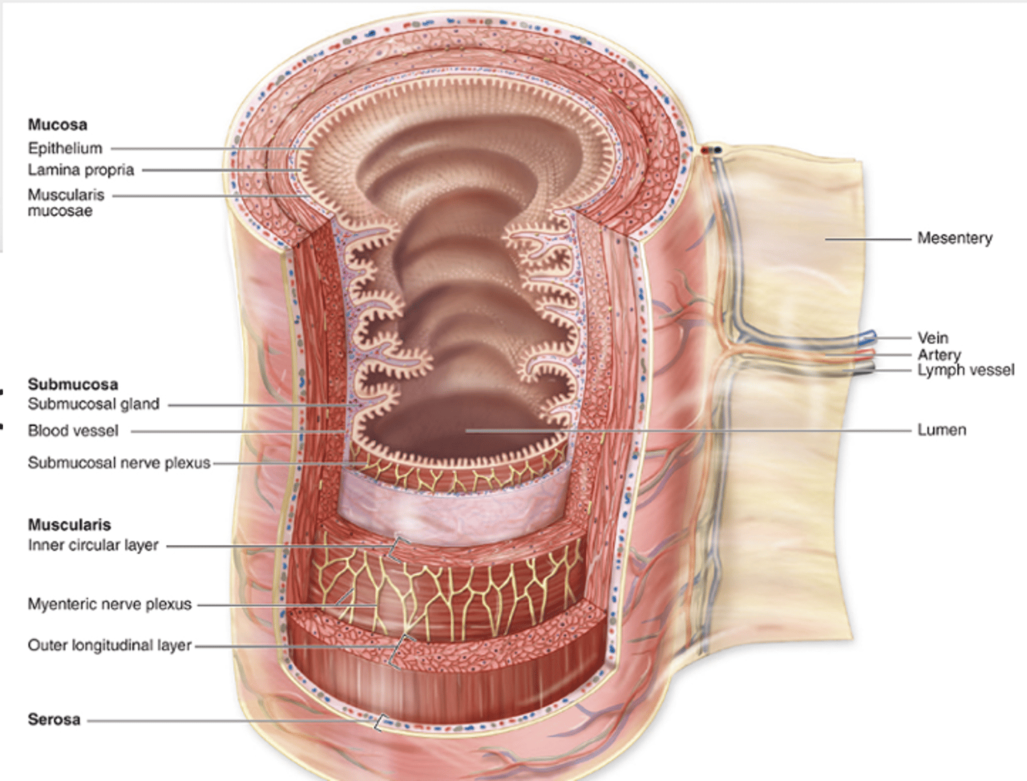
Blood vessels, lymphatics, glands, and lymphocytes.
What structures are found in the lamina propria in the digestive tract? (4)
Denser connective tissue.
What type of connective tissue is found in the submucosa?
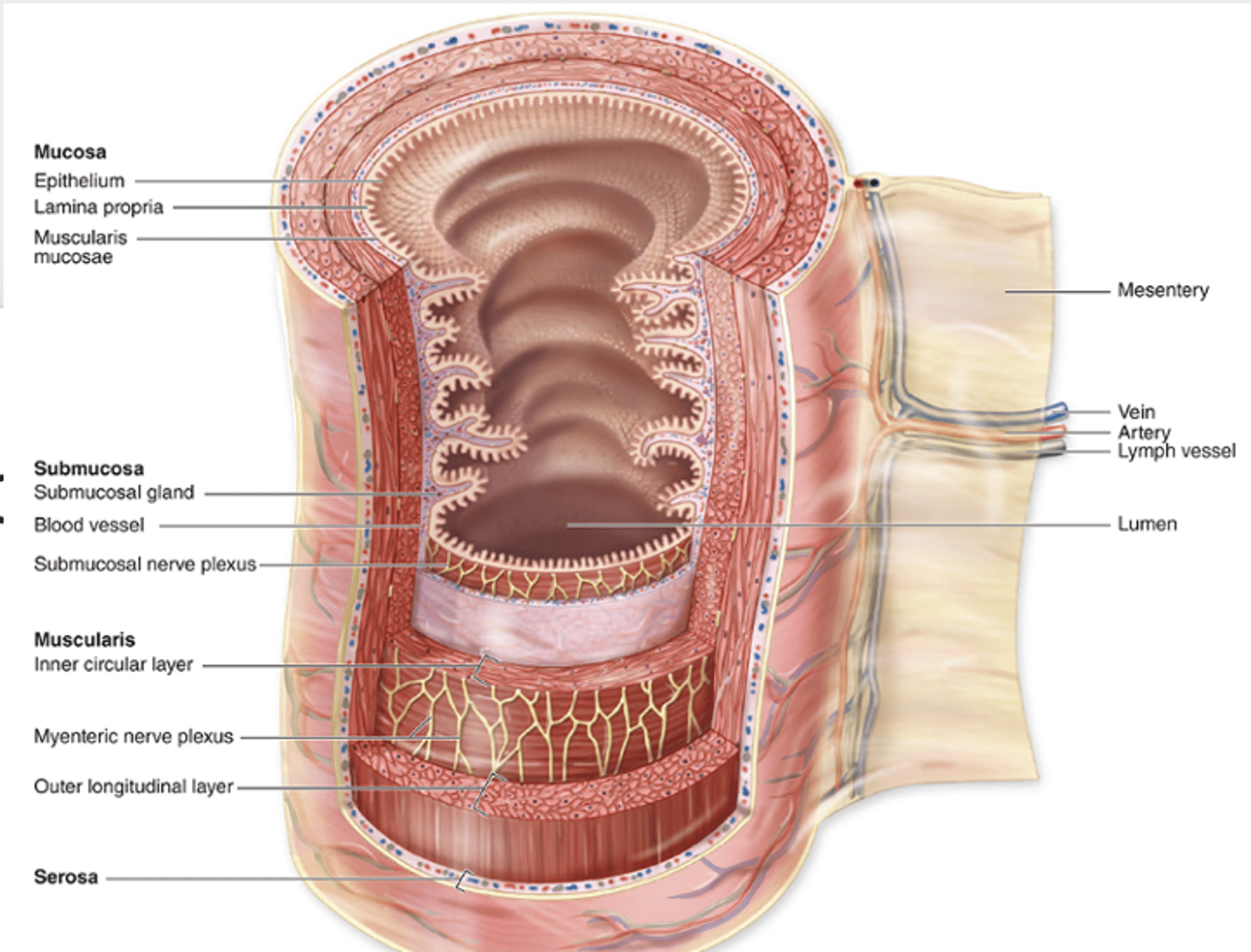
1. Submucosal plexus
2. glands in the duodenum
3. Peyer patches in the ileum
What structures are found in the submucosa of the digestive tract? (3)
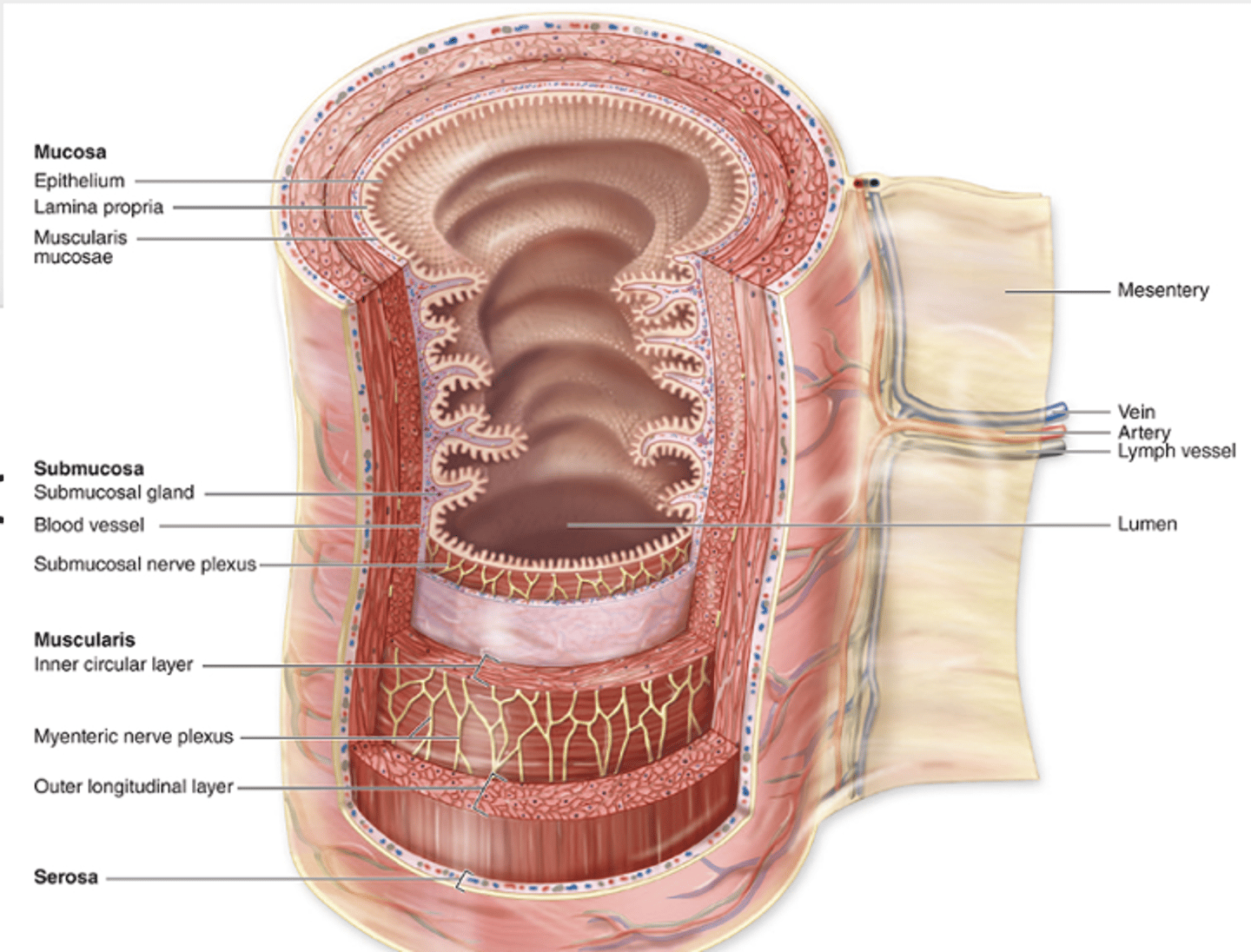
Smooth muscle.
What type of muscle makes up the muscularis externa?
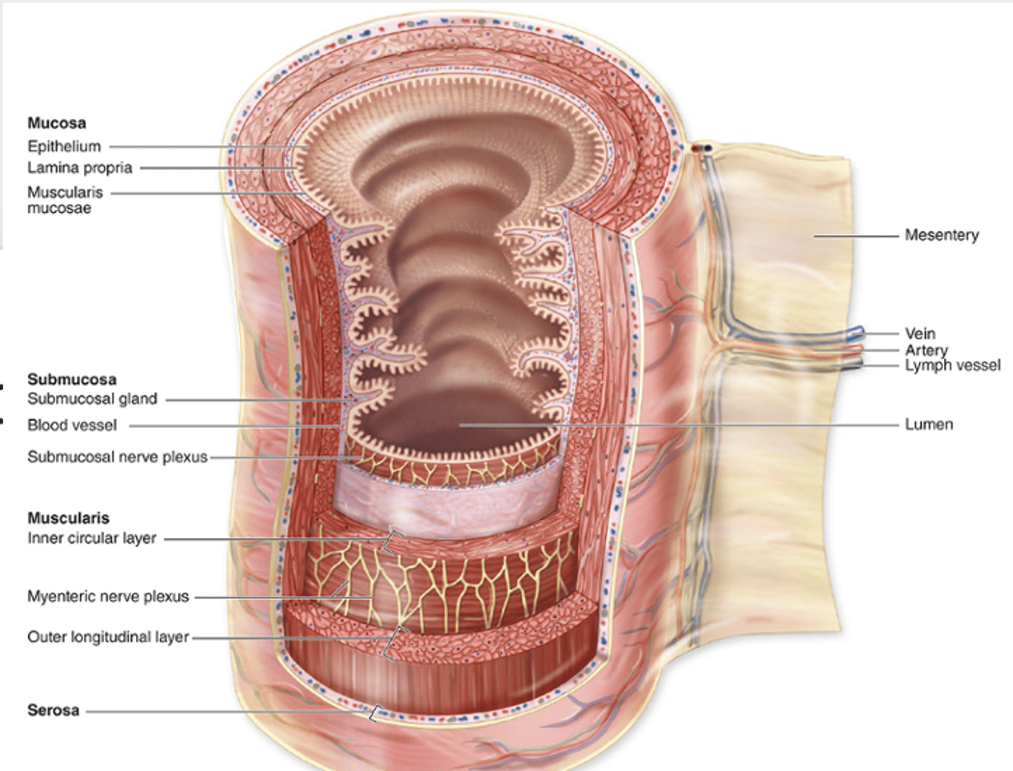
Two layers, except in the stomach where there are three.
How many layers are in the muscularis externa?
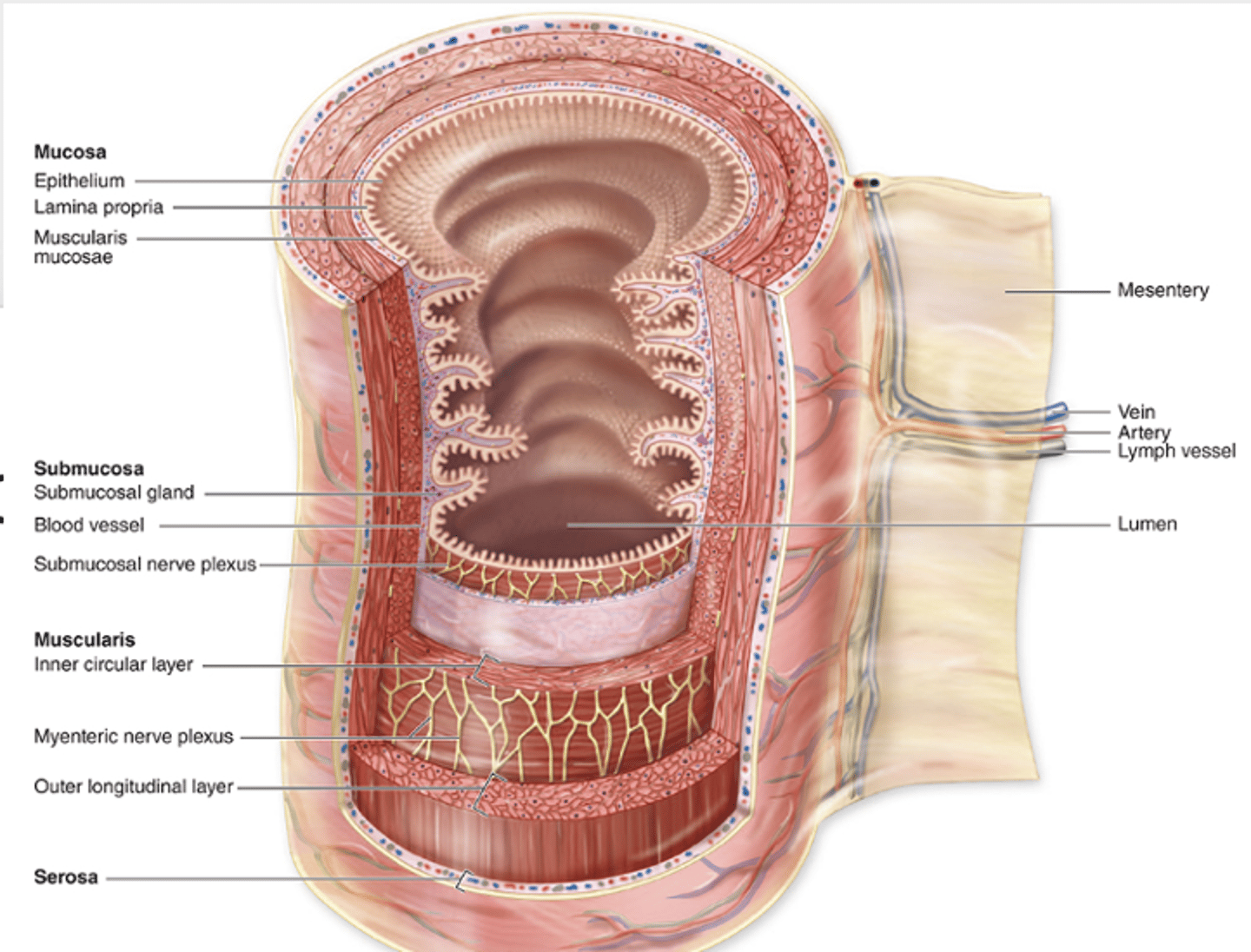
The myenteric plexus.
What neural plexus is found in the muscularis externa?
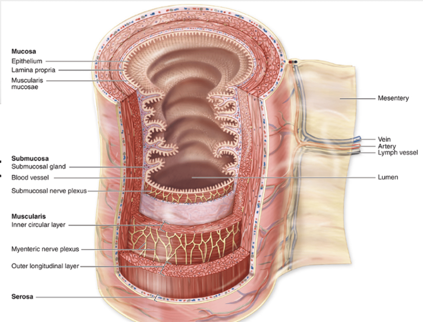
A thin layer of loose connective tissue containing blood vessels, lymphatics, and adipose tissue.
What does the serosa consist of?
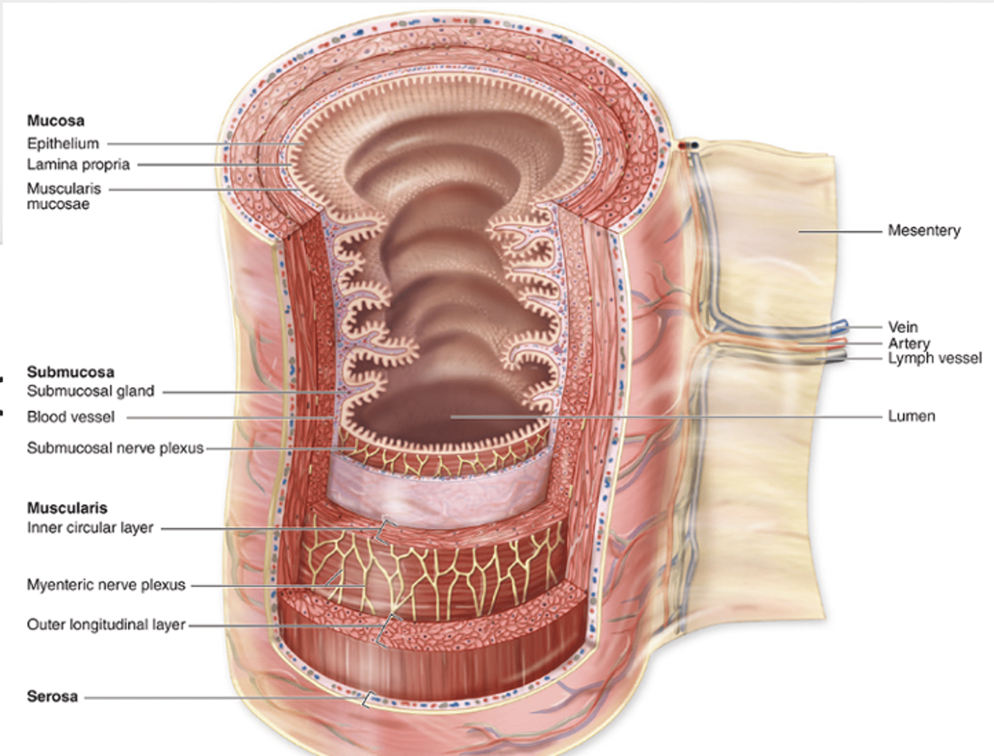
Simple squamous mesothelium.
What lines the serosa?
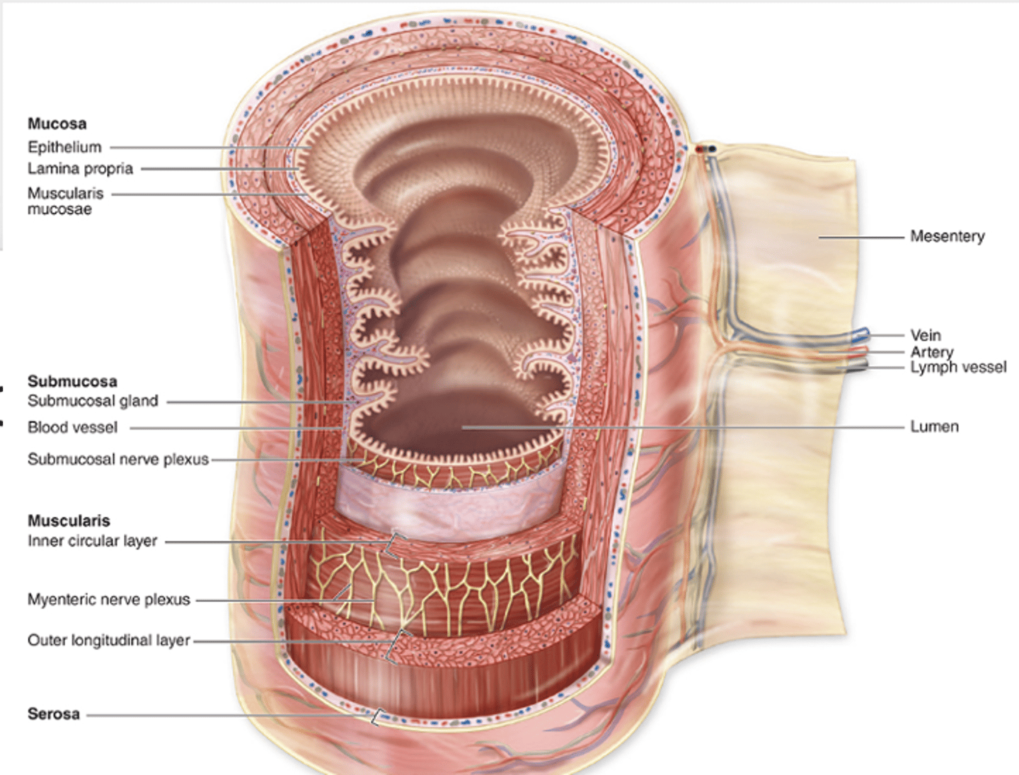
A connective tissue covering found in the esophagus and extraperitoneal organs.
What is the adventitia and where is it found?
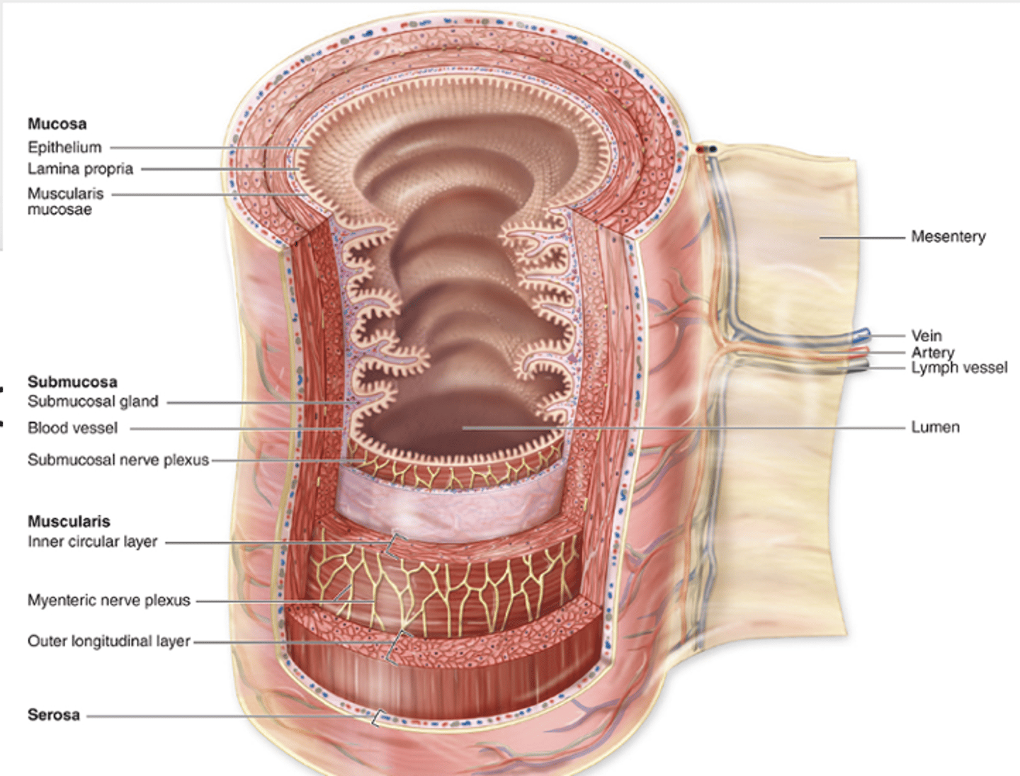
1. Mucosa
2. submucosa
3. muscularis externa
4. Adventita (INSTEAD OF SEROSA)
What are the four well-delimited layers of the esophagus?
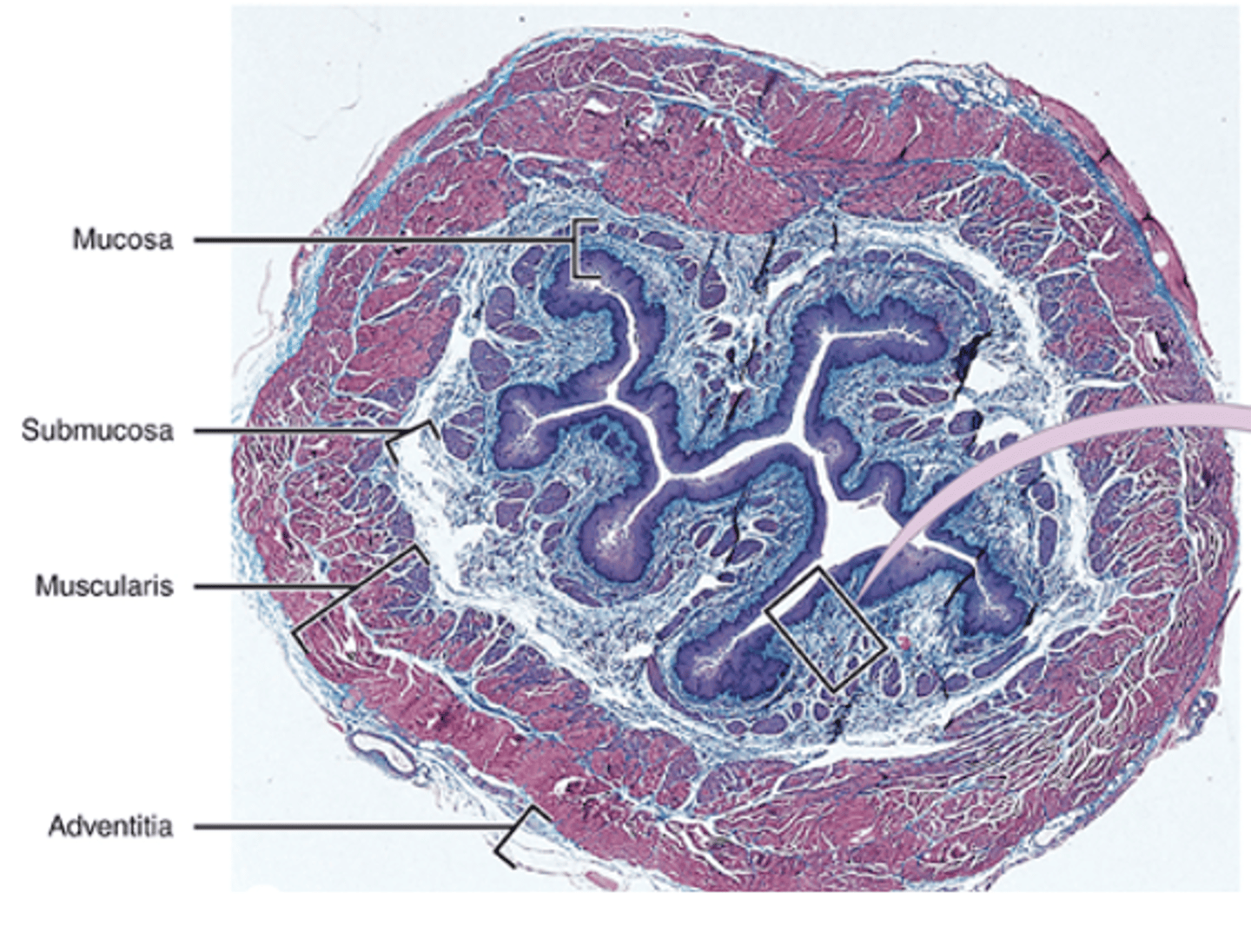
It is folded.
What happens to the esophageal mucosa when the esophagus is collapsed?
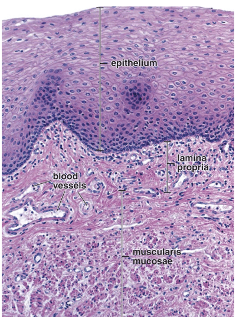
Nonkeratinized stratified squamous epithelium.
What type of epithelium lines the esophagus?
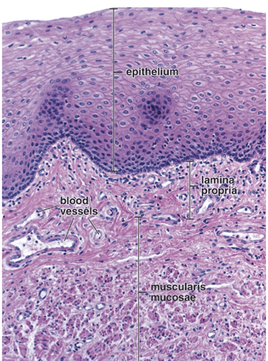
Scattered lymphocytes
some lymphatic nodules
blood vessels
nerves
Lymphatic vessels.
What structures are found in the lamina propria of the esophagus?
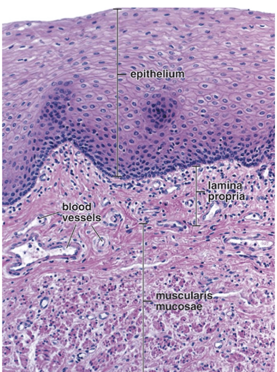
Strands of longitudinal smooth muscle fibers.
What type of muscle fibers make up the muscularis mucosae of the esophagus?
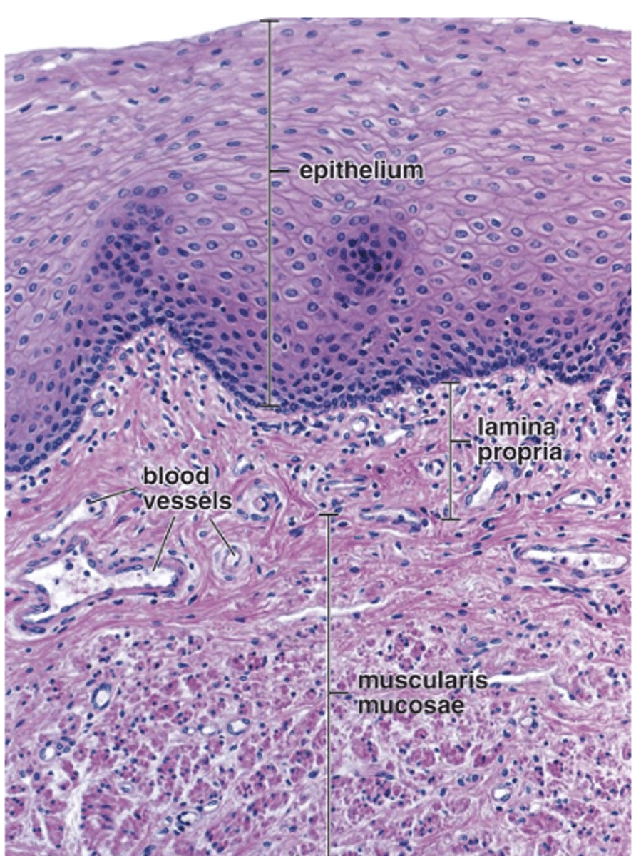
The proximal region.
In which region of the esophagus is the muscularis mucosae particularly thicker?
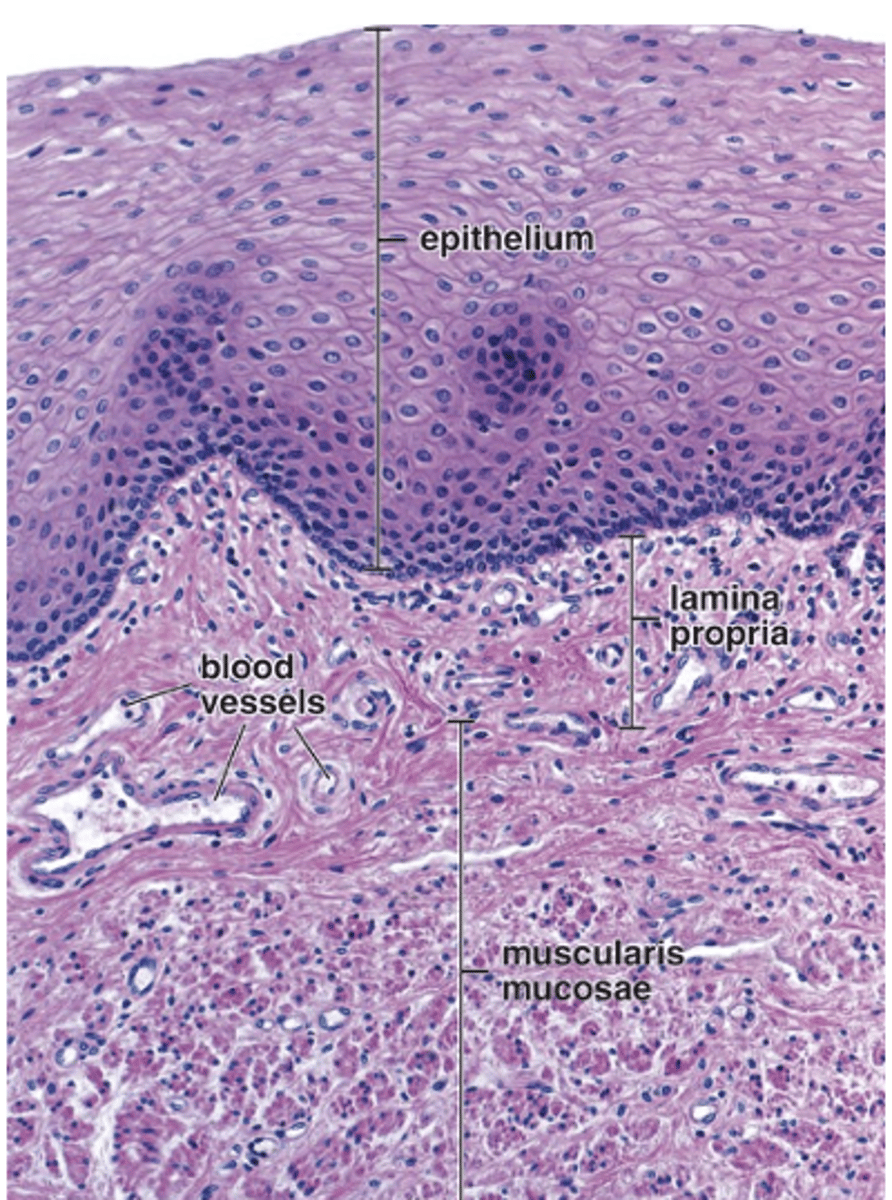
Dense irregular connective tissue.
What type of connective tissue is found in the submucosa of the esophagus?
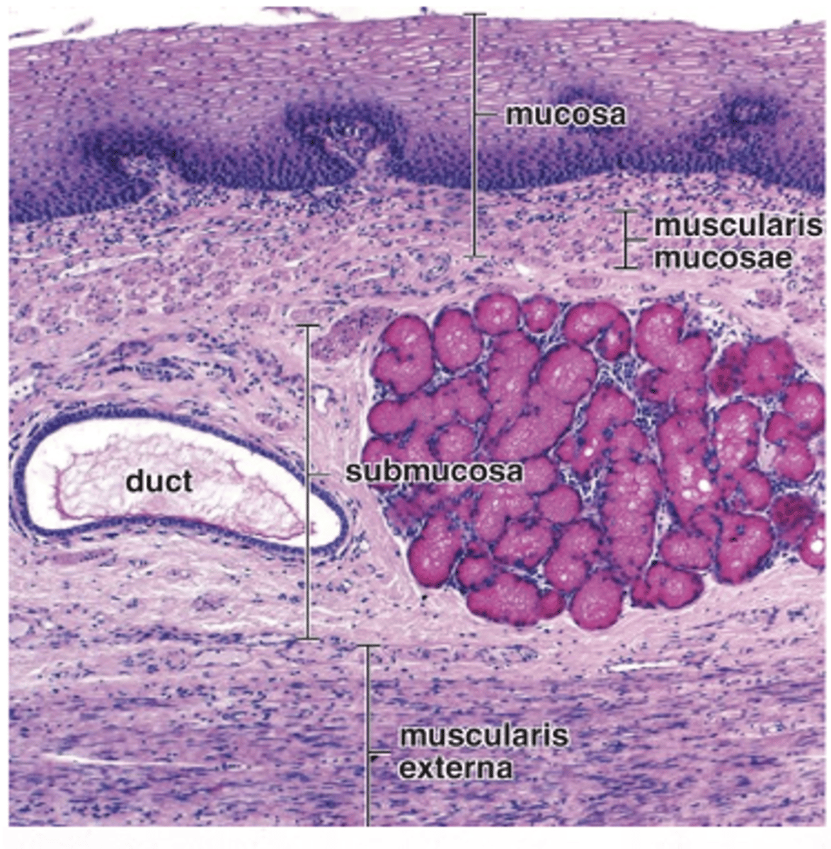
Large blood and lymphatic vessels.
What large vessels are found in the esophageal submucosa?
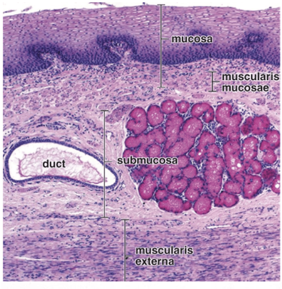
Nerve fibers and ganglion cells (submucosal or Meissner’s plexus).
What neural elements are present in the submucosa of the esophagus?
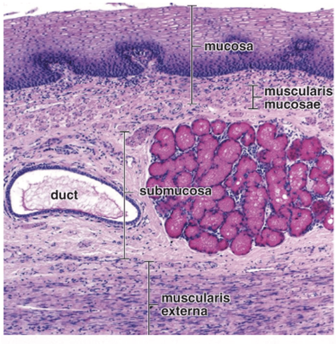
1. Esophageal (mucous) glands
2. Esophageal cardiac glands (near the gastroesophageal junction).
What glands are present in the esophageal submucosa? (2)
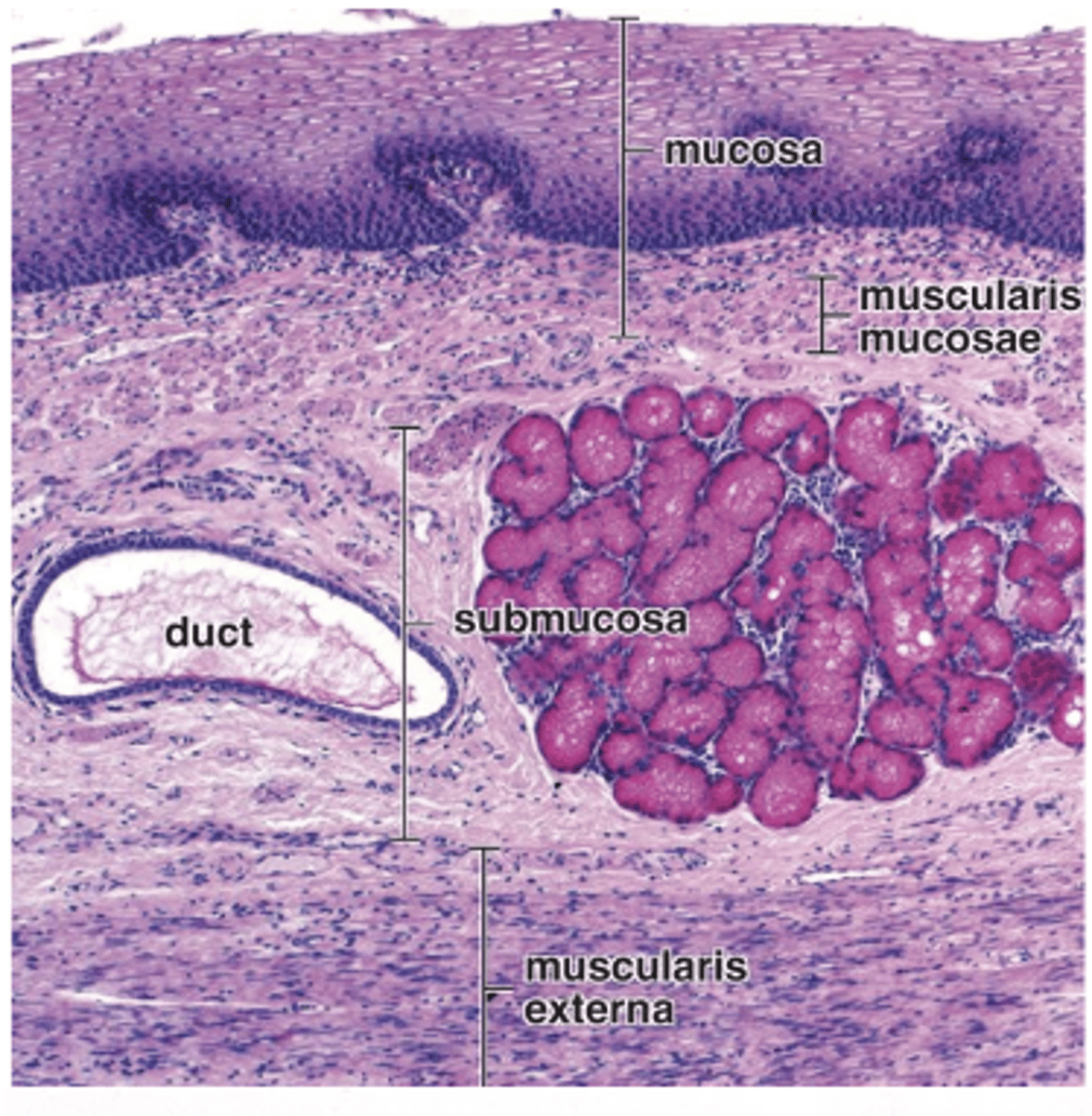
In the upper and lower parts.
Where is diffuse and nodular lymphatic tissue found in the esophageal submucosa?
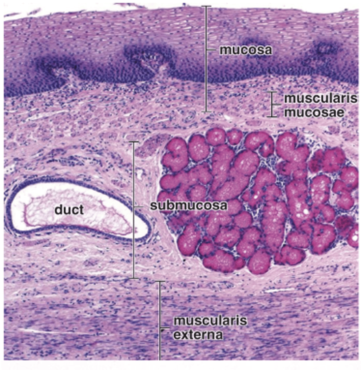
Skeletal muscle.
What type of muscle is found in the upper one-third of the esophageal muscularis externa?
Inner layer skeletal muscle
outer layer smooth muscle.
What muscle types are present in the middle one-third of the esophageal muscularis externa?
Smooth muscle.
What type of muscle is found in the lower one-third of the esophageal muscularis externa?
Simple columnar epithelium.
What type of epithelium lines the mucosa of the stomach?
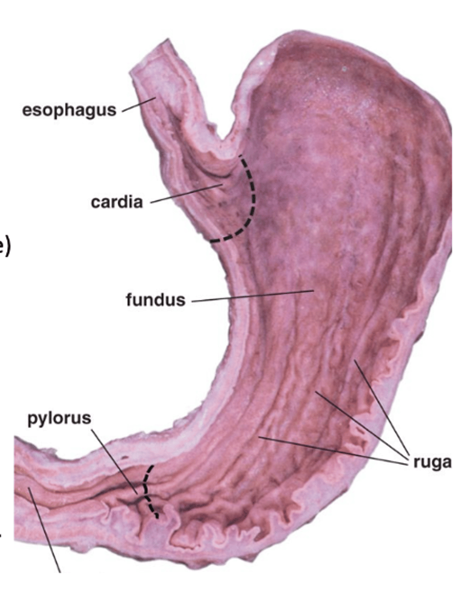
Mucosa and submucosa.
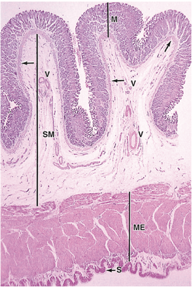
What are gastric rugae composed of? (2)
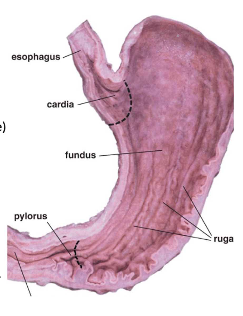
In the mucosa of the stomach.
Where are gastric pits located?
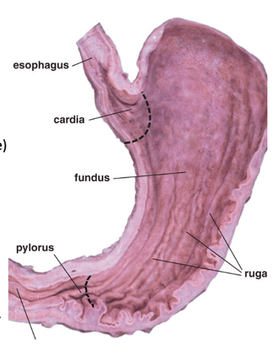
Three layers of smooth muscle.
How many layers does the muscularis externa of the stomach have
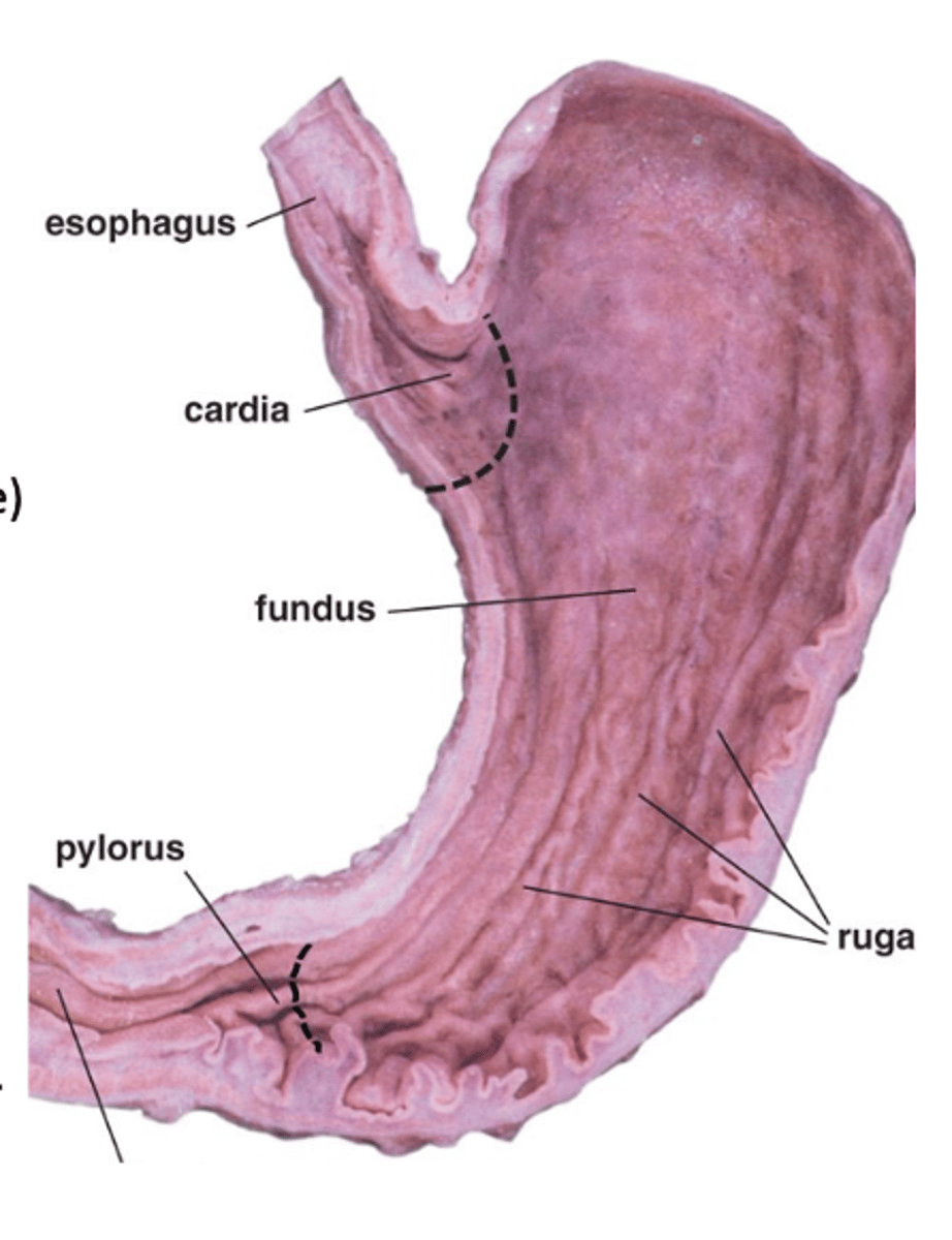
1. Continue the digestion of carbohydrates
initiated by the amylase of saliva
2. Add an acidic fluid to the ingested food and mixing its contents
into a viscous mass (chyme) by the churning activity of the muscularis
3. Begin digestion of triglycerides by a secreted lipase
4. Promote the initial digestion of proteins with the enzyme pepsin.
What are the main functions of the stomach (4)
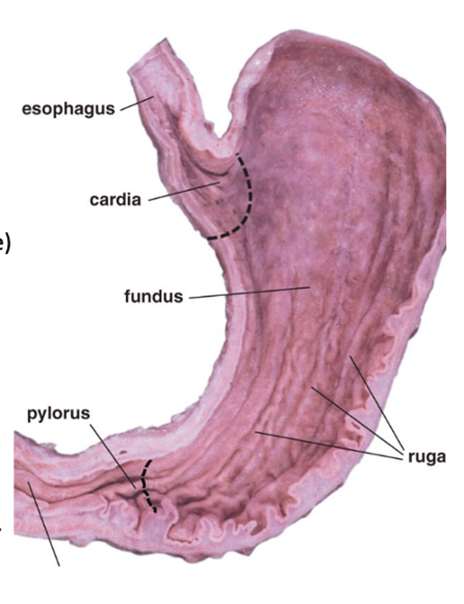
1. Mucosa
2. Submucosa
3. Muscularis externa
4. Serosa
What are the 4 major layers of the gastric wall?
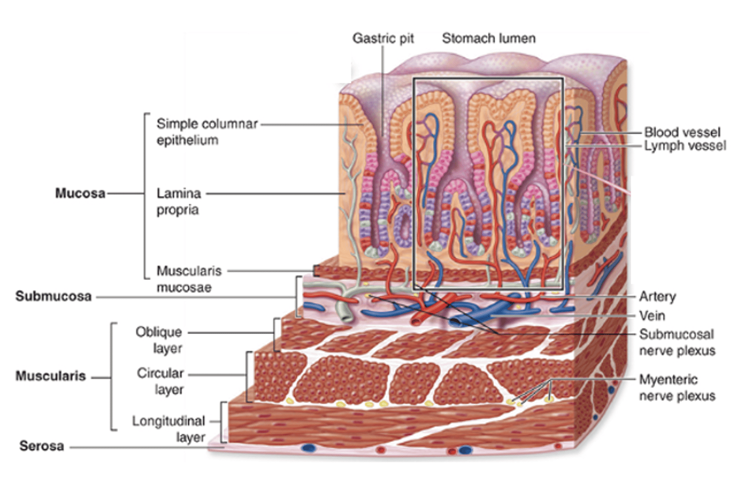
It is a narrow transitional zone.
What characterizes the cardia region of the stomach?
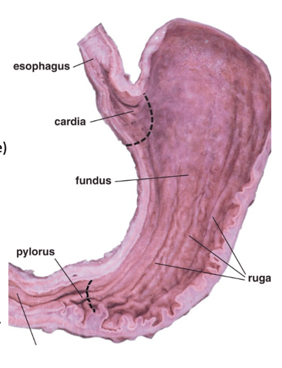
It is a funnel-shaped region with similar histology that produces mucus
What is the shape of the pylorus, and what is its main function?
The fundus and body.
Which regions of the stomach are larger and contain gastric glands?
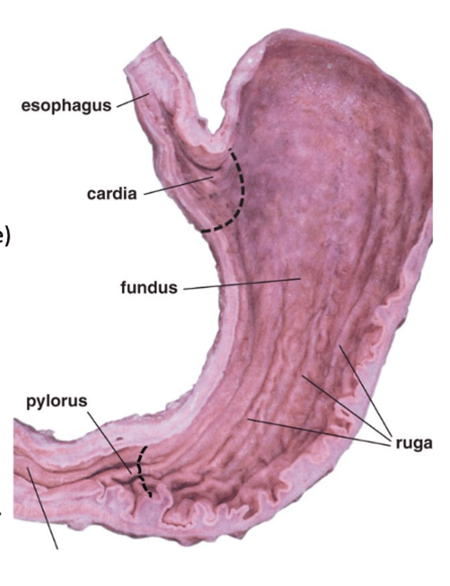
Acidic gastric juice
1. including HCl
2. pepsinogen
3. intrinsic factor
What do the gastric glands of the fundus and body release?
Folds of mucosa and submucosa. Only when the stomach is empty.
What are gastric rugae, and when are they present?
Simple columnar epithelium (mucous cells).
What type of epithelium lines the stomach mucosa?
Gastric pits.
What structures are present on the surface of the stomach mucosa?
Loose connective tissue with reticular fibers.
What type of connective tissue makes up the lamina propria of the stomach?
Lymphocytes, plasma cells, macrophages, and some eosinophils.
What lymphoid cells are present in the lamina propria of the stomach?
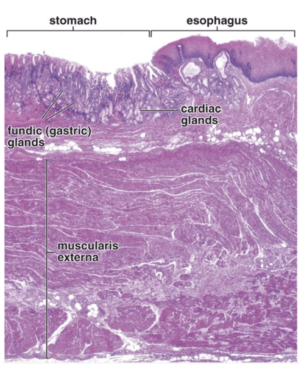
Gastric glands.
What glands are present in the lamina propria of the stomach?
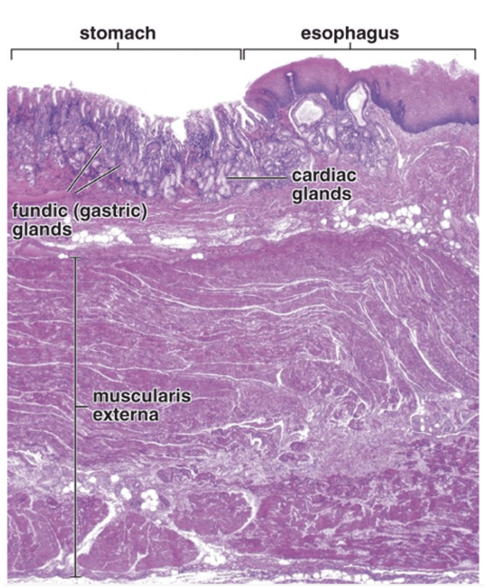
Smooth muscle
What type of muscle composes the muscularis mucosae of the stomach?
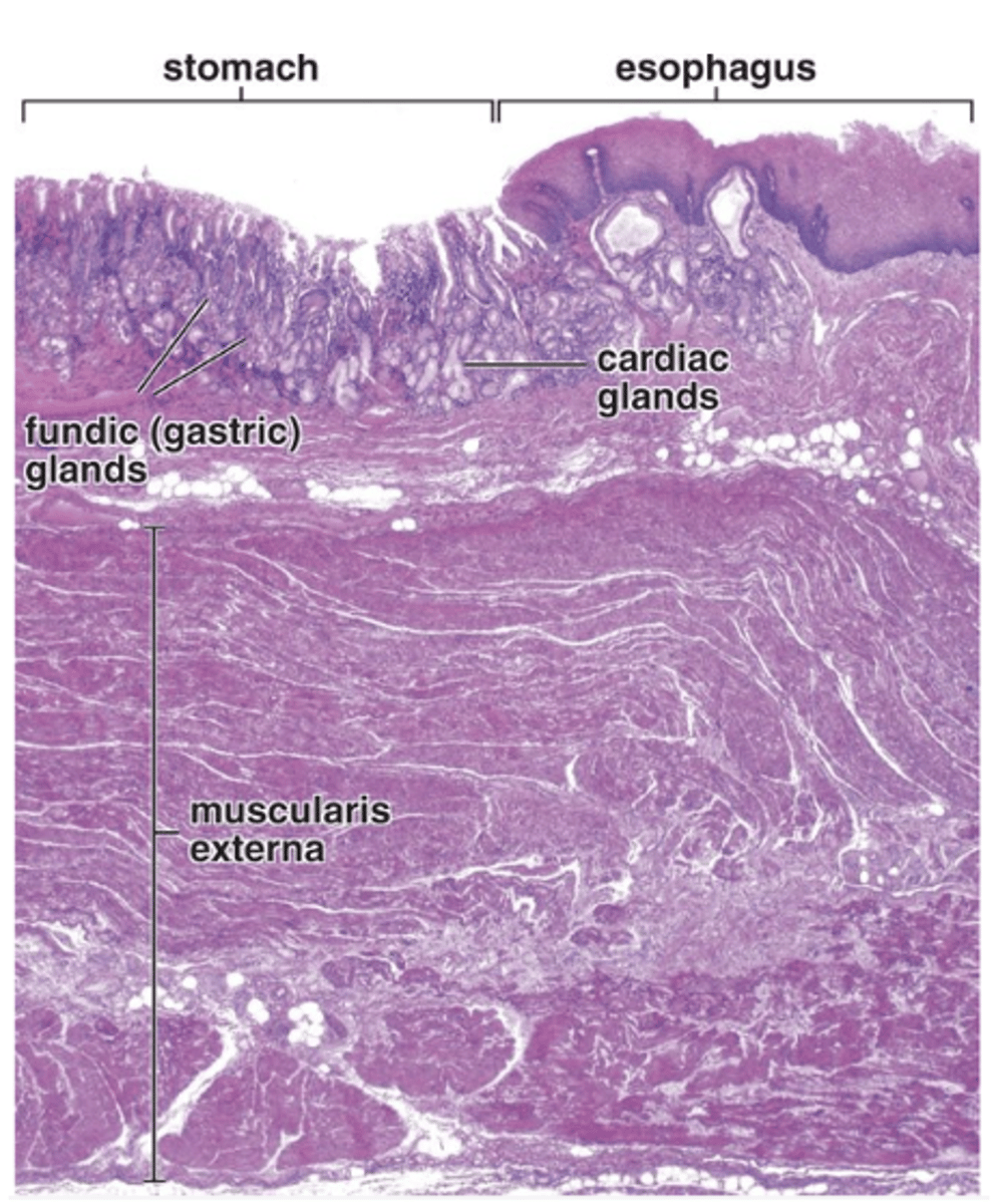
Inner circular and outer longitudinal.
What are the two thin bands of the muscularis mucosae in the stomach?
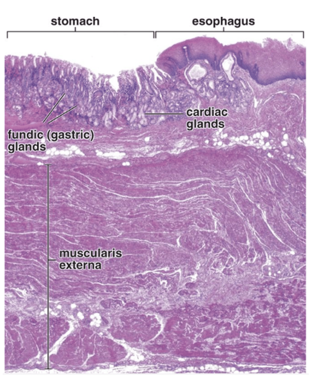
Long, branched, tubular glands.
What do gastric pits lead to?
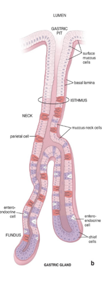
Mucous (columnar epithelial) cells.
What type of cells line the gastric pits?
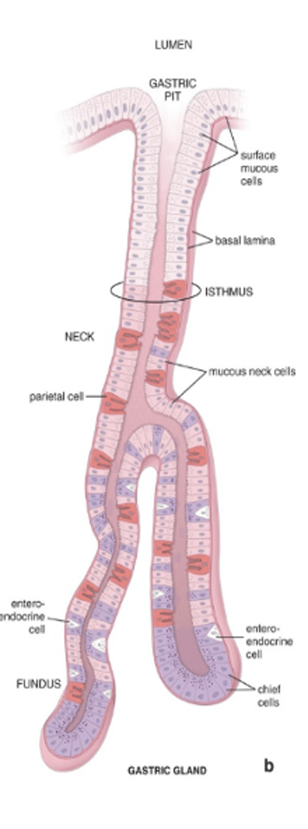
In the isthmus
Where is the stem cell located in the gastric pit–gland junction?
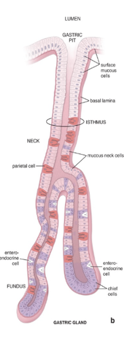
They fill most of the mucosa of the fundus and body.
Where are fundic (gastric) glands located?
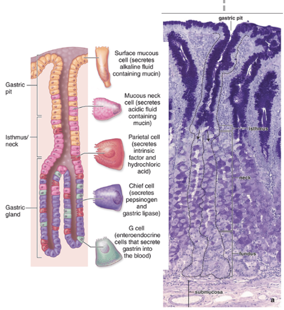
They extend to the muscularis mucosae.
How far do fundic (gastric) glands extend?
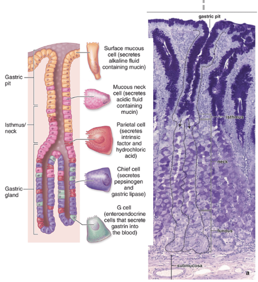
At the isthmus.
Where do most fundic (gastric) glands branch?
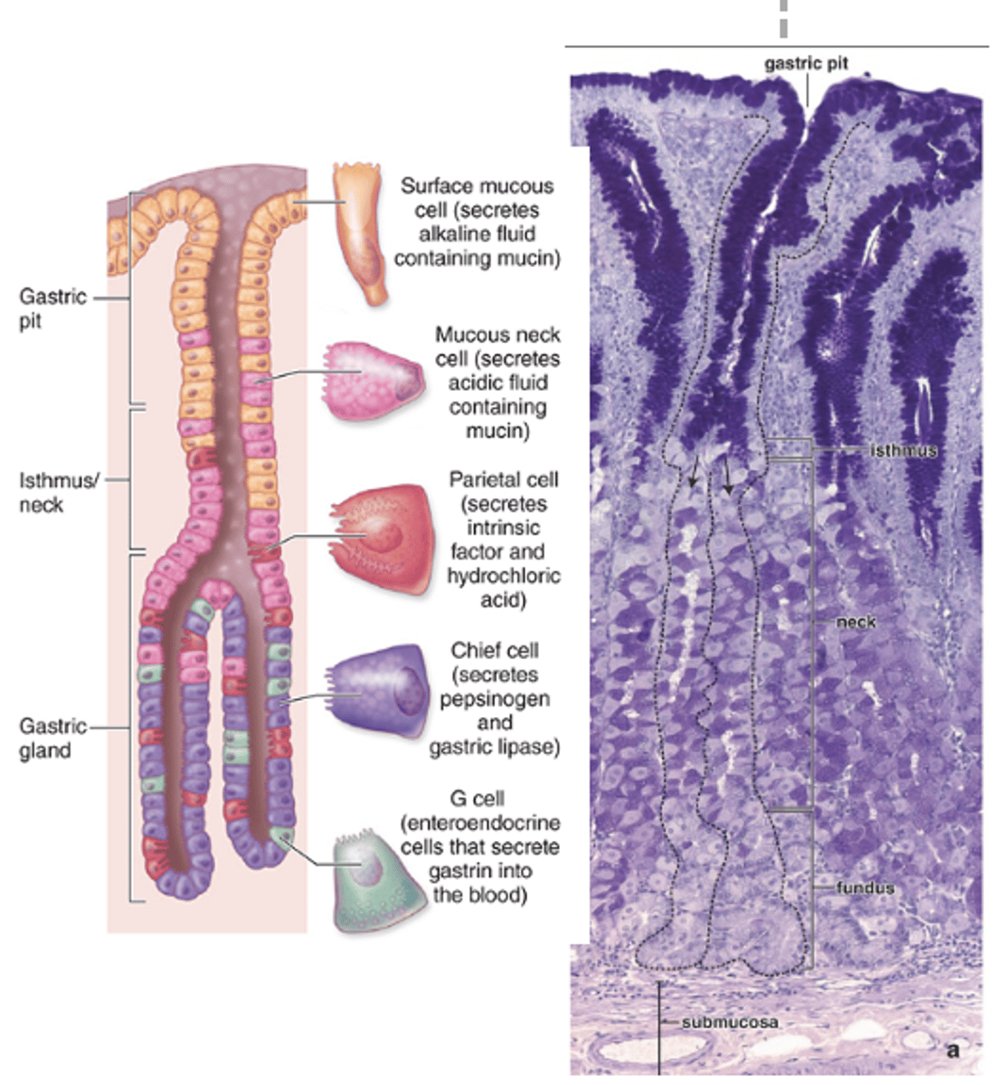
Simple, branched, tubular glands.
What type of glands are fundic (gastric) glands in structure?
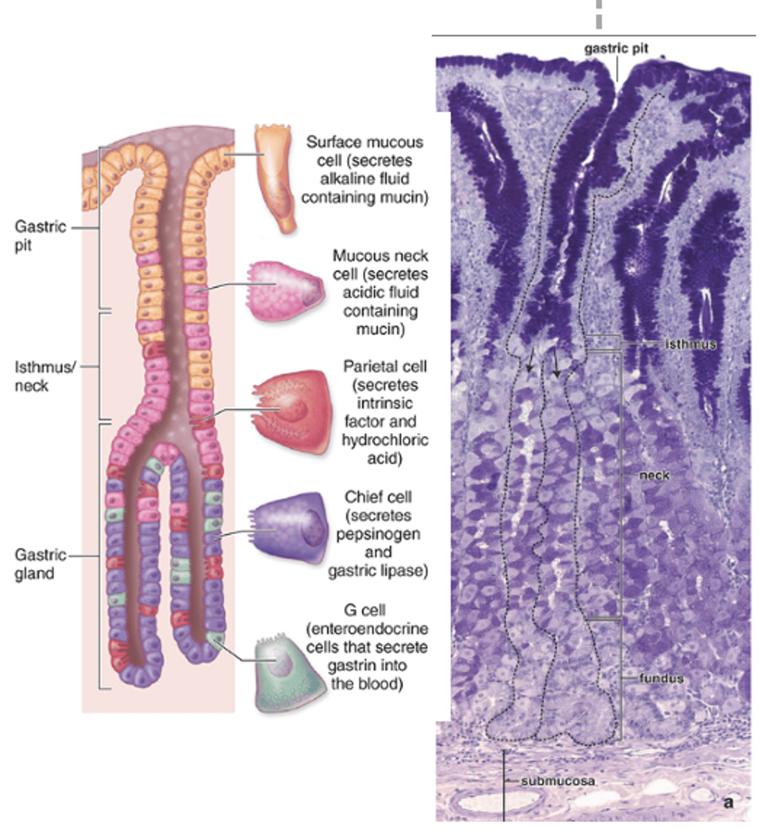
1. Narrow, long neck segment
2. Short and wide base or fundic segment
o Two or three branches
o Slightly coiled near the muscularis mucosae
What are the two parts of a fundic (gastric) gland?
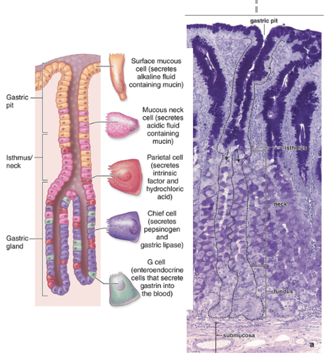
Progenitor and immature surface mucous cells, with some parietal cells in between the clusters.
With what other cell types may mucous neck cells be clustered or interspersed?
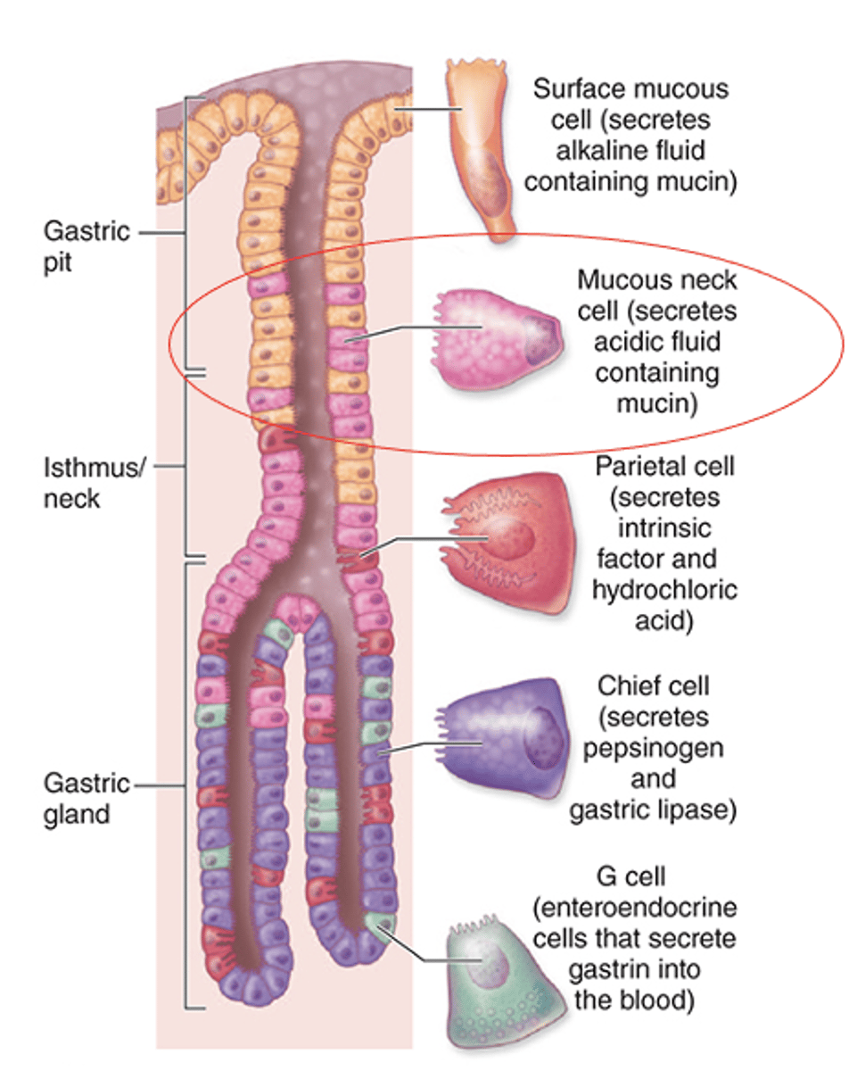
They are less columnar.
How do mucous neck cells differ in shape compared to surface mucous cells?
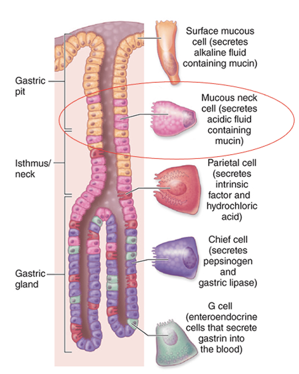
Mucin (considerably less than surface mucous cells).
What do the apical granules of mucous neck cells contain?
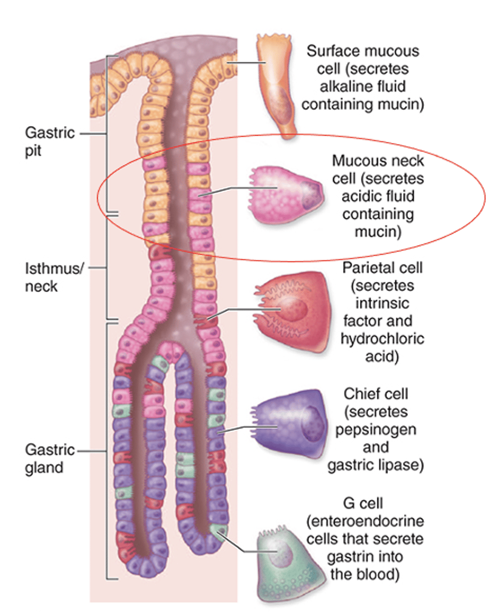
It is more spherical.
How does the nucleus of mucous neck cells compare to surface mucous cells?
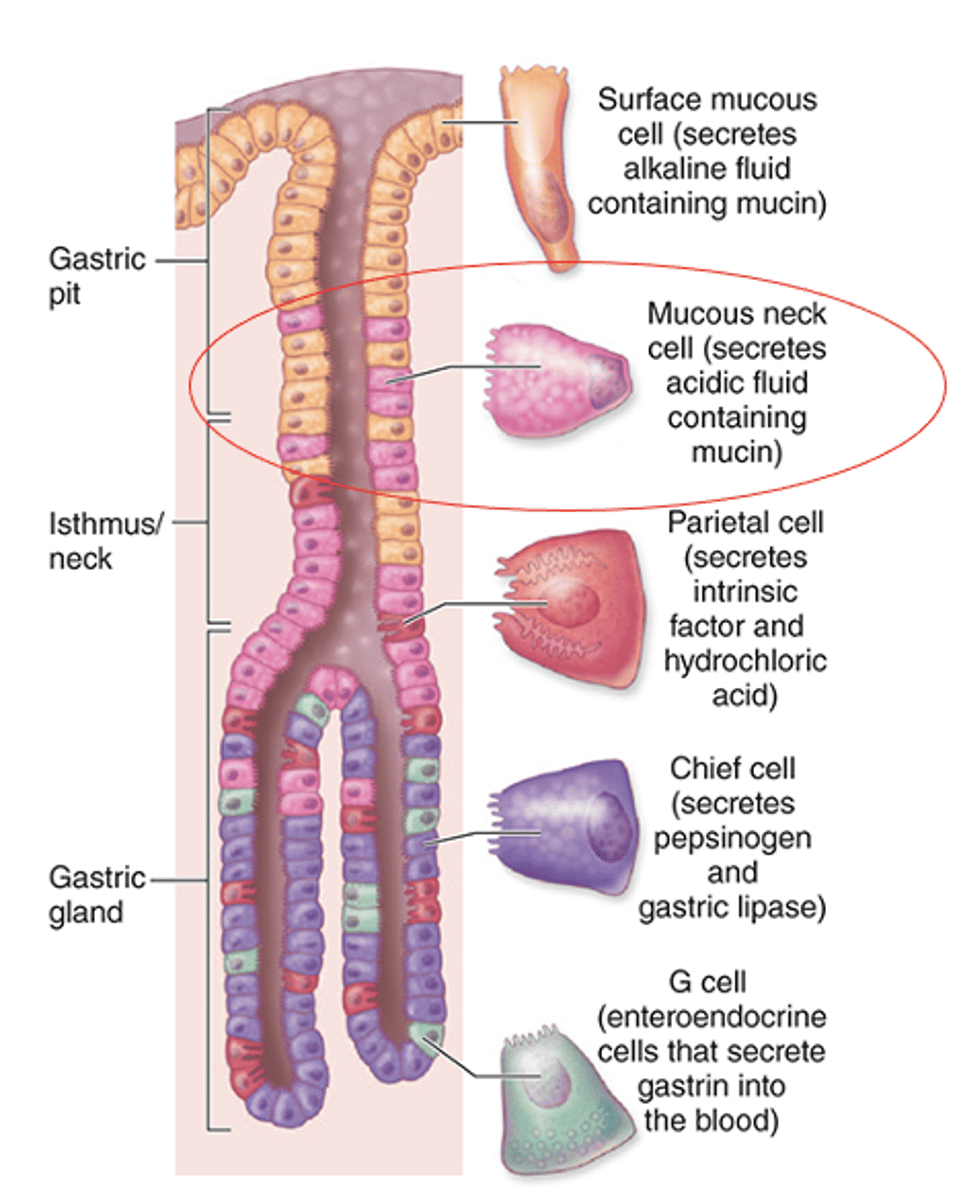
A less alkaline mucus compared to surface mucous cells.
What type of mucus do mucous neck cells secrete?
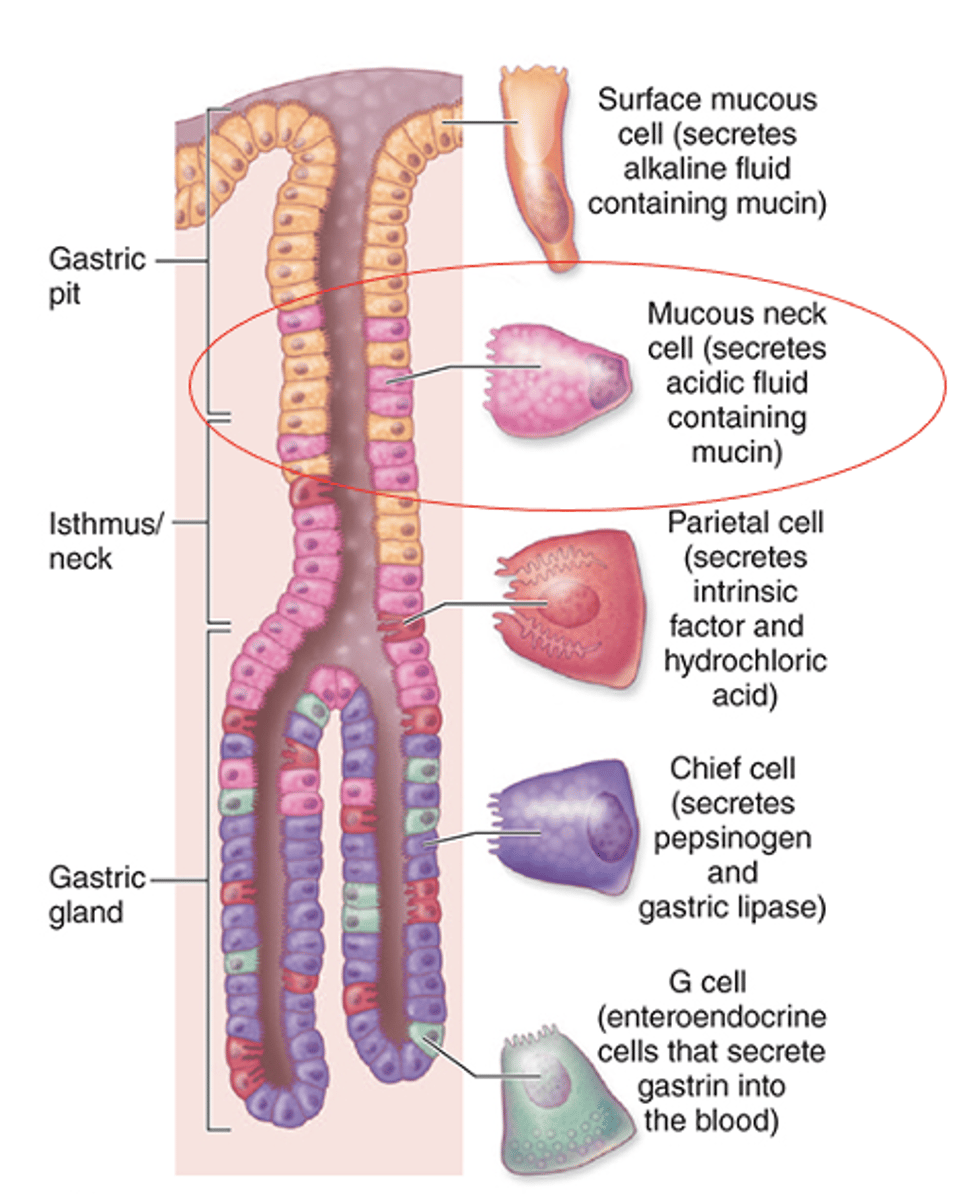
In the lower regions of the glands.
Where are chief (zymogenic) cells located in the gastric glands?
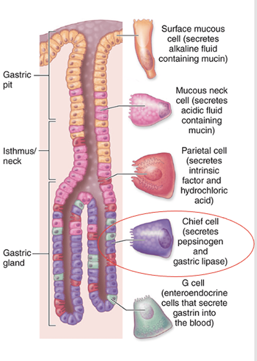
Typical protein-secreting cells.
What type of secreting cells are chief (zymogenic) cells?
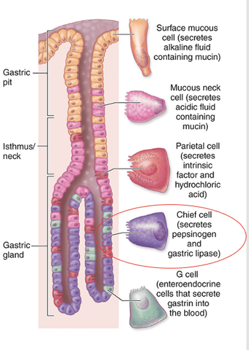
Basal RER.
What gives chief cells a basophilic appearance basally?
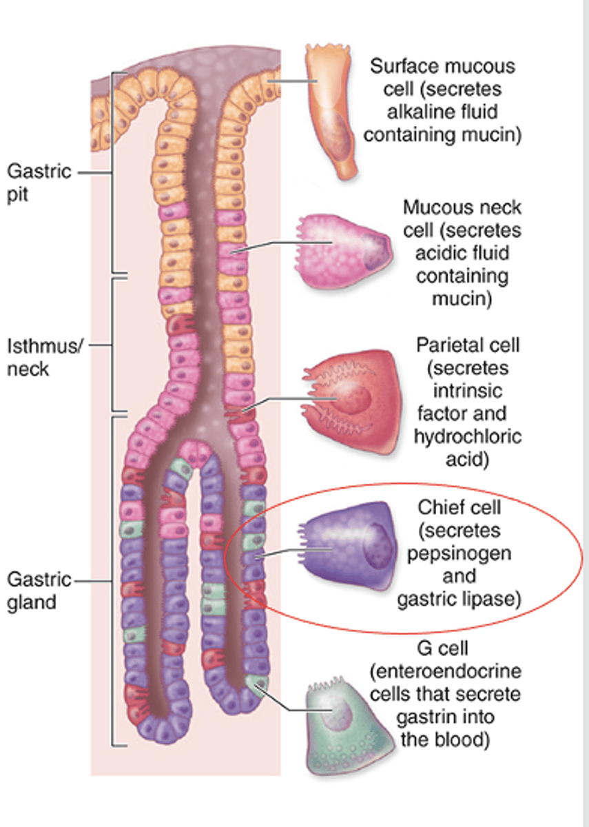
Apical secretory vesicles.
What gives chief cells an eosinophilic appearance apically?
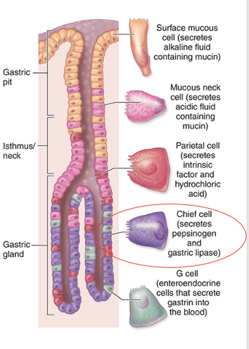
Pepsinogen and a weak lipase (gastric lipase).
What do chief (Zymogenic) cells secrete?
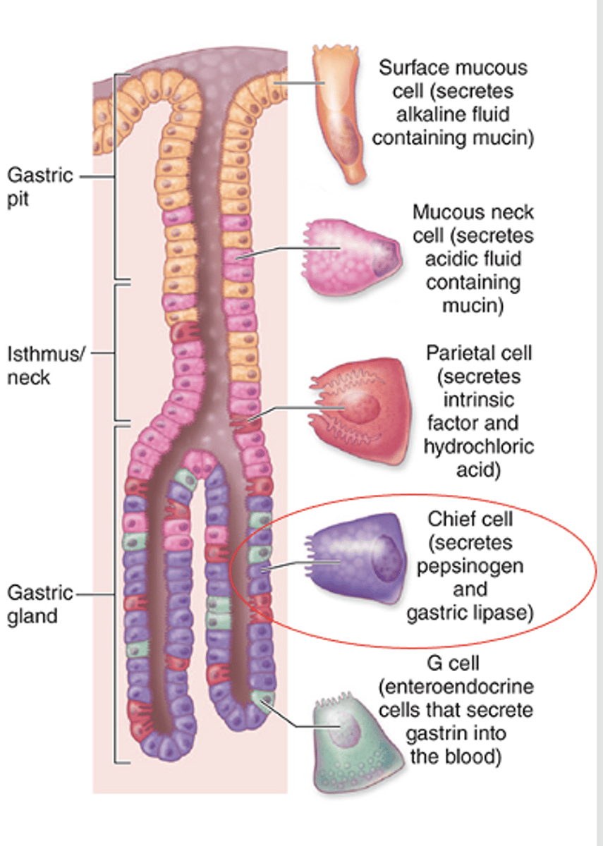
HCl and intrinsic factor.
What do parietal (oxyntic) cells secrete? (2)
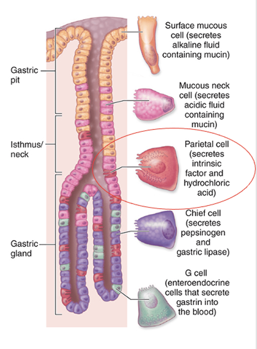
In the neck among the mucous neck cells and in the upper and middle parts of the glands.
Where are parietal (oxyntic) cells located within the gastric gland?
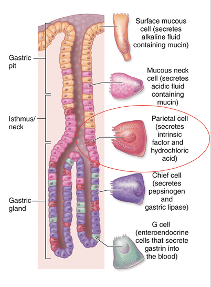
Large, round or pyramidal cells with the apex toward the lumen and the base on the basement membrane.
What is the shape of parietal (oxyntic) cells?
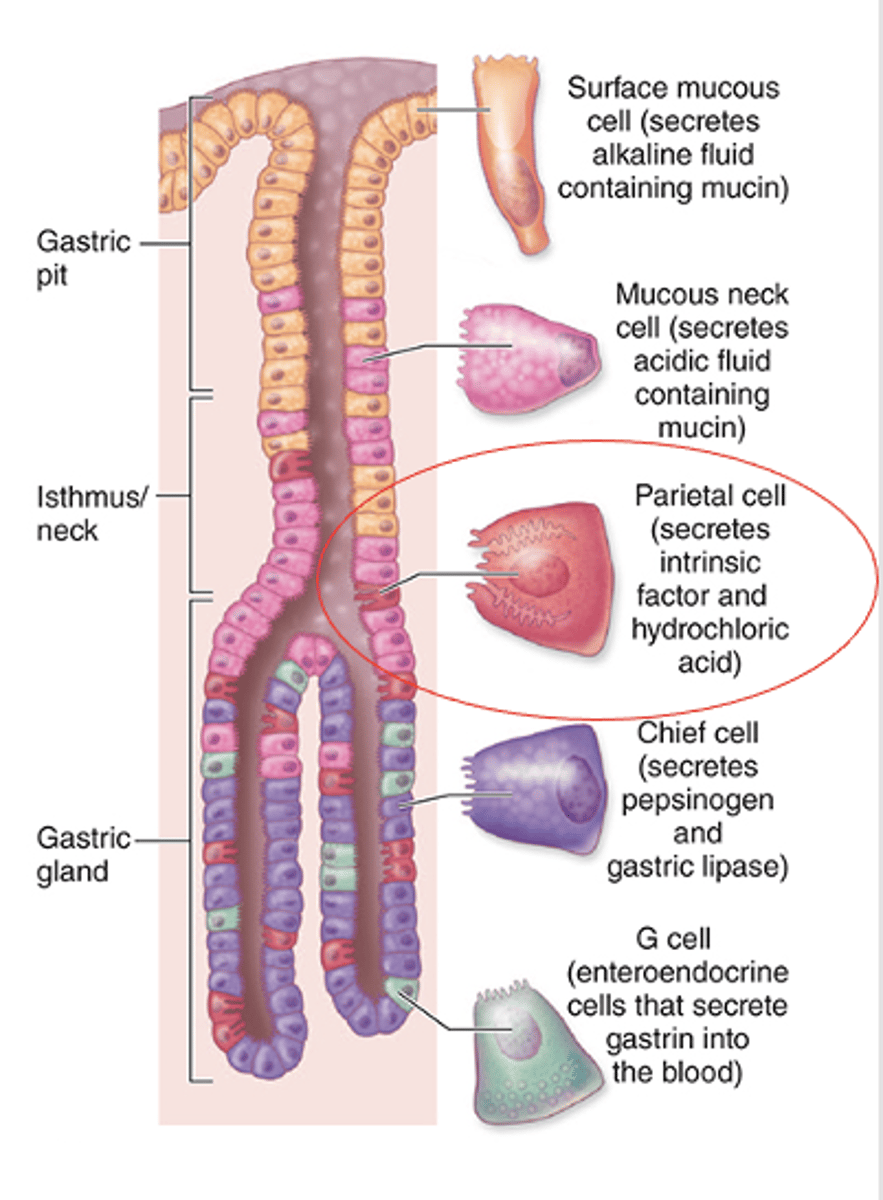
Abundant mitochondria give Intense cytoplasmic eosinophilia.
What gives parietal (oxyntic) cells their intense staining appearance?
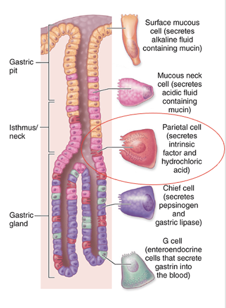
The intracellular canaliculus.
What structure is present in active parietal (oxyntic) cells due to deep apical circular invaginations?
A tubulovesicular membrane system adjacent to the intracellular canalicular system.
What membrane system is found in resting parietal (ocyntic) cells?
A reservoir of plasma membrane with active proton pumps.
What does the tubulovesicular system store in resting parietal (oxyntic) cells?
Throughout the gland, but more abundant in the lower portions
Where are enteroendocrine cells located in gastric glands?
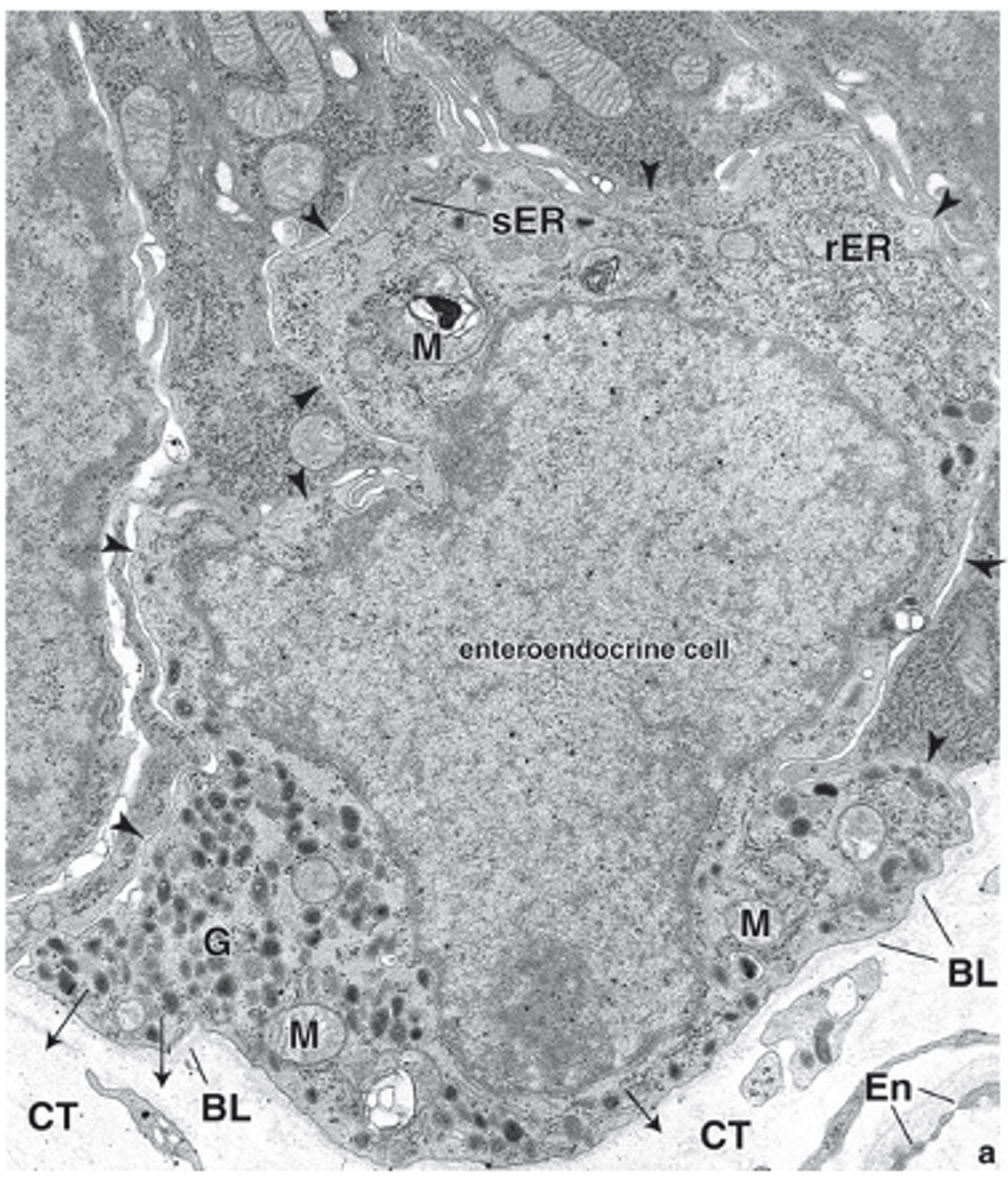
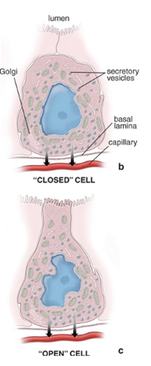
“Closed” enteroendocrine cells and “open” enteroendocrine cells.
What are the two main types of enteroendocrine cells?
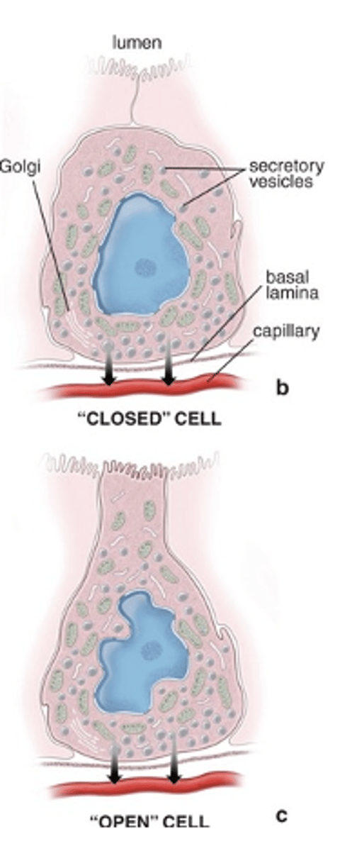
They are small, rest on the basement membrane, and do not reach the lumen.
What characterizes enteroendocrine “closed” cells?
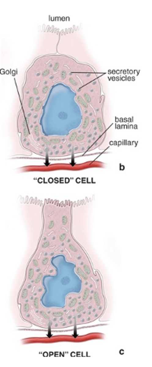
They have a thin cytoplasmic extension with microvilli that reaches the lumen and act as chemoreceptors.
What characterizes enteroendocrine “open” cells?
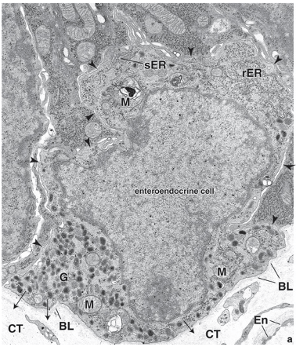
Different hormones or paracrine factors.
What do enteroendocrine cells secrete?
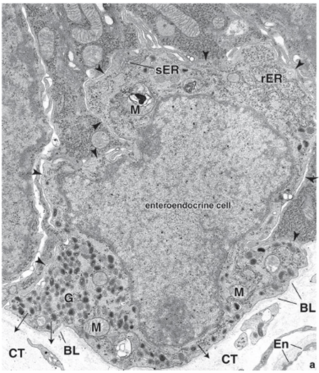
In the gastrointestinal mucosa.
Where are enteroendocrine cells found?
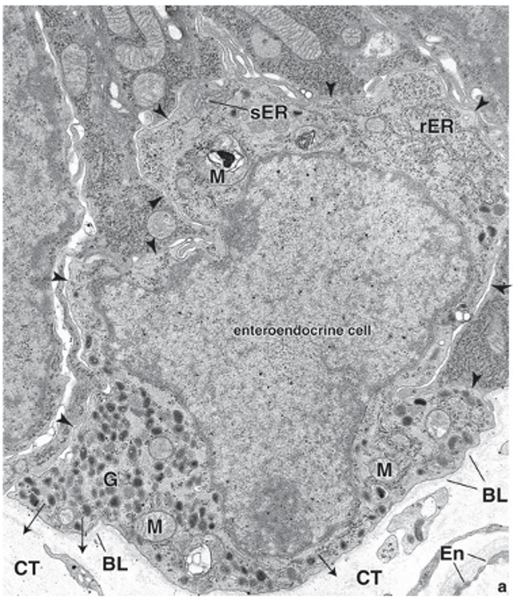
Chromium or silver salts (TEM).
What treatments are used to identify enteroendocrine cells? (difficult to see in routine H&E stains)
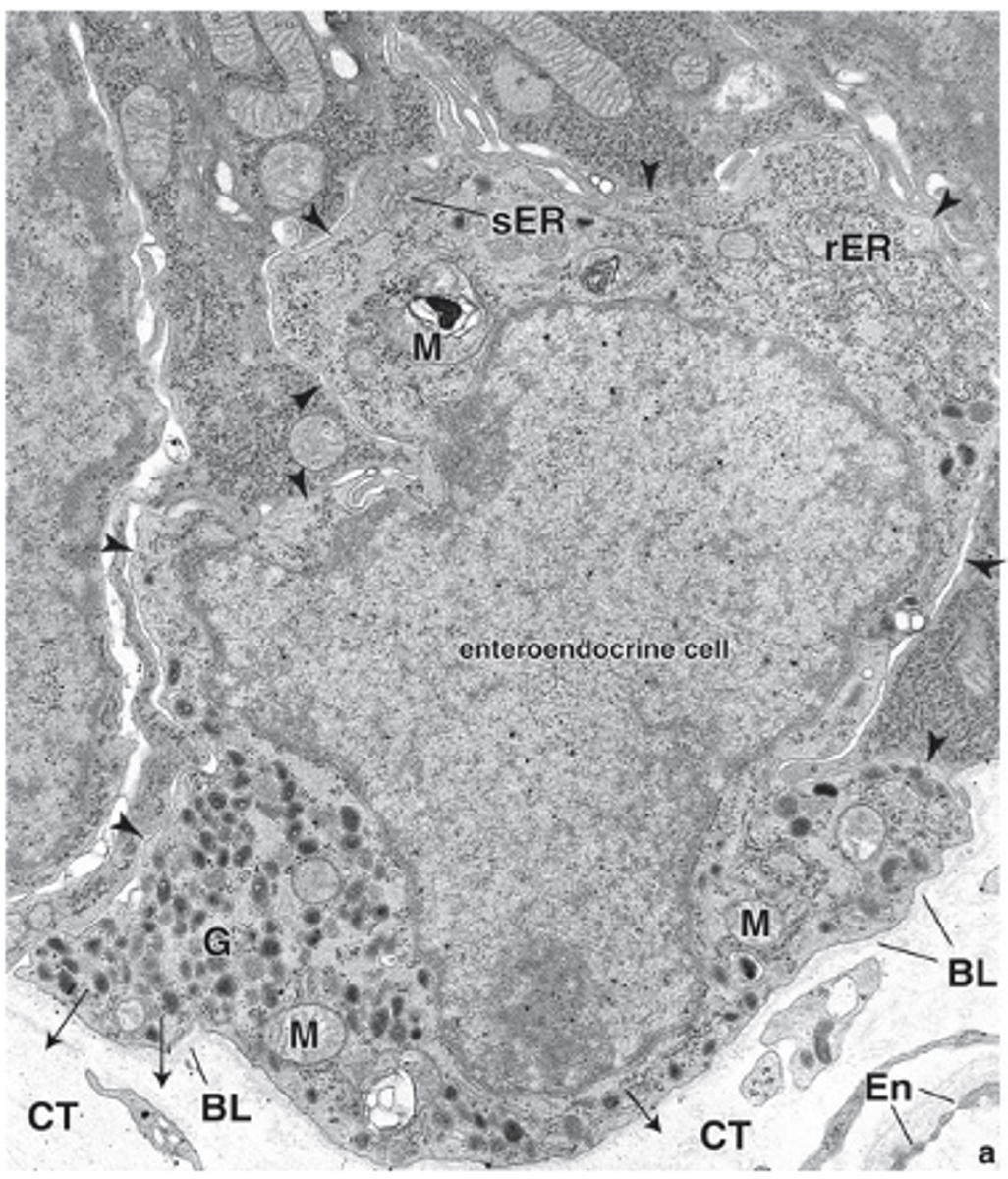
Immunohistochemistry (antibodies against their products).
What modern method is used to identify enteroendocrine cell products?
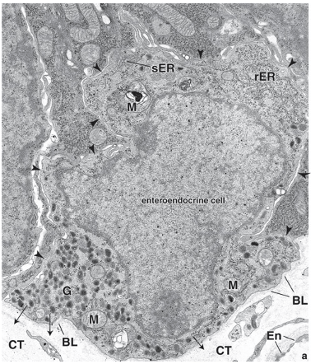
Amine precursor uptake and decarboxylation activity.
What characteristic defines most enteroendocrine cells as APUD cells?
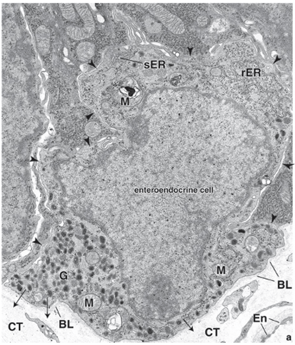
DNES (diffuse neuroendocrine system).
What larger system are all APUD cells considered part of?
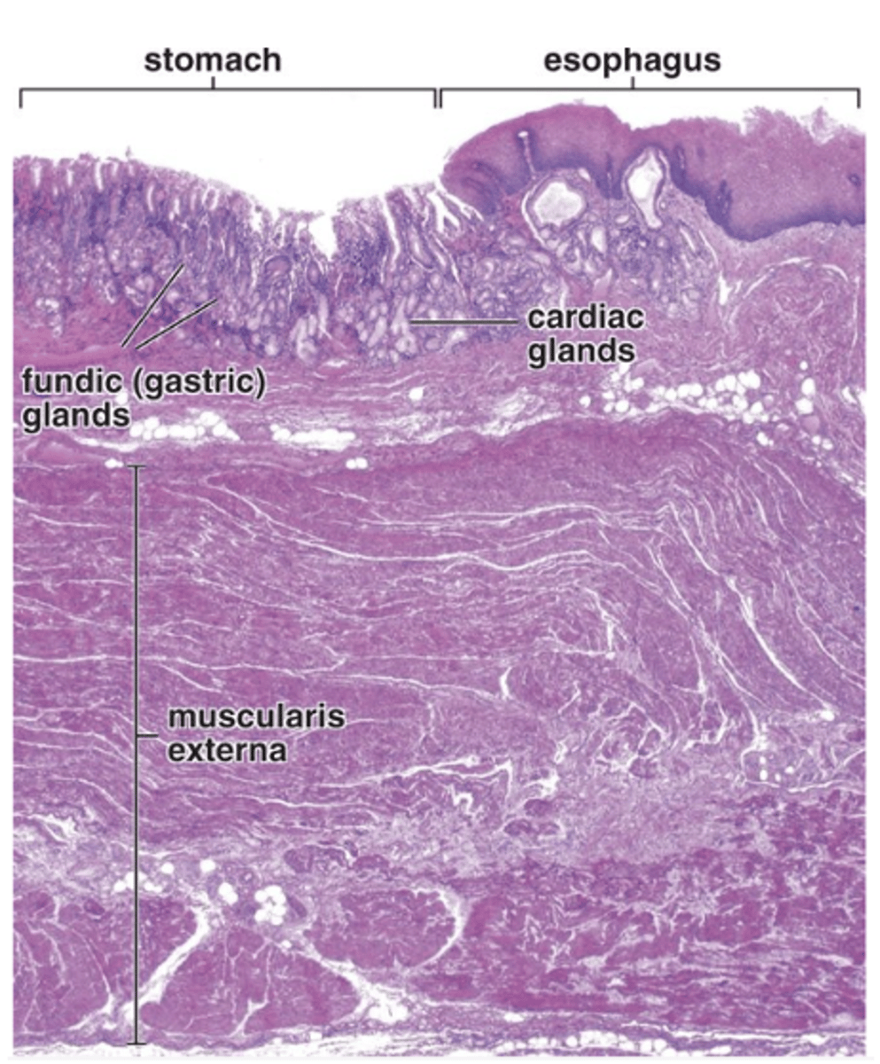
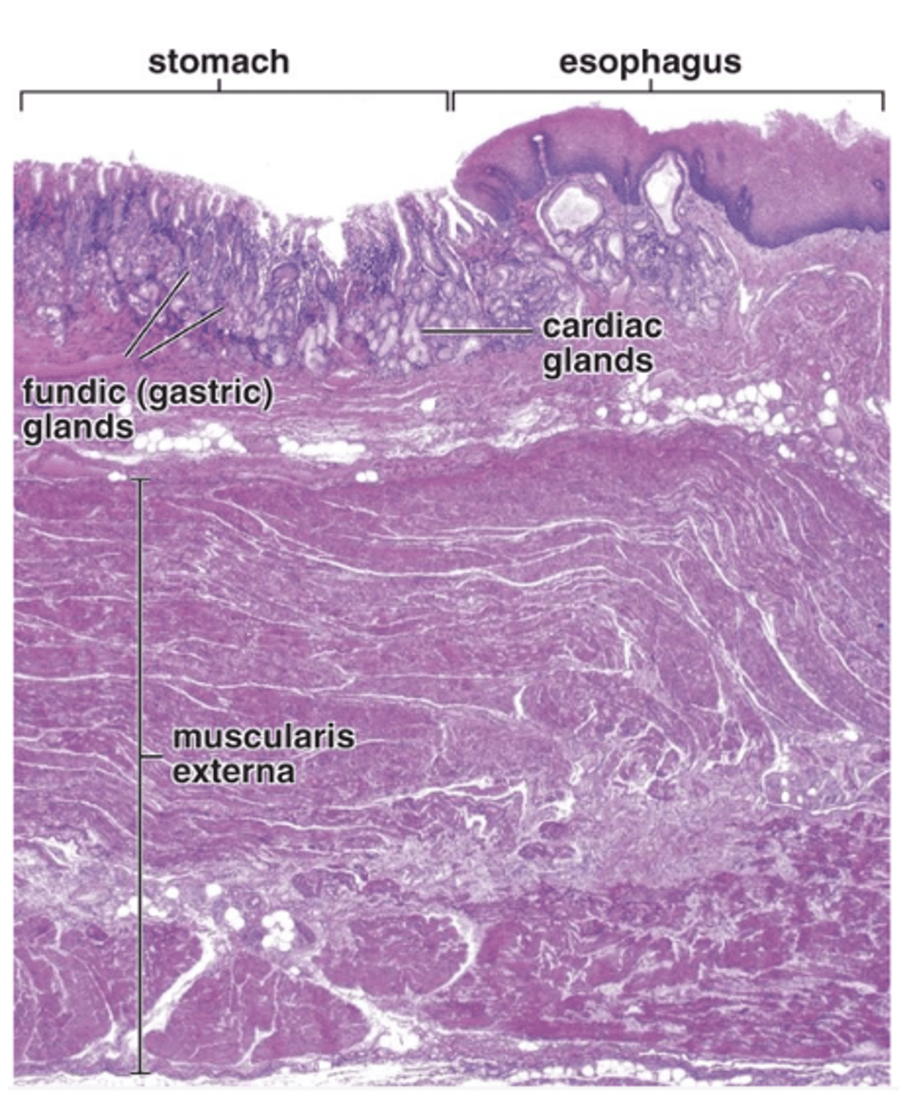
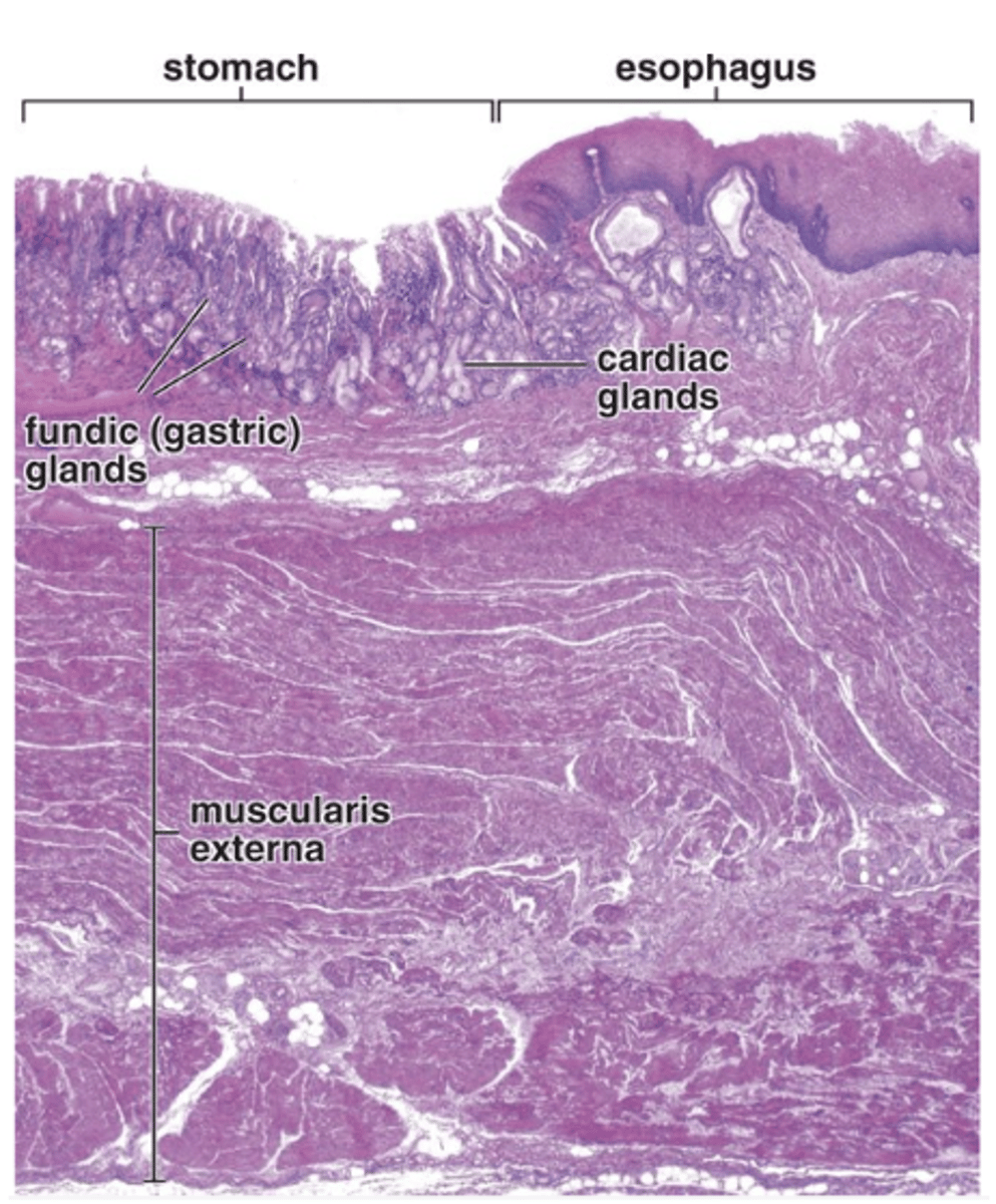
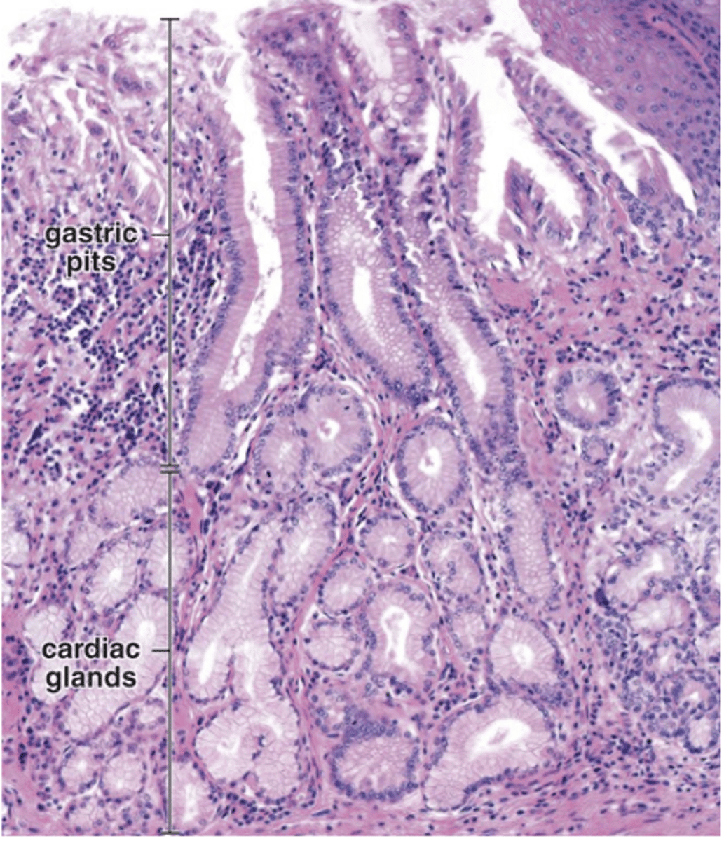
In the cardia around the esophageal orifice.
Where are cardiac glands located?
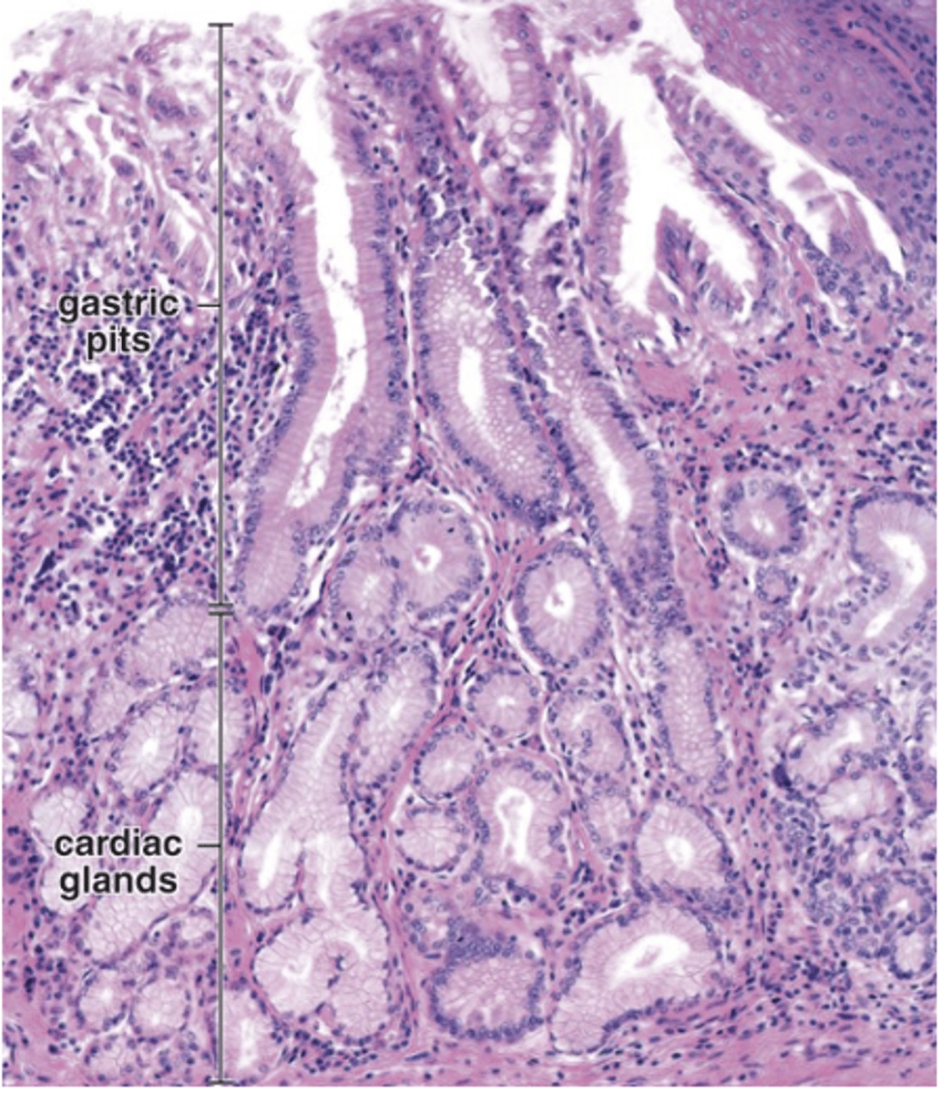
Tubular, tortuous, and occasionally branched.
What is the structure of cardiac glands?
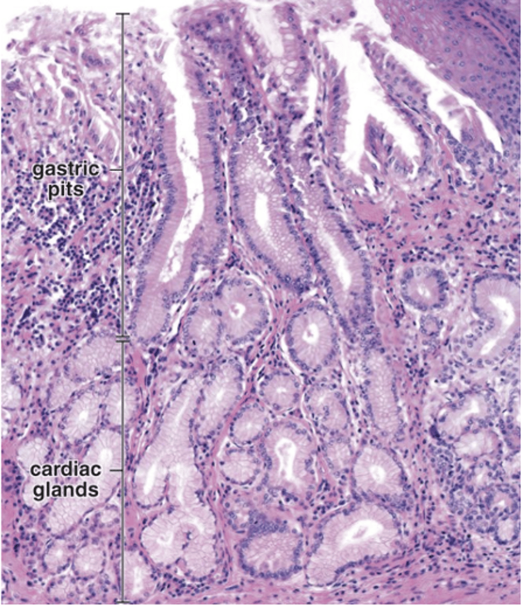
Mucus-secreting cells.
What type of cells mainly make up cardiac glands?
Mucus and lysozyme.
What do cardiac glands secrete?
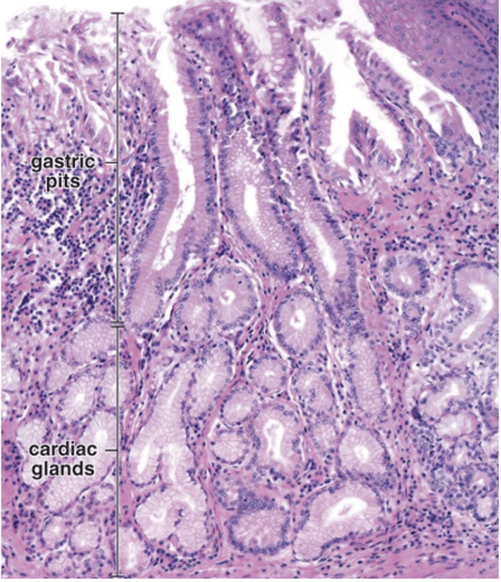
Enteroendocrine cells.
What other cell type may be interspersed in cardiac glands?
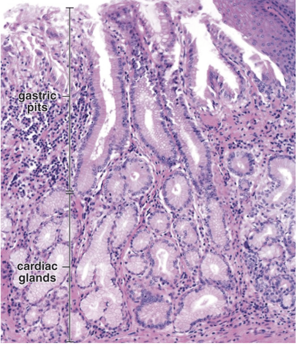
Chief cells and oxyntic (parietal) cells.
Which cell types are absent in cardiac glands?
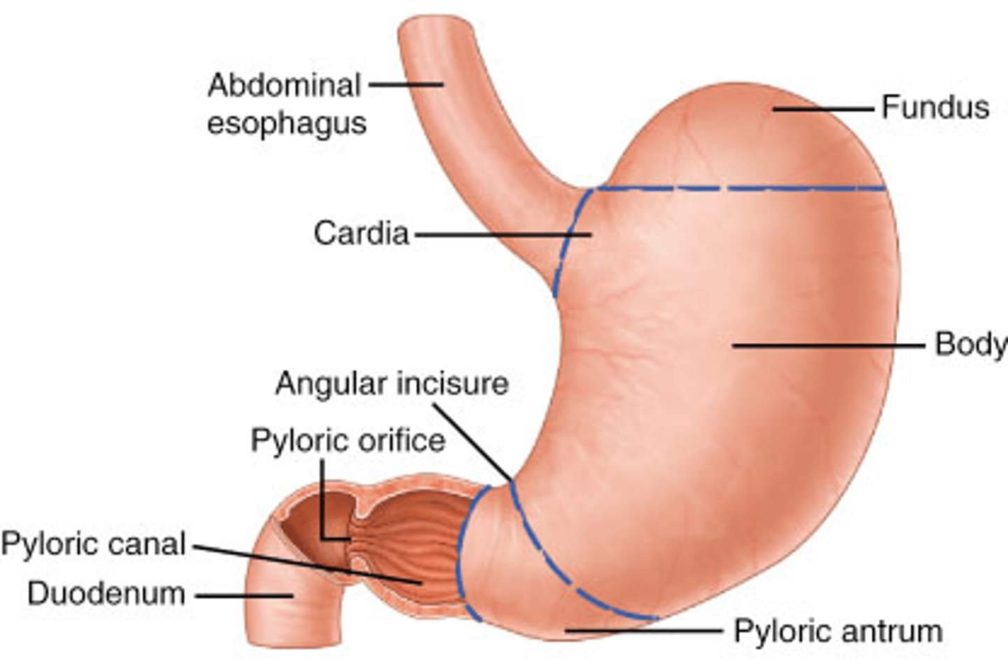
In the pyloric antrum.
Where are pyloric glands located?
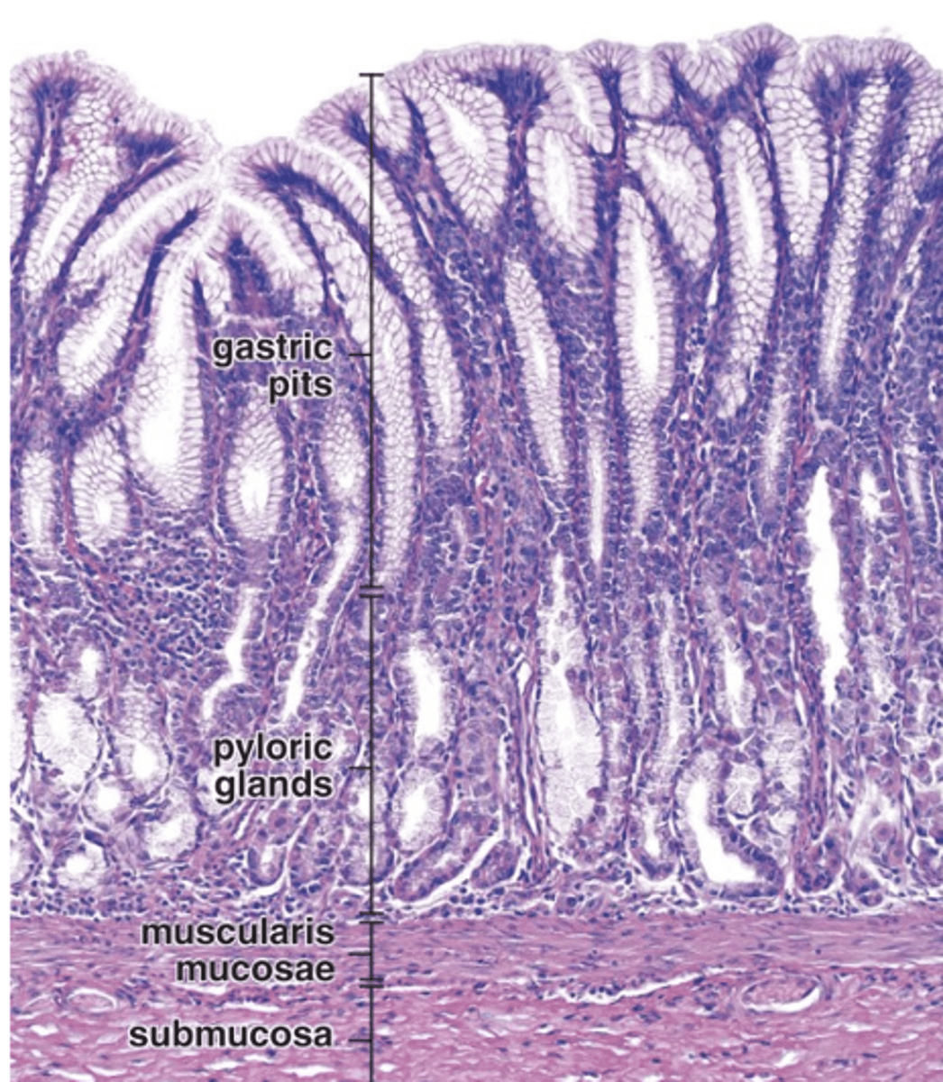
They are branched, coiled, tubular glands.
How are pyloric glands structurally similar to cardiac glands?
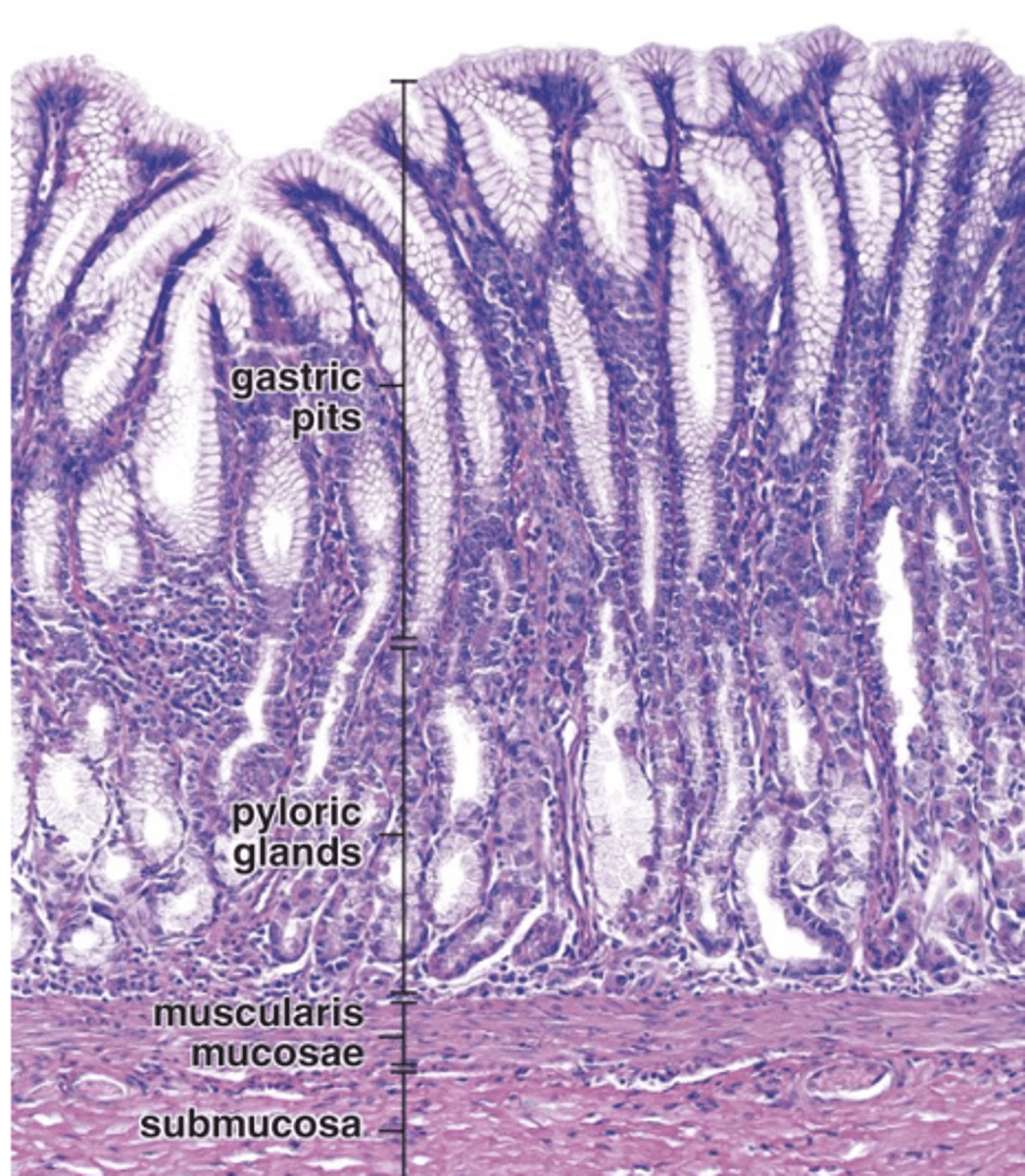
Mucous cells similar to surface mucous cells.
What type of cells line pyloric glands?
Some enteroendocrine cells and occasional parietal cells.
What other cell types are found in pyloric glands?
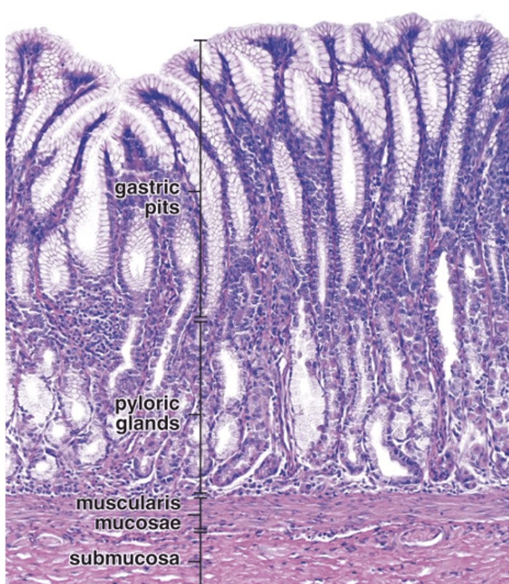
Almost half of the mucosa.
How much of the mucosa is occupied by gastric pits in the pylorus?
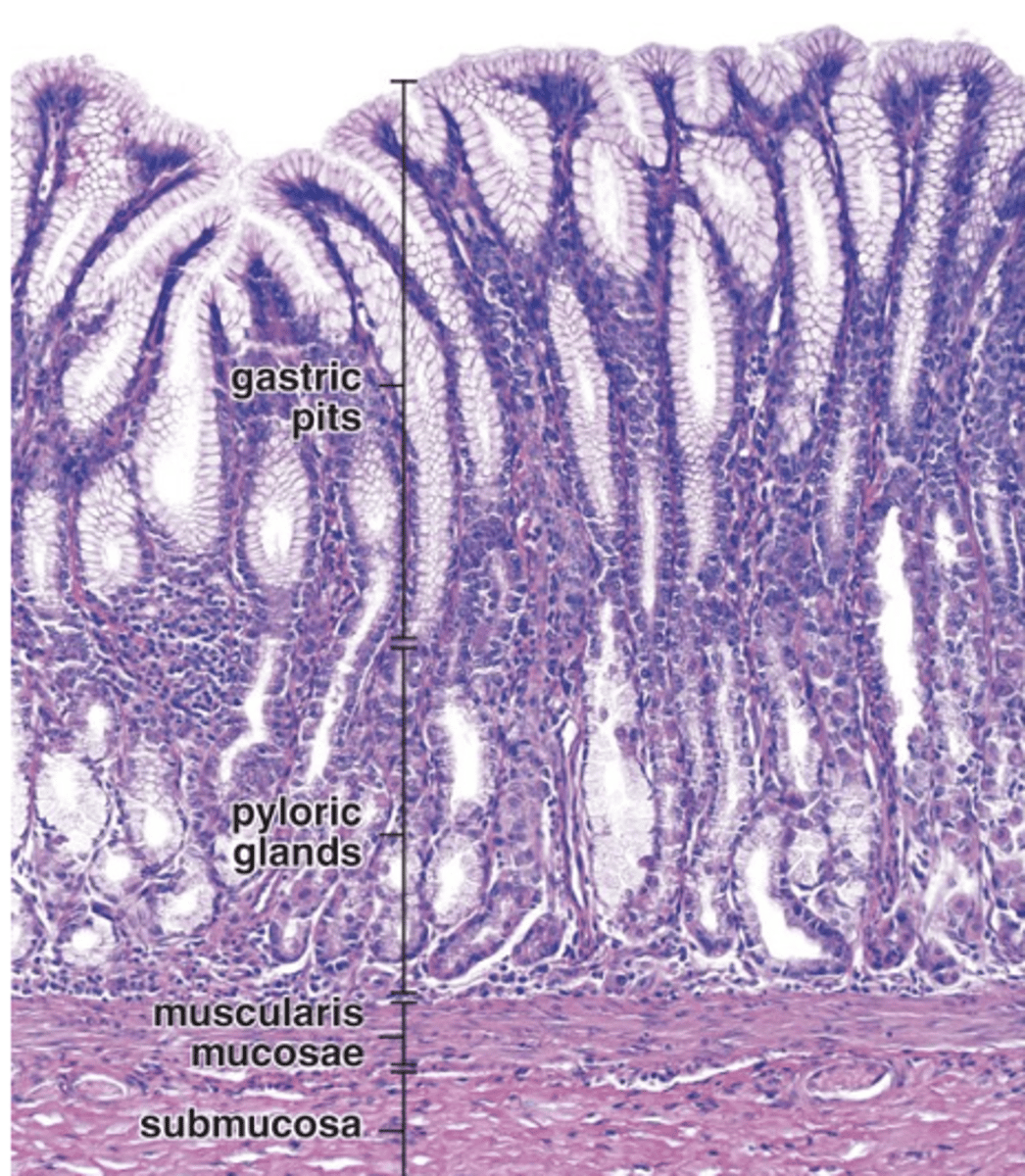
Denser irregular connective tissue.
What type of connective tissue makes up the submucosa of the stomach?
The submucosal (Meissner’s) plexus.
What neural structure is found in the submucosa of the stomach?
Three poorly defined layers:
1. Internal oblique
2. Middle circular
3. External longitudinal
How many layers of smooth muscle are in the stomach’s muscularis externa?
Myenteric (Auerbach’s) plexus.
What neural plexus is found in the muscularis externa of the stomach?
Loose connective tissue.
What type of connective tissue makes up the serosa of the stomach?
It has a rich vascular supply.
What is notable about the vascular supply of the stomach’s serosa?
Simple squamous epithelium (mesothelium).
What epithelium lines the serosa of the stomach?
Greater omentum.
What connects the greater curvature of the stomach to the parietal peritoneum?
Lesser omentum.
What connects the lesser curvature of the stomach to the visceral peritoneum of the liver?
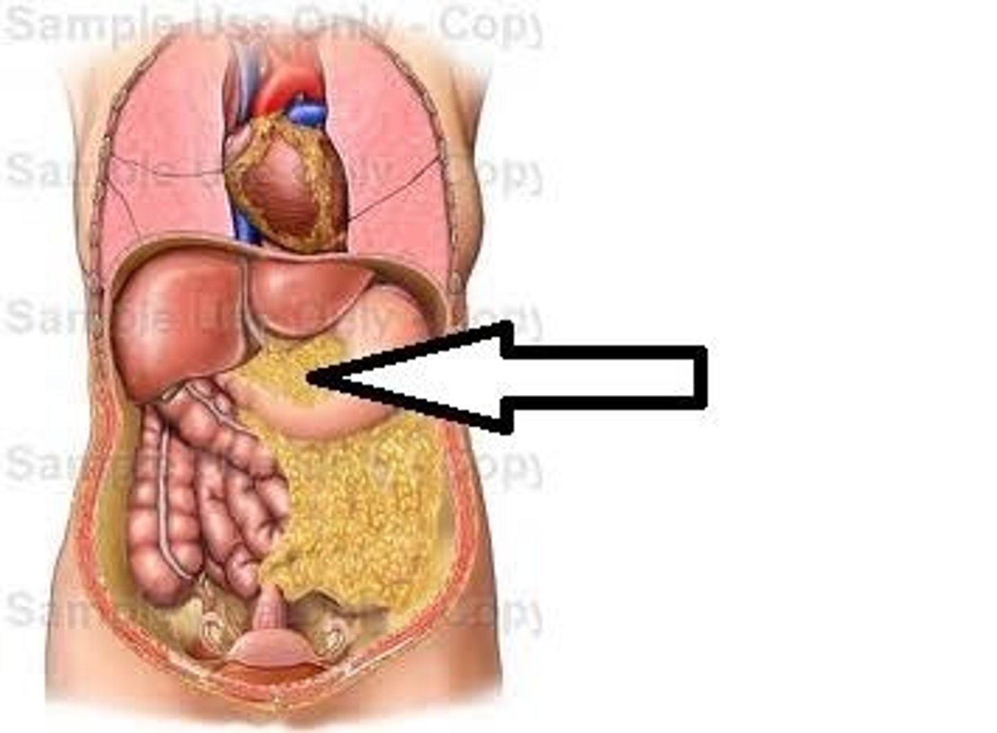
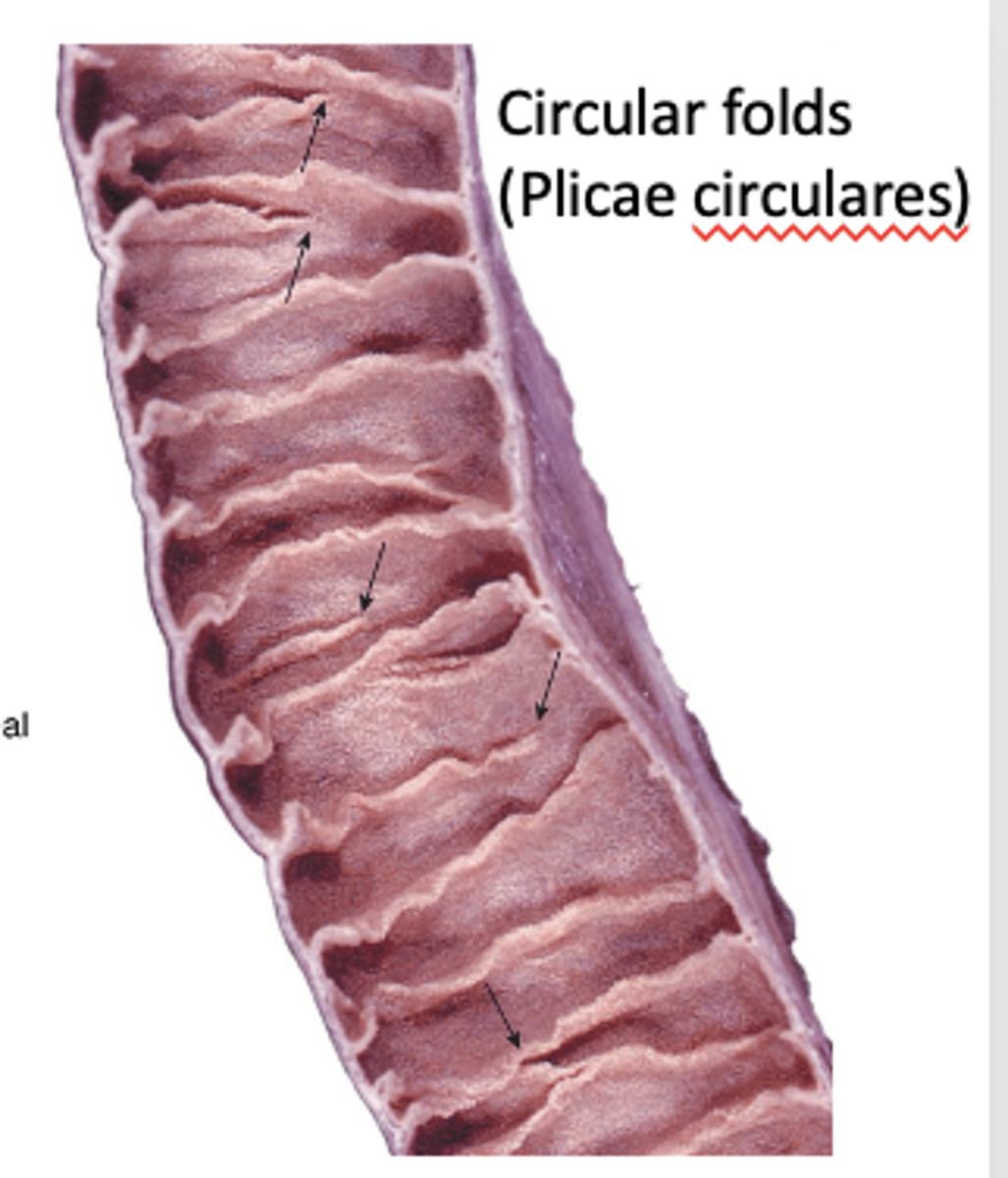
Macroscopic folds of mucosa and submucosa, best developed in the jejunum.
What are plicae circulares and where are they found?