Bovine Respiratory Disease: Primary Pathogens and Case Approach ACC
 Call Kai
 Learn
 Practice Test
 Spaced Repetition
 Match
 Flashcards
 Knowt Play
1/150

There's no tags or description
Looks like no tags are added yet.
151 Terms
What is BRD (bovine respiratory disease)?
-General term for respiratory disease in cattle
- Multifactorial, so involves multiple pathogens, environmental conditions and management decisions
What is pneumonia?
Inflammation of the lungs
What is shipping fever or transit fever?
-Respiratory disease after a stress such as transport or mixing of animals from different groups/sources
-Can be associated with a diet change
most common pathogen for shipping fever/ transit fever
Mannheimia Haemolytica
What is husk?
This is lungworm- Dictyocaulus viviparus
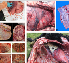
BRD
What is happening in the lungs with BRD?
-inflammation of the lungs causes scarring of the alveoli
-limited ability of lung tissue to oxygenate the blood
-so reduction in oxygen available for aerobic respiration so more lactic acid fermentation
-causes released energy release from the diet so reduced feed conversion efficiency
-leads to less energy for growth and milk production
What is the impact of BRD on dairy?
-reduced DLWG
-reduced milk yeild
-reduced survival
what kind of aetiology does brd have
multifactorial
what factors are involved in the aetiology of brd
environmental, management and nutritional
host
an initial pathogen
what pathogen factors contribute to brd
alter animals defence mechanisms → allows consolidation of LRT by bact
likely be an opportunisitic comemnsal translocating
usually primary pathogen is virus
reduced immunity allows to travel from URT to LRT
what bacteria may consolidate the lrt
m. haemolytica
p. multocida
h. somni
mycoplasma bovis
trueperella pyogenes
what host factors contribute to brd
level of immunity
what environmental, management and nutritional factors contribute to brd
transport
weaning
crowding
dust and poor air quality
incelment weather
inadequate ventialtion
poo quality milk replacers
partial or complete failure of passive transfer
What are the viral pathogens of BRD?
Infectious Bovine Rhinotracheitis (IBR) |
Parainfluenza 3 PI3 |
Respiratory Syncytial Virus (RSV) |
Bovine Viral Diarrhoea Virus (BVDV) |
Malignant catarrhal fever (MCF) |

What are the bacterial pathogens of BRD?
-mycoplasma bovis
-mannhaemia haemolytica
-pasteurella multicoda
-TB
-histophilus somni
-truperella pyogenes
-contagious bovine pleuropneumonia

What are the parasitic pathogen of BRD?
Dictyocaulus viviparus
What is Infectious Bovine Rhinotracheitis?
Herpesvirus - means there is a latent infection
Latent infections will recrudesce and shed after stress → carries for life
Highly contagious and infectious - so multiple animals will present with signs (group level)
Not notifiable or zoonotic
incubation period and prevalence of infectious bovine rhinotracheitis
incubation period: variable but ~1 to 2 weeks so disease seen quickly after introduction
Prevalence: common - estimated 40% of cattle in the UK have been exposed
transmission of infectious bovine rhinotracheitis
Transmission: direct contact/shared airspaces
what are the clinical presentations of ibr
resp disease,
peracute death
reproductive
What is the respiratory disease clinical presentation of IBR?
-Marked pyrexia (commonly >40C)
-Bilateral conjunctivitis with epiphora (they look they have been crying)
-Milk drop in the adult herd
-Hyperpnoea of the nose (red nose)
-Later: Nasal discharge which can become purulent

What is the peracute death clinical presentation of IBR?
-Very high fever and death in 24 hours
-Get secondary bronchopneumonia

What is the reproductive clinical presentation of IBR?
-Early embroynic death (decreased conception rates across herd)
-Abortions (can be up to 100 days after respiratory signs)
-Genital lesions (IPV)
How do we diagnose IBR?
-Ag detection= Sites: conjunctivia or nasal passageway, Sample: plain swab, Test: IFAT or PCR
-Paired serology=rising antibody levels, Issue: wait for 3 weeks for results
-PME= necrotising tracheitis including erosions on the mucosal surface
IBR Diagnosis: Surveillance
-detect antibodies with ELISA test
-Use blood for over 8 months old→ maternal abodies
-can use individual or bulk milk tank
-If the herd vaccinates we need DIVA test
how does diva differentiate for ibr
identifies wild type/natural infection from vaccinated cattle when using marker vaccines (gE-deleted)
what should you do on clinical exam for ibr
palp and ausc trachea →harsh urt sounds
What is IBR treatment?
-viral so cant use antibiotics
-give NSAIDS
-Auscultate around the trachea when doing clinical exam
-give meloxicam or carprofen or flunixin (NSAIDS)
-give oral fluids
what nsaids to give to cows
ketoprofen- 0 withdrawal
meloxicam in calf→ linger duration of action
carprofen
flunixin
(not phenyl butazone in farm!!)
Describe IBR control
VACCINATE
For outbreak =Intranasal - quick acting; lasts 3-6 months; from 2 weeks old→ mucosal immunity
For endemic =Parenteral which needs primary course of 2x; lasts ~12 months; from 3 months old, cannot be done in really young so need to rely on IgG mucosal response
Manage stress especially when housed and consider air quality
Buying in: accredited herds
Test and quarantine any bought in animals
What is RSV? (respiratory syncitial virus)
Can be primary sole pathogen or in combination with others
Primarily affects calves but can be seen at adults in times of stress → eg. calving
Often associated with outbreaks and can be reinfected
transmission of rsv
Transmission: respiratory secretions through aerosol and close contact
What are the RSV clinical signs?
-Severe viral interstital pneumonia
-Lung damage such as pulmonary oedma, interstitial/subcutaneous emphysema
-Damage to respiratory defences -> secondary bacterial infection
-Severe cases will deteriorate rapidly and die
-Dyspnoea showing abdominal effort, open mouth breathing, extended head/neck
-Fever (40-42C), pepression, decreased appetite, cough, nasal discharge elevated RR and effort
How do we diagnose RSV?
-Ag detection= Sites: nasal passageway, Sample: plain swab, Test: PCR
-Paired serology=rising antibody levels, Issue: wait for 3 weeks for results
-PME=severe consolidation of CrV lobe with striking caudodorsal emphysema and odema - "checkerboard" appearance as alternating areas of consolidation, oedma and emphysema

emphesema
→ rsv
What is the treatment of RSV?
-Antibiotics may be used as athough viral may have secondary bacterial infection. Use Oxytetracycline.→ broad spec, penetrates lungs
-oral fluids
-NSAIDS
what are the 3 first line treatment abx for farm
amoxicillin
oxytetracycline
tmps
What is the RSV control?
-Vaccinate= Prior to risk of disease, Parental or Intranasal
•Manage stress especially when housed and consider air quality
•Quarantine and Test any bought in animals
•Colostrum management for calves
•Minimise stress
What is Pi3? (parainfluenza 3)
common
Not notifiable nor zoonotic
Predominantly calves affected
Acute upper and lower RTI
Recover in 7-10 days
clinical signs of Pi3 if subclinical
Often subclinical signs and generally NOT associated with clinical syndrome (Damages cells of the respiratory tract and impairs pulmonary clearance -> secondary infection)
clinical signs of Pi3 if clinical
If clinical: Fever >40C, depression, anorexia, nasal and ocular serous discharge, increased RR, tracheal rales +/- in LRT
How do we diagnose PI3?
-Age detection =nasopharyngeal swabs, tracheal washes, Test: PCR, iFAT
-Paired serology=rising antibody levels, Issue: wait for 3 weeks for results. not calves
-PME=Start: commonly apical and cardiac lobes - atelectasis and emphysema, Later on: dark red coloured consolidations, Acute inflammation of the nasal mucosa and mucopurulent exudate

Pi3
dark red consolidation of lungs
How do we treat PI3?
-Antibiotics as acts in conjunction with other pathogens
-NSAIDS
-Oral fluids
How do we control PI3?
-Vaccinate= Parental or Intranasal
•Manage stress especially when housed and consider air quality
•Colostrum management for calves
•Minimise stress
What is Contagious Bovine Pleuropneumonia?
Notifiable
Not currently in the UK
Pathogen: Mycoplasma mycoides mycoides
describe the epidemiology of contagious bovine pleuropneumonia
Susceptible cattle become infected by inhaling droplets disseminated by coughing in affected cattle.
Cattle that recover from the disease can become chronic carriers with chronic sequestra of infection in the lung.
Of recovered animals, 25% may become carriers with chronic lung lesions in the form of sequestra of variable size.
Viability of the organism in the environment is poor.
describe the transmission of contagious bovine pleuropneumonia
found in saliva, urine, fetal membranes, and uterine discharges. Transplacental infection of the fetus can occur.
CBPP: Clinical Signs...
Clinical Signs include fever up to (41.5°C [107°F]); anorexia; and painful, difficult breathing.
head lowered and extended, its back slightly arched, and its elbows turned out.
Percussion of the chest is painful; respiration is rapid, shallow, and abdominal. If the animal is forced to move quickly, the breathing becomes more distressed, and a soft, moist cough may result.
The disease progresses rapidly, animals lose condition, and breathing becomes very labored, with a grunt at expiration. '
The animal becomes recumbent and dies after 1-3 weeks.
Susceptible herds may show up to 70% morbidity. Mortality is likely to be ~50% in herds experiencing the disease for the first time.
How do we diagnose CBPP?
NOTIFY APHA
-Age detection= nasal swabs, tracheal washes, BAL, Test: PCR, culture
-Ab detection= Complement fixation test, competitive ELISA, Latex agglutination test
-PME =Thoracic cavity: up to 10 L of clear yellow or turbid fluid mixed with fibrin flakes; thoracic organs covered by thick deposits of fibrin.
Largely unilateral, only one lung. The affected portion is enlarged and solid. Typical marbled appearance of pleuropneumonia due to widened interlobular septa and subpleural tissue that encloses gray, yellow, or red consolidated lung lobules.

cbpp
yellow turbidy fluid, fibrin, unilateral, marbled appearance of pleural pneumonia
How do we treat CBPP ?
-Only treat in endemic areas
-antibiotics Tylosin (marcolide) every 12 hours for 6 injections or 3 days danofloxacin (fluroquinolone)
-NSAIDS
-Oral fluids
-euth- welfare
Describe CBPP control
-Vaccinate=Transmission limited but only if high herd level coverage within country
•Tracing source of infected cattle at abattoirs
•Strict cattle movement regulations
•Blood testing
(rare to do these)
What is mycoplasma bovis?
§Common in the UK, especially for a cause of respiratory disease
§Not notifiable nor zoonotic
§One of many species of Mycoplasma found associated with cattle; not all species of Mycoplasma are pathogenic in cattle
§Can be an upper respiratory tract commensal
describe the infection of mycoplasma bovis
Haematogenous spread in the animal
Intracelluar pathogen + can change surface proteins -> evades immune system
Lacks cell wall -> pencillins ineffective
Can produce a biofilm (sugar matrix) -> hide from immune system and antibiotics
—
eyes nose vagina rectum and milk
calf infected from others and dam
close repeated contact
4 syndrome: respiratory, contagious mastitis, polyarthritis, asymtomatic
What are the clinical signs of mycoplasma bovis?——————————————————————————————————————————————————————————————————————
commonly causes otitis media → to a head tilt and/or droopy ears in calves
abortions, mastitis, lame cows with swollen legs (vv bad), conjunctivits
resp signs in young: cough
head tilts, bc otitis media
how can mycoplasma bovis act, and whats it pathology
Can act as a sole pathogen, or more commonly in combination with other pathogenic respiratory bacteria, viruses or parasites.
Pathology: caseonecrotic pneumonia is recognised commonly in dairy calves
describe transmission of mycoplasma bovis
shed in respiratory surface secretions and can be transmitted by aerosols through coughing; there is initial colonization of the tonsils
M.bovis diagnosis...
Antigen detection=
Antibody detection= detects active infection
-PME= Pulmonary lesions in naturally infected calves comprise exudative bronchopneumonia and extensive foci of coagulative necrosis surrounded by inflammatory cells
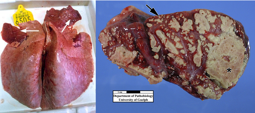
m.bovis
options for antigen detection in m.bovis diagnosis and how
-Option 1: culture on selective media and prolonged incubation (few days upto 21 days)
MUST species differentiate through PCR, DNA sequencing, MALDI-TOF MS→ as some are not pathogenic
-Option 2: PCR but only detect M. bovis so limited in terms of broad species scan
- bulk milk tank culture (BMT) and PCR potential for screening but high risk of contamination
-Individual animals on swabs, BAL, milk, joint fluid
but lives normally in the throat so no nasopahryngeal swab
Examples of when to take which samples of M.bovis?

Describe M.bovis antibiotic treatment
-tetracyclines
-florfenicols
-macrolides
-ALSO USE NSAIDS AND ORAL FLUID
macrolides for m bovis treatment
Protein synthesis inhibition - static,
highly variable response with substantial resistance in some populations,
favourable tissue penetration,
prolonged persistence
florfenicols for m bovis treatment
Protein synthesis inhibition - static,
useful systemic and respiratory tissue,
evidence of some resistance but less than tetracyclines
tetracyclines for m. bovis treatment
Protein synthesis inhibition - static,
can be used but variable clinical response with resistance documented
what abx can we NOT use for treating m. bovis
>Lacks a cell wall so beta-lactams are ineffective (penicillins, amoxicillins, cephalosporins)
What other considerations for M.bovis treatment
1.M.bovis causes caseous bronchopneumonia so makes antibiotic penetration harder
2.If chronic we see irreversible damage already present and contribution to Clinical signs→ is it worth treating
3.Often mixed infection
4.Clinical improvement does not mean the bacteria has gone (bc is caseous bronchopneumonia)
How can we control M.bovis?
-BUYING IN CATTLE:
-VACCINATE
-WASTE MILK: Feeding of waste milk to calves is not recommended where Mycoplasma bovis has been diagnosed. → transmit from dam to calf
how buy in cattle for m. bovis control
•clinically or subclinically infected
•Maintaining a strictly closed herd policy is the best
•If cows or heifers have to be purchased can be minimise risk:
a detailed history,
only purchasing from low SCC herds
by screening individuals quarantined before they enter the main herd, by serology
What is Mannheimia and Pasteurella species?
•Mannheimia haemolytica serotype 1: bacterial pathogen most frequently isolated from the lungs of recently weaned cattle with BRD and in dairy, beef or veal calves with enzootic pneumonia.
•Although less frequently cultured, Pasteurella multocida is also an important cause of bacterial pneumonia
•Histophilus somni is also recognized as an important pathogen in enzootic pneumonia and in some cases of BRD.
•These are all commensals of the upper respiratory tract so in the nasopharynx
Mannheimia and Pasteurella species

Mannheimia causing BRD
-Stress or viral replication in URT causes mannheimia replication and gets inhaled into lungs causing colonisation and pneumonia

What are the clinical signs of Mannheimia and Pasteurella species?
-Often preceded by viral respiratory disease signs→ off feed, pyrexia
-With the onset of bacterial pneumonia, clinical signs increase in severity.
-A combination of clinical signs of depression and fever (>39.4°C) without any signs attributable to other body systems, are the classic components of a case definition for early cases of BRD
-Serous to mucopurulent nasal discharge; moist cough; and a rapid, shallow respiratory rate
-Auscultation of the cranioventral lung field reveals increased bronchial sounds, crackles, and wheezes.
where will you find most resp diseae in cow lungs
cranioventral lung lobe→ ausc on clinical exam!!!
what is seen in severe cases of mannheimia and pasturealla infection
-In severe cases, pleurisy may develop, characterized by an irregular breathing pattern and grunting on expiration. The animal will become unthrifty in appearance if the pneumonia becomes chronic, which is usually associated with formation of pulmonary abscesses.
what is the importance of catching and treating viral respiratory diseases early in terms of mannheimia and pasturella
usually have a viral infection → allows mannheimia and pasturella make their way down
if catch early and treat → stop this
pathology of abscesses
Pulmonary abscessation can occur as the pneumonia becomes chronic.
Abscesses develop in ~3 weeks but do not become encapsulated until 4 weeks.
T pyogenes is frequently cultured from these abscesses.

pathology of h somni
Bronchopneumonia that may be followed by septicemia and infection of multiple organs
Associated with extensive fibrinous pleuritis in feedlot calves.
pathology of p. multocida
Associated with only small amounts of fibrin exudation,
some thromboses,
limited lung necrosis,
suppurative bronchitis and bronchiolitis.
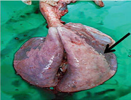
pathology of m. haemolytica
Severe, acute, haemorrhagic fibrinonecrotic pneumonia, with a bronchopneumonic pattern.
Extensive reddish-black to grayish-brown cranioventral regions of consolidation with gelatinous thickening of interlobular septa and fibrinous pleuritis.
Extensive thromboses, foci of lung necrosis, and limited evidence of bronchitis and bronchiolitis.
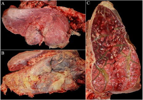
How do we diagnose Mannheimia and Pasteurella species?
often relies on gross necropsy findings and bacterial culture, immunohistochemistry or PCR assay.
Lung specimens can be collected for culture at necropsy.
if poss specimens should be collected from animals that have not been treated with antimicrobials
A multiplex PCR assay has been used to identify a number of bacterial agents implicated in bovine respiratory disease, including M haemolytica
pme best way
why do we want lung specimens from untreated animals
to permit determination of antimicrobial sensitivity patterns.
what is the issue with bacterial culture for mannheimia and pasturella
how can this be overcome
the bacteria involved are normal inhabitants of the upper respiratory tract,
the specificity of culture can be increased by collecting antemortem specimens from the lower respiratory tract by deep nasopharyngeal swab, transtracheal wash, or bronchoalveolar lavage.
How do we traet BRD?
Early recognition of bacterial infection followed by prompt treatment with antimicrobials is essential for successful treatment.
First line antibiotic: Oxytetracycline (cat D)
Long-acting antimicrobials such as tulathromycin, tilmicosin, florfenicol can be used as second line treatment to treat BRD (based on treatment failures and culture and sensitivity).
-NSAIDS and oral fluids
What is the Mannheimia and Pasteurella species control?
-minimise stress
-vaccinate (Parental vaccine vs Pasteruella and Mannheimia)
vacc dam → boost colostrum abodies
Diagnosing BRD...
-There is no gold standard
-pathogen detection
-clinical signs detected
-lung pathology
-epidemiology detection
what does us treating an animal mean for pme
it will have changes the pathogens present and the ability to find the causative agent
so if doing pme → want one that hasnt been treated
Describe diagnosing BRD with lung pathology
Any dead or moribund animals should be sent for post-mortem
Treatment, widespread mucosal destruction and secondary bacterial infections may reduce the likelihood of identifying the causative agent(s).
If full PME not possible, examine the abdomen, lungs and trachea on farm.
distribution and severity of lesions: cranio-ventral lung lobes typically worst affected.
Take samples that include the margin between normal and affected tissue, send fresh tissue for culture and sensitivity and fixed tissue for histopathology.
Describe pathogen detection for BRD diagnosis
-done for acutely unwell animals before secondary bacterial infections have become established.
- Conjunctival swab submitted for an immunofluorescent antibody test (IFAT) or PCR can be used for IBR diagnosis.
- Nasopharyngeal swab and broncho-alveolar lavage are usually not utilized as the first course of action a result of treatment failure, to allow more specific directed treatment.
-Unlike a nasopharyngeal swab, a BAL ensures that samples are obtained from the lower respiratory tract and hence reduces the chances of isolating commensal bacteria
does pathogen detection change our treatment choice?
no but most likely not change out treatment
but might change our vacc choices
ibr if not vacc already
pi3 and isv are ubiquous on diary farms
Elaborate lung pathology for BRD diagnoses
Any dead or moribund animals should be sent for post-mortem
Treatment, widespread mucosal destruction and secondary bacterial infections may reduce the likelihood of identifying the causative agent(s).
If full PME not possible, examine the abdomen, lungs and trachea on farm.
Take samples that include the margin between normal and affected tissue, send fresh tissue for culture and sensitivity and fixed tissue for histopathology.
which lung lobe might be worse affected on pme
cranioventral
advantages of detecting antigen
Rapid test results possible
Multiple animals can be sampled including cases and controls
Identification of viral pathogens can be highly significant depending on the nature of the herd
disadvantages of detection of antigen
• Viral infections are transient
• Identification is impaired by rising antibody titers
• Bacterial isolates from some samples are of dubious significance due to many of them being commensals
adv of serology to detect rising ab
• High sensitivity as most respiratory pathogens induce a strong antibody response.
• Less time-dependent as it can be effective in the relatively late stage of the disease
• Multiple animals tested makes the result relevant to the herd problem
disadv of serology to detect rising ab
• Requires convalescent serum so results are not available for >3 weeks
• Testing multiple samples can be expensive
• Correlation of seroconversion with clinical disease may be impossible in situations when viral infections are excepted to be common
adv of pme
• Gross and histopathologic examination usually suggests a cause and may be pathognomonic.
• Comprehensive and robust investigation
• Easy to sample the lung and trachea and are ideal sites for sampling for further tests