Acquired Cardiac Diseases in Dogs Pt. 2
 Call Kai
 Learn
 Practice Test
 Spaced Repetition
 Match
 Flashcards
 Knowt Play
1/27

There's no tags or description
Looks like no tags are added yet.
28 Terms
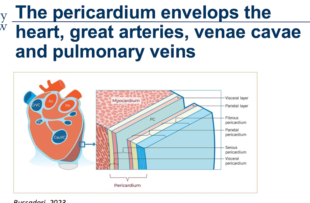
What is the anatomical location and function of the pericardium?
Acts as a protective barrier for the heart, allows friction free movement, allows different parts of the heart to work together, provides support and structure, metabolic effects.
What does the term “pericardial effusion (PE)” mean?
• An increase in the physiological amount of fluid in the pericardial sac, which may result in the clinical signs of cardiac tamponade.
• Cardiac tamponade:
• Critical condition, heart is not able to fill effectively and cannot pump blood
• Sudden and/or excessive accumulation of PE, pericardial does not have time to adapt
If chronic accumulation, the pericardium can stretch to accomodate
• Restricts appropriate filling of the cardiac chambers
Reduced CO
How does the length of time that the pericardium fills affect the pressure within the pericardium?
Acute
Raises pressure very quickly
Chronic
Slower pressure rise, adapts
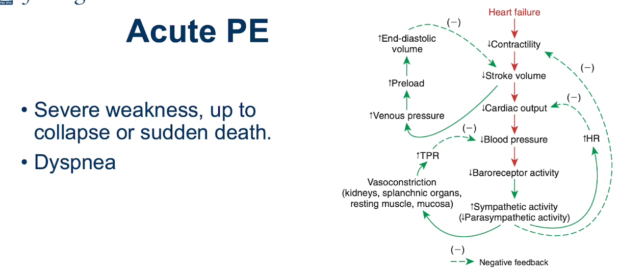
What are the consequences of acute PE?
Output and diffusion is low - which is why weakness, sudden death and dyspnea may occur
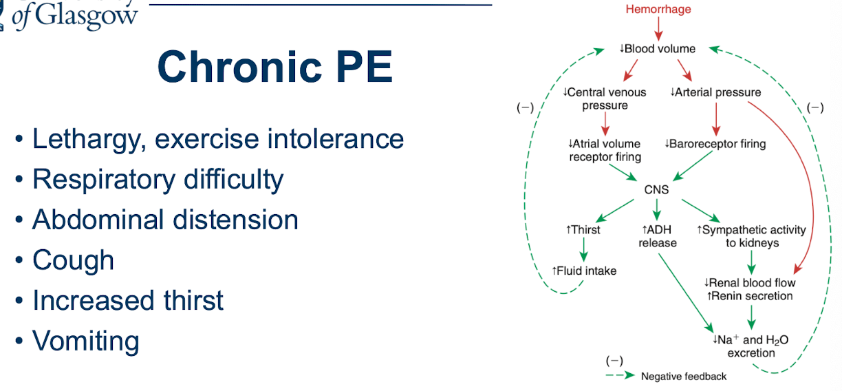
What are the consequences of Chronic PE?
Chronic - allows for adaptation of additional mechanisms to adapt to condition
Thirst - to increase volume
Kidneys - increase urine retention, to lose less fluid
Increased pressure in right side raises hydrostatic pressure, pushing fluid out, causing resp. difficulties and abdominal distension
What are other common additional signs associated with acute and chronic PE?
Muffled cardiac sounds - due to effusion within the pericardial sac
May also auscultate a higher heart rate (To try and increase CO due to reduced SV caused by effusion pressure)
What is pulsus paradoxus?
Pulsus paradoxus is an abnormally large drop in systolic blood pressure (greater than 10 mmHg) that happens when you breathe in (inspiration)
Increased Right Ventricular Filling: When you breathe in, chest pressure drops, pulling more blood into the right side of the heart.
Ventricular Interdependence (Chamber Competition): Normally, the heart expands to accommodate this extra blood. However, trapped inside a stiff, fluid-filled pericardial sac, the right ventricle cannot expand outward.
Septal Bowing: To make room, the muscular wall separating the two ventricles (the interventricular septum) bulges directly into the left ventricle.
Reduced Left Ventricular Output: With the left ventricle squeezed smaller, it receives and pumps out significantly less blood. This causes the systolic blood pressure to plummet by more than 10 mmHg.
How does a heart with PE appear on ECG?
Electrical alternans of the QRS
Electrical Alternans: This is a beat-to-beat variation in the height or amplitude of the QRS complexes (one beat is taller, the next is shorter, and so on). It is classically caused by the "swinging heart" phenomenon—because the heart is surrounded by a large pool of fluid, it physically rocks or oscillates back and forth with every heartbeat, changing its angle relative to the ECG electrodes from moment to moment.
Complexes may be present on ECG.
The QRS may be small.
When there is a lot of fluid in the pericardial sac (the membrane surrounding the heart), that fluid acts as an electrical insulator or buffer, muting the electrical signal before it reaches the skin electrodes.
Not all dogs have these ECG findings.
How does PE appear on ultrasound?
Free fluid present around the ventricles, causes collapse of the right atria (due to pressure)
Right ventricle also more effected since thinner and more compliant
How is a pericardiocentesis performed?
Left lateral recumbency, clip skin, use needle to remove fluid to allow the heart to expand
What are the most common causes of PE?

After removal of fluid from the pericardium, how can the fluid be analyzed and categorized?
Most of neoplastic origin - are hemorrhagic

What are the main congenital pericardial diseases?

What are the main cardiac neoplasias as causes of PE?
• Hemangiosarcoma
• Neuroendocrine tumors (aortic body tumors)
Chemodectoma
Paraganglioma
Ectopic thyroid carcinoma
• Pericardial Mesothelioma
Lymphosarcoma (rare in dogs, more in cats)
What may happen to the pericardium after inflammation or multiple episodes of PE?
The pericardium may become stiff after an inflammation, multiple episodes of PE, etc.
Ventricular interdependence is the phenomenon where the size, shape, and mechanical behavior of one cardiac ventricle directly influence the filling and function of the other.
May cause right sided heart failure
Without fluid (Dry Form) → Constrictive pericarditis
Small amount → Effusive constrictive pericarditis

What are the main features of pericardial mesothelioma?
• Originates from the mesothelium (cell lining present on the pericardial, pleural and peritoneal cavities)
• Rare (less than 0.2% of all tumors)
• Often no clear mass present, only PE.
• Potentially concomitant pleural involvement.
What are the most frequent neoplasias of the heart?
• Hemangiosarcoma (Mainly german shepherds, and labrador retriever) Chemodectoma (Common in brachycephalic breeds, originates from chemoreceptors in branch or aortic arch) most commonly identified.
In mice - if increasing CO2, hypercapnia, tumour was more common
Brachycephalics due to airway - more predisposed due to the hypercania state caused by their anatomy
• Breed predispositions present.

What is the main pathology caused by cardiac hamangiosarcoma?
• Usually, mass arising from right atrium and/or right auricle.
Hemangiosarcoma is a tumour of the blood vesels
• Frequently located in the spleen.
But can be primary in heart, or primary in spleen and spread to heart
• Metastasis and invades
• Tend to break and bleed
• German Shepherd Dog, Golden and Labrador retriever
Prognosis very poor, once there, already spread, cannot easily be removed surgically, chemo not available.
What is the main pathology caused by chemodectomas?
• Originates from chemoreceptor cells from the aortic root
• Hypoxia as trigger → present in brachycephalic breeds.
• Usually does not invade or produce metastasis - benign
• Problematic if too big and compresses the heart → Right sided heart failure.
What is pulmonary hypertension (PH)?
• Pathological hemodynamic condition defined by an increased systolic/mean pulmonary arterial pressure (PAP).
• Chronic increased PAP leads to right sided concentric hypertrophy → potentially can cause right sided heart failure.
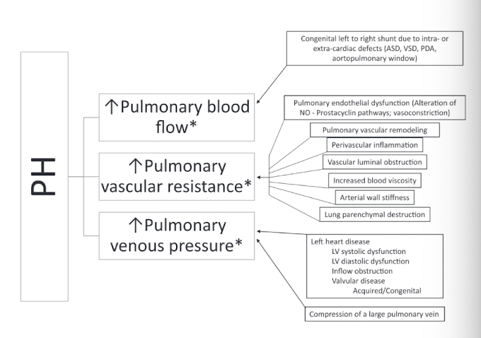
What are the two main categories pulmonary hypertension can be split into?
Pre-capillary PH happens before blood reaches the lung capillaries (the tiny blood vessels where oxygen is exchanged).
What it means: The problem originates inside the lung's own arteries or tissue, rather than from the heart backing up.
Post-capillary PH happens after blood passes through the lung capillaries. It is caused by pressure backing up from the left side of the heart.
What it means: The left side of the heart cannot pump blood out effectively, causing blood and pressure to back up into the pulmonary veins and capillaries.

What are the main causes of PH in dogs?
1. Pulmonary arterial hypertension (includes cardiac shunts).
2. Left heart disease
3. Secondary to respiratory disease
4. Thromboembolic
5. Parasitic (Heartworms, Lungworms)
6. Miscellaneous
Describe how a heartworm infection may contribute to PTH?
Right-Sided Strain: Because resistance is elevated in the pulmonary arterial tree, the right ventricle has to work harder to pump blood through the lungs, which can eventually lead to right-sided heart strain

What symptoms are caused by heartworm?
How is it diagnosed?
**Need two confirmatory tests

What is the protocol for treating a heartworm infection?
Macrocyclic lactone and doxycycline
Injection of melarsomine as an adulticide drug
Describe the infection caused by angiostrongylus vasorum.
AKA french heartworm/lungworm
• Infection via slugs and snails
• Get into the bronchial walls → Coughed out → Swallowed → Expelled in the faeces.
Intermediate hosts: Slugs and snails act as intermediate hosts. Dogs can become infected by eating them or potentially taking in larvae from slime trails on grass, outdoor toys, or puddles.
Paratenic hosts: Animals such as frogs can carry larvae and serve as a route of infection if eaten.
Life cycle: Larvae penetrate the gut wall, travel to the heart and pulmonary arteries, mature, and lay eggs. Hatched larvae move into the airways, are coughed up and swallowed, and pass out in feces—where slugs or snails can pick them up.
Can cause PH
Heart and lungworms in cats and dogs:
Dirofilaria immitis
Angiostrongylus vasorum

What are the main features of endocarditis?
How does it present?
• Infection of the valves (bacterial)
• Aortic and Mitral valve most common.
• Potentially all valves can be targeted, but on the left side is most common.
• Presence of sub-aorticstenosis, having aortic regurgitation, using corticosteroids may predispose to it.
Presentation:
• Dogs often: Fever, new murmur, lameness
• Cats may not have fever and/or murmurs
• Requires to find origin of infection
• Blood cultures
• Urine culture
• Imagining (Chest, Abdomen) Pediatric
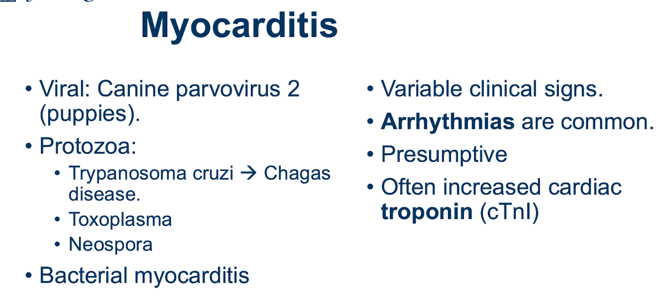
What are the main features of myocarditis?
How does it present? What is the aetiology?
• Pathological infiltration of the myocardium by inflammatory cells.
• Various infection agents or non-infectious may be involved.