MMI 188B Midterm 2
 Call Kai
 Learn
 Practice Test
 Spaced Repetition
 Match
 Flashcards
 Knowt Play
1/153

There's no tags or description
Looks like no tags are added yet.
154 Terms
Similarities of B and T Cell Development
receptor gene rearrangements provide diversity for both
both originate in bone marrow
both undergo negative selection to remove self-reactive clones
Differences in B and T Cell Development
T cells mature in thymus, B cells mature in secondary lymphoid tissues
Self MHC
set of MHC Class I and II molecules encoded by an individuals own genes
ensures T cells function effectively within the host’s genetic and immunological framework
T-Cell Precursor
common progenitor cell that gives rise to both alpha:beta and gamma:delta T-cell lineages within the thymus
T Cell Lineages
when developing, they differentiate into 2 different linages
alpha:beta T cells (the majority)
gamma:delta T cells (the minority)
Thymus
primary lymphoid organ where thymocytes mature
involutes as we get older
Thymocytes
developing immature T cells derived from bone marrow progenitors
Thymic Stroma
a network of epithelial cells that forms the structural framework supporting thymocyte development
Thymic Anlage
rudimentary thymic structure formed during embryogenesis from ectodermal cortical and endodermal medullary epithelial cells
Complete DiGeorge Syndrome
a genetic disorder in which the thymus fails to develop, resulting in a complete lack of T cells and severe immunodeficiency
Involution of the Thymus
the age-related degeneration of thymic tissue beginning about one year after birth, with fat gradually replacing thymocytes
Immunosenescence
the gradual decline of immune function with age, associated with reduced thymic activity and diminished production of new T cells
Thymic Cortex
above the medulla
contains many immature thymocytes
Thymic Medulla
underneath the cortex
many mature thymocytes
Hassall’s Corpuscles
primary source of thymic stromal lymphopoietin (TSLP) which promotes T-reg production
plays a vital role in central tolerance, helping the body prevent autoimmune diseases by suppressing immune responses against self antigens
involved in removal of apoptotic thymocytes (developing T cells that failed selection)
TSLP
cytokine essential for activating myeloid dendritic cells within the thymus
activate dendritic cells which induce the differentiation of developing T-cells into regulatory T-cells (T-regs)
Double-Negative Thymocytes (DN Thymocytes)
early thymic T-cell precursors that lack both CD4 and CD8 surface markers
Thymocyte Commitment to T-Cell Lineage
this is done before the thymocyte rearranges their T-cell receptor genes
involves changes in
gene expression
cell surface markers
Notch 1
surface membrane-associated receptor on thymocyte that drives T-cell development
binds to its ligand on thymic epithelium and turns on the expression of genes essential for T-cell development that:
REMOVE REPRESSIVE TRANSCRIPTION FACTORS
RECRUIT CO-ACTIVATING TRANSCRIPTION FACTORS
Double-Positive Thymocytes (DP Thymocytes)
immature T cells in the thymus that express both CD4 and CD8 co-receptors on their surface
T-Cell Development from Progenitor to Double Positive T Cell (step-by-step)
T-cell precursors that enter the thymus express the hematopoietic stem-cell markers CD34 but none of the characteristic markers of mature T cells
As soon as they produce a complete receptor, gamma:delta cells can leave the thymus and travel to other tissues via the blood
Following beta-chain-rearrangement in the thymus, rearrangement of the alpha-, gamma-, and delta- chain genes resumes, and a productive alpha-chain gene rearrangement produces double-positive CD4 CD8 alpha:beta T cells
A minority of the thymocytes that resume rearrangement give rise to additional gamma:delta T cells
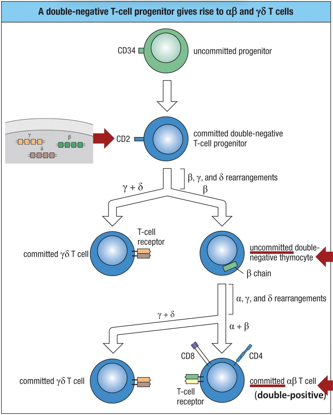
Result of Gene Rearrangement in Double-Negative Thymocytes
leads to assembly of either a gamma:delta receptor or a pre-T-cell receptor
Pre-T Alpha Chain
an invariant polypeptide that serves as a surrogate alpha chain in developing thymocytes
Pre-T-Cell Receptor (pre-TCR)
formed when a beta chain pairs with pT-alpha, creating a heterodimer that dimerizes further into a superdimer
Lymphocyte-Specific Protein Tyrosine Kinase (Lck)
a cytoplasmic tyrosine kinase that transduces signals from the pre-TCR through phosphorylation cascade
Pre-T Cell
a thymocyte that has successfully assembled a pre-TCR and received survival signals progresses to the pre-T cell stage
Fate of Apoptotic Immature T Cells
ingested by macrophages in the thymic cortex
TCR Diversity
generated by gene rearrangement during T cell development
Beta-Chain TCR Gene Rearrangements
beta-chain REARRANGES FIRST
has V-D-J Segments
Pre-T-Cell receptor contains a mature TCR-Beta Chain
two attempts at recombination are possible for each chromosome (total of 4 attempts)
nonproductive beta-chain rearrangements can be rescued by further rearrangement made possible by the two C-beta genes and their associated D-beta and J-beta segments

The Alpha:Beta Advantage
commitment to alpha:beta lineage requires only one productive gene rearrangement
commitment to the gamma:delta lineage requires a minimum of two productive rearrangements
Alpha-Chain TCR Gene Rearrangement
rearranges after beta-chain
only occurs in pre-T-Cells
TCRalpha lacks D segments
can undergo several sequential rearrangements
rearrangement of the alpha-chain gene leads to the loss of the delta locus
delta-chain gene is eliminated
Double Negative T Cell Development Steps
committed T-cell progenitor
Rearrange beta, gamma, delta
first checkpoint
pre-TCR receptor
dividing pre-T cells
pTalpha receptor
Double Positive T Cell Development Steps
rearrange alpha, gammam delta chain
CD8 + CD4 receptor
second checkpoint
TCR
single positive T cell
Positive Selection
ensures only thymocytes with T-cell receptors (TCRs) capable of recognizing and effectively interacting with self MHC molecules survive
occurs in the thymus
determines expression of CD4 or CD8
does not occur to gamma:delta cells
MHC Restriction
cell recognizes antigen only when it is presented by the individual’s own MHC molecules
does not occur to gamma:delta cells
Negative Selection
eliminates autoreactive T cells with TCRs that bind too strongly to self-peptide: self-MHC complexes
ensures central tolerance, preventing self-reactive T cells from entering circulation and causing autoimmune disease
removed in the thymus
does not occur to gamma:delta cells
Thymoproteasome
a specialized form of the proteasome found exclusively in cortical epithelial cells of the thymus
generates self-peptides optimized for positive selection of developing CD8+ T cells that recognize peptides presented by MHC Class I molecules
Single-Positive Thymocytes
express only one co-receptor, CD4 or CD8, following positive selection in the thymus
CD4 single-positive thymocytes → precursors of helper T cells
CD8 single-positive thymocytes → precursors of cytotoxic T cells
Steps and Results of Positive Selection
Selects TCRs that bind to self MHC
called MHC restriction
selection made by cortical epithelial cells in the thymus
express both MHC I and MHC II
Instructs expression of either CD4 or CD8 and other genes
interact with self MHC I → CD8
interact with self MHC II → CD4
other co-receptor is downregulated
mature into single positive thymocytes
Steps and Results of Negative Selection
dendritic cells present antigen
removal of T cells with autoreactive TCRs is central tolerance
autoimmune regulator (AIRE) induces expression of multiple tissue-specific genes in the thymus to allow negative selection
mutations in AIRE result in autoimmunity
macrophages engulf dead T cells
Autoimmune Regulator (AIRE)
a transcription factor expressed by a specialized subset of medullary thymic epithelial cells (mTECs)
extends negative selection to include tissue-specific antigens that are not normally expressed in the thymus
Autoimmune Polyendocrinopathy-Candidiasis-Ectodermal Dystrophy (APECED)
results from loss-of-function mutations in the AIRE gene
without AIRE, tissue-specific antigens are not expressed in the thymus, so self-reactive T cells specific for those antigens escape negative selection
these T cells enter the periphery and attack multiple organs and tissues, causing widespread autoimmune damage
Regulatory T Cells
specialized subset of CD4 T cells that help maintain self-tolerance and immune homeostasis
recognize self antigens presented by MHC class II molecules but do not initiate immune responses
suppress the activation and proliferation of self-reactive naive T cells, preventing autoimmune damage
suppress autoreactive T cells in the periphery (peripheral tolerance)
create a backup line of defense against autoimmunity
produce suppressor cytokines
IL-10, TGF-Beta
FoxP3
transcription factor expressed by regulatory T cells
mutation in this transcription factor causes autoimmunity
Location and Process of T Cell Development
Proliferation and differentiation of double-negative CD3- thymocytes and double-positive CD3+ thymocytes
double-negative CD3- thymocytes in the subcapsular zone
Positive Selection
double-positive CD3+ thymocytes in the thymic cortex
Negative Selection
double-positive CD3+ thymocytes throughout cortex and especially at the cortico-medullary junction
Entry to the Circulation
mature self-restricted, self tolerant, single positive CD4 or CD8 T cells leave the thymus in the blood venules
X-Linked Severe Combined Immunodeficiency (SCID)
a deficiency in T and B cells
SCID Thymus
very small
loss of lymphocytes
no cortico-medullary differentiation
Hassall’s corpsucles are absent
SCID Diagnosis
Enumeration of TRECs (T cell receptor excision circles, due to gene segment rearrangements)
Enumeration, phenotypic characterization, and functional analysis of circulating lymphocytes
Mutation analysis is used to provide definitive diagnosis
Defects That Result in SCID
Defects that impair lymphocyte survival
Defects in cytokine-mediated signals for lymphocyte maturation and proliferation (ex: mutation of IL2RG gene which encodes the common gamma chain, shared by IL-2R and other cytokine receptors)
Defects in machinery for somatic gene rearrangement that assembles the immuhnoglobulin and T-Cell Receptor genes during lymphocyte development, the so-called V(D)J recombination process
Mutations in genes that encode the CD3 molecules that participate in the formation of the CD3:TCR complex. This complex allows signaling through the pre-T-cell receptor
Basic 3 Roles of Naive T Cells
Meet specific antigen in secondary lymphoid tissu e
Undergo clonal expansion
Differentiate into effector T cells
Basic 3 Facts of Effector T Cells
Migrate to sites of infection
Antigen stimulates effector T cells to function
Two main kinds: CD8 T cells and CD4 T cells
Naive T Lymphocytes
T cells that have not yet been stimulated by antigen
Recirculate through peripheral lymphoid organs
visit lymph nodes
express L-selection that binds to ligands on High Endothelial Venules (HEV)
Express TCR, but do NOT engage in effector functions (ex: cytokine production, cytolysis)
.
Effector T Lymphocyte
T cells that have differentiated in response to antigen
Perform effector functions required to eliminate pathogens
cytokine production
cytotoxins
cytolysis (cytotoxic T cells)
Migrate to peripheral sites of infection or antigen entry
VLA-4
expressed by ONLY effector T cells that enables them to enter infected tissue
LFA-1, CD2, and CD44
adhesion molecules secreted by effector T cells and naive T cells
in effector T cells, they are made in larger quantities and it allows them to make stronger interactions with target cells
CD45O
secreted ONLY by effector T cells to increase their capacity to interact with target cells
Memory T Lymphocytes
T cells that have differentiated in response to antigen and are
long-lived as clones
functionally silent
capable of mounting enhanced responses to antigen challenge
migrate to lymphoid tissues and peripheral sites of antigen entry
survive without antigen recognition
clones can remain for decades, replicating to maintain numbers
T-Cell Activation (T-Cell Priming)
first stage of the primary adaptive immune response in which a naive T cell encounters and responds to its specific antigen for the first time
marks the transition from innate to adaptive immunity
establishes a pool of antigen-specific effector and memory T cells, essential for both pathogen clearance and long-term immune protection
Activation of Naive T Cells by Antigen
mature dendritic cell carries an antigen from infection site to secondary lymphoid tissue where it encounters a naive T cell
naive T cell is primed encountering an antigen for the first time, initiating its adaptive immune response
after encountering a specific antigen, they
proliferate
differentiate
leave lymph node in efferent lymph and enter the circulation
Immature Dendritic Cell
functions as sentinels that capture, uptake, and process antigens from pathogens through phagocytosis, macropinocytosis, and receptor-mediated endocytosis
located in tissues
known as Langerhans cells in skin
Dendritic Cells Changing Function and Morphology
in lymphatic circulation, they stop phagocytosis so MHC II moves out of the endocytic vesicles onto the cell surface
when reaching T-cell area of lymph node, the maturing cell halts uptake and processing of antigens and focuses on presenting the to T cells
Micropinocytosis
a receptor-mediated endocytic process used by dendritic cells to capture small volumes of extracellular fluid containing bacteria or viral particles
a method for dendritic cells to internalize pathogens
Macropinocytosis
a NON-receptor-mediated process that captures larger volumes of extracellular fluid, including pathogens not recognized by endocytic receptors
a method for dendritic cells to internalize pathogens
Dendritic Cell MHC Class II Processing and Presentation
used in
receptor-mediated endocytosis of bacteria
macropinocytosis of bacteria or viruses
then they’re presented to CD4 T cells
Dendritic Cell MHC Class I Processing and Presentation
used in
viral infection
viral proteins from viral DNA get broken down
cross-presentation of exogenous viral antigens
then they’re presented to CD8 T cells
T-Cell Area (T-Cell Zone)
outer cortex region of the lymph node where naive T cells and antigen-bearing dendritic cells interact
primary site of T cell activation during adaptive immune responses
Homing
the process by which naive T cells exit the bloodstream and enter the T-cell zone of lymph nodes
L-Selectin
adhesion molecule on naive T cells ONLY that binds to vascular addressing on HEV surfaces
Vascular Addressins
mediate the initial tethering and rolling of T cells along HEV walls during homing
include CD34 and GlyCAM-1
Integrins
activated upon TCR and CD28 signaling
help stabilize TCR:MHC interactions and cell-cell contact
help activated T cells leave the vasculature to go to sites of inflammation
ICAM-3
binds to DC-SIGN and promotes initial stable contact between naive T cells and dendritic cells for antigen scanning
CD2 and LFA-3
an adhesion molecule on the T cell surface and its ligand on dendritic cells and other antigen-presenting cells
Cognate Pair (or Conjugate Pair)
a long-lived complex between a mature dendritic cell and an antigen-specific naive T cell
S1P
a lipid molecule that creates a chemotactic gradient within the lymph node
Functions of T Cell Integrins like LFA-1
Adhesion of T cells to APCs
T cell activaiton
Binding of T cells to endothelium
T cell migration
Ligands for integrins are induced on APCs and endothelial cells by inflammatory cytokines and microbes
these ligands promote T cell responses to infectious agents
Affinity of integrins for their ligands is increased by
chemokines (rapid response during inflammation)
TCR ligation (amplifies response to antigens on APCs)
LFA-1 (Lymphocyte Function-Associated Antigen 1)
integrin adhesion molecule that is expressed by all leukocytes
binds ICAM-1, ICAM-2, ICAM-3
promotes adhesion and cytoskeletal reorganization during T cell activation
promotes stable TCR:MHC interaction
Transient Adhesive Interaction Between a Naive T Cell and a Dendritic Cell
naive T cell binds to dendritic cell cell with low affinity LFA-1-ICAM-1 interactions, but through recognition of a specific antigen, a signal is sent to LFA-1 that induces a conformational change which increases affinity and prolongs cell-cell contact

Co-Stimulatory Signal
the second signal required for activation, survival, and proliferation of naive T cells, in addition to antigen recognition by the T-cell receptor (TCR) and co-receptor (CD4 or CD8)
Co-Stimulatory Receptor
binds to a corresponding co-stimulator on an antigen-presenting cell to deliver the co-stimulatory signal
CD28 and B7 molecule
CD28
co-stimulators receptor present on naive T cells
the only B7 receptor on naive T cells
expressed as a homodimer on
90% of CD4+ T cells
50% of CD8+ T cells
binds B7 expressed on APCs (including B cells)
when bound, it activates a T cell to divide
B7 Molecule
co-stimulator expressed on professional antigen-presenting cells
CTLA4
acts as a negative regulator, suppressing further T-cell activation and proliferation when bound to B7 to maintain immune balance and prevent overreaction
binds B7 20x stronger which makes it act as a brake on CD28
CD28 would otherwise allow the T cell to divide
Signals for T Cell Activation
Antigen
determines specificity of response*
mediated by TCR:MHC+peptide interactions
Co-stimulators
surface molecules expressed by APCs and T cells
provide amplification*
determine type of response*
Cytokines
growth and differentiation factors for T cells
produce by APCs and T cells themselves
provide amplification*
determine type of response*
CD28:B7
necessary for effective co-stimulation of antigen-specific naive T cells
induce activation of PI-3 kinase and the NFkappaB signal transduction pathway
NFkappaB is a transcriptional response element
promote cell survival through upregulation of Bcl-xL
increase production of IL-2
promote IL-4 production and T cell differentiation
Bcl-xL (B-cell lymphoma-extra large)
transmembrane molecule in mitochondria
anti-apoptotic protein that prevents release of mitochondrial contents like cytochrome c
leads to caspase activation and apoptosis
Activation of Naive T Cell (Two Signals)
requires BOTH an antigen-specific signal AND a co-stimulator signal
B7 co-stimulators signal delivered by the APC
CD28 acts as an accelerator for the naive T cell that binds B7 in APC
CTLA4 acts as a break on CD28 on activated T cells, binding B7 20 times stronger than CD28
T-Cell Synapse
a specialized region of contact between a T cell and an antigen-presenting cell (such as a dendritic cell) where ligand-receptor interactions occur
facilitates communication and signal integration during T-cell activation
ITAMs
short amino acid sequences located in the cytoplasmic tails of CD3 proteins and the zeta chain of the TCR complex
Summary of Intracellular Signaling Pathways
Initiated by T-Cell Receptor Complex + CD4 or CD8 co-receptor + CD28 co-simulatory receptor
When naive T cell interacts w dendritic cell presenting its antigen, the ZAP70 kinase activates, initiating a series of reactions
NFkappaB, NFAT, and AP-1 change the pattern of gene expression
cell division, proliferation, and differentiation to effector T cells
T-Cell Signaling Defects
abnormalities in TCR signaling pathway that impair activation, proliferation, or effector function of T cells
can result in immunodeficiency disorders
Severe Combined Immunodeficiency (SCID)
results in a complete failure of T cell activation and severe impairment of adaptive immunity
Distal TCR Signaling Defects
cause partial or variable immune dysfunction depending on which pathway is disrupted
IL-2
cytokine crucial for T-cell proliferation and differentiation, originally called T-cell growth factor
acts as a central regulator of adaptive immune response, promoting clonal expansion of antigen-activated T cells
Autocrine Action
when a cytokine acts on the same cell that secreted it
Paracrine Action
when a cytokine acts on nearby but different cell types
IL-2 Receptor on Naive T Cells
express the low-affinity receptor, a heterodimer of the beta chain and the gamma chain
IL-2 Receptor on Activated T cells
express the high-affinity receptor, a heterodimer comprising an alpha-chain, a beta-chain, and a gamma-chain
How IL-2 Affects Activated T Cells
IL-2 is synthesized and secreted by the activated T cell
IL-2 binds to the IL-2 receptor on the same T cell
co-stimulation through CD28 increases the synthesis of IL-2 by 100-fold in the T cell
T cell proliferates and differentiates from signaling from IL-2
some immunosuppressive drugs inhibit cytokine IL-2 or the IL-2 receptor to block T cell activation and differentiation
Antigen Recognition Without Co-Stimulation
may result in
no response (ignorance)
apoptosis
anergy (functional inactivation)
T-Cell Anergy
an irreversible, tolerance-inducing mechanism that prevents activation of self-reactive T cells in the absence of co-stimulation
Adjuvants
substances grant enhance the immunogenicity of an antigen by stimulating the innate immune system