3001: Cholesterol, Fatty acids
 Call Kai
 Learn
 Practice Test
 Spaced Repetition
 Match
 Flashcards
 Knowt Play
1/139

There's no tags or description
Looks like no tags are added yet.
140 Terms
What is cholesterol, and what are its main functions?
A sterol (steroid with an -OH group).
Maintains cell membrane fluidity and stability;
precursor for bile acids, steroid hormones, and vitamin D.
Makes up approximately 25% of animal membrane lipids.

How is cholesterol stored, and where does it come from?
Sources: diet, bile, and endogenous synthesis.
Free cholesterol is mainly found in membranes.
Stored as cholesterol esters formed with fatty acids.
Approximately 150 mg/day of unabsorbed cholesterol is excreted.
NPC1L1 transports dietary and biliary cholesterol and phytosterols into enterocytes through the apical brush-border membrane.
Absorption depends on dietary fat, fiber, plant sterols, and intestinal transit time.
Plant sterols that compete with cholesterol absorption. Enriched foods or supplements can reduce blood cholesterol by 10% or more.
ABCG5/ABCG8 transport sterols back into the intestinal lumen.
What happens to cholesterol after entering an enterocyte?
Some enters membranes; most is esterified by ACAT2.
MTP and ApoB-48 help package it into chylomicrons → lymph → blood near subclavian vein.
Triglycerides are removed, and chylomicron remnants travel to the liver.
What happens to cholesterol in the liver, and what is NPC1L1's role?
The liver receives cholesterol from chylomicron remnants and bile.
Cholesterol is reused for bile acids and steroid synthesis or stored.
NPC1L1 is also in hepatocyte (cell lining of liver) canalicular membranes and may reabsorb cholesterol from bile, but its exact role is uncertain.
What are NPC1 and NPC2, and what happens when they are defective?
NPC1 is a lysosomal transmembrane protein;
NPC2 is a soluble lysosomal protein.
Both transport cholesterol out of lysosomes. Defects cause cholesterol accumulation and Niemann-Pick type C disease.

Why does the brain synthesize its own cholesterol?
The blood-brain barrier prevents dietary and peripheral cholesterol from entering.
The brain synthesizes its own cholesterol and converts excess into 24S-hydroxycholesterol for removal across the barrier.
Why is cholesterol homeostasis important?
Cholesterol synthesis requires ATP and is energetically expensive, so excess Cholesterol cannot always be completely degraded for energy.
Excess is converted into derivatives such as bile acids for excretion. Endogenous cholesterol availability regulates synthesis.
What are oxysterols, and how are they produced?
Cholesterol generates oxysterols to help regulate cholesterol homeostasis, especially when cholesterol levels become excessive.
They are oxygenated cholesterol derivatives that act as key metabolic intermediates and signaling molecules that convert cholesterol into water-soluble compounds and accelerate its removal from the body.
Produced enzymatically or through oxidation by reactive oxygen species (ROS).
Examples: 27-, 24S-, 25-, and 4β-hydroxycholesterol; 7-ketocholesterol and cholesterol epoxides.
How are oxysterols produced from cholesterol, and what are their functions and effects?
Oxysterols are oxygenated cholesterol derivatives produced through two pathways:
1. Enzymatic pathway: Specific enzymes convert cholesterol into oxysterols:
CYP27A1 → 27-hydroxycholesterol
CYP3A4 → 4β-hydroxycholesterol
CYP46A1 → 24S-hydroxycholesterol
CH25H → 25-hydroxycholesterol → CYP7B1 → 7α,25-dihydroxycholesterol
2. Non-enzymatic pathway: Reactive oxygen species (ROS) oxidize cholesterol, producing 5,6-epoxycholesterol, cholestane-3β,5α,6β-triol, and 7-ketocholesterol (also shown with CYP7A1 in the diagram).
Functions and effects:
Oxysterols act as metabolic intermediates and bioactive signaling molecules.
Some activate LXR to regulate cholesterol homeostasis.
Different oxysterols have different biological effects.
Physiological levels: Safe and necessary for normal functions.
Excessive production: Can cause oxidative damage, lipid damage, and cellular toxicity.

What are Liver X Receptors (LXR)?
Nuclear receptors that act as transcription factors.
Two forms: LXRα and LXRβ. Activated by oxysterols to regulate cholesterol homeostasis by changing expression of lipid-metabolizing genes.
How does LXR regulate cholesterol levels?
Increases ABCA1/ABCG1 → cholesterol efflux to HDL for reverse cholesterol transport.
Decreases NPC1L1 → less intestinal absorption.
Suppresses cholesterol synthesis genes (HMG-CoA reductase).
Has anti-inflammatory effects. It modulates gene expression to reduce atherosclerosis risk.
Why can excess oxysterols be harmful?
Over production of Oxysterols can be harmful to cells as their 1000s of oxidative products are accumulated in the body and oxidizes other cellular metabolites and membrane lipids
Some of these derivatives are not soluble and but toxic and accumulate in the body (oxidative products of oxysterols)- these are highly reactive species
Etiology for cancer, atherosclerosis, Alzherimer’s disease, cataracts, age related neurological other diseases, osteoporosis, inflammatory diseases, etc.,
Reducing agents such as Vit E, Vit C and Glutathione can reduce the oxysterol loads and oxidative products of oxysterols
What is membrane transition temperature (Tm)?
The temperature at which a membrane changes phases. Involved in membrane fluidity.
Below Tm → rigid, solid-like gel phase.
Above Tm → fluid, liquid-crystalline phase.

How is cholesterol positioned in the phospholipid bilayer?
Its polar/hydrophillic -OH group lies near phospholipid heads.
Hydrophobic steroid rings and hydrocarbon tail lie within the membrane interior.
Rigid rings improve packing and strength; the flexible alkyl chain interacts with phospholipid tails, including unsaturated chains.

How does cholesterol's amphipathic structure provide dual functions in phospholipid membranes?
Rigid hydrophobic sterol ring: Embeds among phospholipid tails and interacts with saturated fatty acid chains, filling gaps → ↑ membrane density, ↓ defects, and ↑ mechanical strength.
Flexible hydrophobic alkyl chain: Interacts with unsaturated fatty acid tails, reducing disorder caused by their kinks → prevents excessive membrane fluidity.
Overall: Cholesterol improves membrane packing, stability, and mechanical strength while regulating fluidity.
How does cholesterol regulate membrane fluidity at different temperatures?
Tm (transition temperature): Temperature at which a membrane transitions between a rigid gel phase and a fluid liquid-crystalline phase.
Below Tm: Pure PL membranes are rigid (gel phase). Cholesterol disrupts tight PL packing → prevents excessive rigidity.
Above Tm: Pure PL membranes are fluid (liquid-crystalline phase). Cholesterol restricts vigorous PL movement, especially lateral motion → prevents excessive fluidity.
Near Tm: Cholesterol buffers and smooths the phase transition, preventing sudden changes in fluidity.
Extreme temperatures: Cholesterol's sterol rings hinder orderly PL packing, helping prevent rigid gel formation at low temperatures and stabilize membrane structure.
Overall: Cholesterol acts as a fluidity buffer, stabilizing the liquid-crystalline phase over a broader

How do certain antifungal drugs use membrane sterols to kill fungi?
Fungal membranes contain ergosterol, similar to cholesterol.
Certain antifungals bind ergosterol and disrupt membrane function by reducing the ergosterol function as it is no more available for membrane function, therby, decreasing the fluidity of fungal cell membrane – more rigid not favor and break easily
They preferentially target ergosterol rather than human cholesterol, so they don’t harm us.
Sex hormones (progesterone, testosterone, estradiol); corticosteroids (cortisol); bile acids (cholic acid); and vitamin D3 (formed through 7-dehydrocholesterol).
What happens to cholesterol during bile synthesis and recycling?
The liver converts cholesterol into bile acids that help digest and absorb fats.
Bile components can be reabsorbed in the intestine and returned to the liver for reuse.
The lecture identifies bile formation as accounting for approximately 50% of cholesterol excretion.
How are vitamin D2 and vitamin D3 produced?
Vitamin D2 (ergocalciferol) forms from ergosterol through UV irradiation.
Vitamin D3 (cholecalciferol) forms in skin: 7-dehydrocholesterol → UVB → previtamin D3 → thermal isomerization → vitamin D3. UV exposure can also form lumisterol and tachysterol.

How is vitamin D transported in the body?
Vitamin D travels through blood in chylomicrons or bound to vitamin D-binding protein (DBP), reaching the liver for further metabolism.
How do vitamin D (calcitriol) and PTH regulate blood calcium levels?
Skin: UVB sunlight converts 7-dehydrocholesterol into vitamin D3 (cholecalciferol).
Liver: Converts vitamin D3 into 25-OH vitamin D (calcidiol)
In response to low calcium levels:
Parathyroid glands: Release parathyroid hormone (PTH).
Kidneys: PTH stimulates conversion of 25-OH vitamin D into active calcitriol (1,25-(OH)₂ D) via renal hydroxylase
Kidneys: PTH and calcitriol increase Ca²⁺ reabsorption through increased calbindin D28k synthesis.
Intestines: Calcitriol increases Ca²⁺ absorption by increasing calbindin D9k, calcium channels, Ca²⁺-ATPase, and claudins.
Bones: PTH and calcitriol stimulate bone resorption, releasing Ca²⁺ and phosphate into the blood.
Final result: Increased blood Ca²⁺ levels, restoring calcium homeostasis.

What are the two pathways of bile acid synthesis in the liver?
Classical pathway: Major pathway; rate-limiting enzyme CYP7A1 (cholesterol 7α-hydroxylase), found in the liver.
Acidic pathway: Minor pathway involving CYP27A1 and CYP7B1; contributes approximately 6% in humans and 25% in rodents. Both produce the bile acids cholic acid and chenodeoxycholic acid.

CYP7A1: Cholesterol → 7α-hydroxycholesterol (classical pathway).
CYP27A1: Sterol 27-hydroxylase (acidic pathway).
CYP7B1: Oxysterol 7α-hydroxylase.
CYP8B1: Sterol 12α-hydroxylase (cholic acid synthesis).
HSD3B7: Participates in both pathways. Reactions involve NADPH and O₂, including side-chain breakdown.
Cholic and chenodeoxycholic acids are conjugated with glycine or taurine before secretion into bile canaliculi.
Produces glycocholic, taurocholic, glycochenodeoxycholic, and taurochenodeoxycholic acids. Conjugation increases water solubility, decreases toxicity, and makes secretion easier.

What are the main properties and functions of bile acids?
Amphipathic molecules with hydrophilic and hydrophobic regions.
Emulsify fats; improve absorption of lipids, cholesterol, and fat-soluble vitamins;
Regulate cholesterol homeostasis; via excretion & signaling, predominant biliary cholesterol excretion
Act as signaling molecules controlling bile acid, lipid, and energy balance;
Influence gut microbiome composition; influence AAs metabolism via diversifying gut microbiome, certain bile acids influence growth of certain bacteria that may or may not be beneficial for host organ.
What are inborn errors of bile acid synthesis? (Failed to produce physiological levels of bile acids or Accumulation of bile acid intermediates in the blood)
Rare genetic disorders causing insufficient bile acid production and accumulation of intermediates.
Cause neonatal cholestasis and fat-soluble vitamin deficiencies.
Serum bile acid levels may be normal or low.
Untreated cases can cause chronic liver disease or liver failure.
Early diagnosis and oral bile acid therapy help treat some forms.
What is betaine, and why is it important?
Betaine is a glycine-derived nutrient found in foods and synthesized from choline.
It acts as a methyl donor, converting homocysteine back into methionine.
Important methyl donor for Phosphatidylcholine (PC), a phospholipid (PL) synthesis (PL are major components of cell membranes)
Use in methionine-homocysteine cycle, produces SAM (S-Adenosine Methionine)
How does betaine participate in the methionine cycle and phospholipid/VLDL synthesis?
Methionine → SAM (S-adenosylmethionine), the universal methyl donor.
SAM donates methyl groups for phosphatidylcholine (PC/phospholipid) synthesis, DNA methylation, creatine synthesis, and other reactions.
After donating methyl groups, SAM → SAH → homocysteine.
Elevated plasma homocysteine is associated with cardiovascular disease.
Betaine donates a methyl group to homocysteine → regenerates methionine, maintaining the cycle.
Synthesized phospholipids are incorporated into VLDL and HDL in the liver.
VLDL lipid core:
Triglycerides: 50–70%
Cholesterol esters: 10–25%
Fatty acids: less than 10%
VLDL function: Transports cholesterol, triglycerides, and proteins to peripheral cells for essential biological activities

In the high-fat diet context discussed, betaine upregulates
HMG-CoA reductase (HMGCR), increasing cholesterol synthesis, and CYP7A1, increasing bile acid synthesis.
This was associated with decreased liver fat accumulation.
What is the cholesterol biosynthesis pathway and its rate-limiting enzyme?
Acetyl-CoA → HMG-CoA → mevalonate → mevalonate-5-phosphate → lanosterol → 7-dehydrocholesterol (7-DHC) → cholesterol.
HMG-CoA reductase (HMGCR) is rate-limiting.
HMG-CoA synthase forms HMG-CoA; mevalonate kinase phosphorylates mevalonate (defects cause mevalonic aciduria); lanosterol 14α-demethylase is targeted by ketoconazole;
DHCR7 converts 7-DHC into cholesterol.
All are derived from cholesterol through pregnenolone.
1. Androgens (testosterone).
2. Estrogens (estradiol).
3. Progestins (progesterone).
4. Glucocorticoids (cortisol).
5. Mineralocorticoids (aldosterone).
How is pregnenolone synthesized from cholesterol?
Cholesterol is transported from the outer to inner mitochondrial membrane, where P450scc (CYP11A1) converts it into pregnenolone.
The lecture identifies this as the rate-limiting step of steroidogenesis. Defects can impair cortisol, aldosterone, and sex hormone production, causing adrenal insufficiency.
Glucocorticoids (Cortisol): CYP21A2/CYP11B1 → regulates metabolism and immune responses.
Mineralocorticoids (Aldosterone): CYP11B2 → regulates kidney Na⁺ and K⁺ balance.
Androgens (Testosterone): testes/adrenal cortex → male sexual characteristics and muscle growth.
Estrogens (Estradiol): testosterone → aromatase (CYP19A1) → female reproductive regulation.
Progestogens (Progesterone): pregnenolone → 3β-HSD → menstrual cycle and pregnancy maintenance.
An inherited developmental disorder caused by mutations in DHCR7, which converts 7-DHC into cholesterol.
Causes low cholesterol and accumulation of 7-DHC, resulting in abnormal development, reduced bile acid synthesis, and impaired steroid hormone production (Adrenal insufficiency).

7-DHC is the precursor for both cholesterol and vitamin D3. Its accumulation in SLOS provides more substrate for vitamin D production.
7-DHC → UVB → previtamin D3 → thermal isomerization → vitamin D3.
UV exposure also forms lumisterol and tachysterol. Increased vitamin D metabolites have been observed in some SLOS patients.

Liver: vitamin D3 → 25-OH vitamin D (calcidiol).
Kidney: calcidiol → 1,25-(OH)₂ D (calcitriol), the active form.
Alternative pathway: 24,25-(OH)₂ D.
Further metabolism produces 1,24,25-(OH)₃ D and calcitroic acid.
Increased vitamin D production can increase metabolic derivatives and by-products.
7-DHC is highly susceptible to oxidation, producing reactive oxysterols that damage cells. Plasma 7-DHC can exceed normal concentrations by more than 1,000-fold, with accumulation also occurring in the brain.
Oxidation products cause cytotoxicity, reduced cell proliferation, altered cell differentiation, and changes in gene transcription.

7-DHC inhibits HMG-CoA reductase, further decreasing cholesterol synthesis.
DHCR7 deficiency disrupts cholesterol homeostasis.
Accumulated precursors may enter alternative pathways, producing abnormal oxysterols, sterols, and bile acid derivatives.
Reactive products damage cellular lipids and contribute to disease pathology.
Elevated plasma 7-DHC is used for biochemical diagnosis.
Prenatal diagnosis can measure 7-DHC in amniotic fluid.
Maternal urine sterol measurements may provide a non-invasive method.
Disease severity varies, with some patients surviving longer and others dying in early childhood.
Dietary cholesterol: Supports membranes, bile synthesis, and steroid hormone production but provides limited neurological benefit because dietary cholesterol cannot cross the blood-brain barrier.
Antioxidants: Reduce lipid peroxidation and toxic 7-DHC oxidation products.
Gene therapy: Experimental DHCR7 replacement in rodent models aims to increase cholesterol and decrease 7-DHC.
Alzheimer's disease involves extracellular amyloid-beta (Aβ) accumulation. Cholesterol alters neuronal membrane fluidity and lipid packing, affecting Aβ binding, aggregation, and pore formation.
Increase in cholesterol levels exerted a protective effect against the toxicity induced by Aβ oligomers in neuroblastoma cells
MβCD (methyl-β-cyclodextrin) was used to remove cholesterol from membranes.
Adding cholesterol increased membrane cholesterol.
Graphs showed decreasing cholesterol with increasing MβCD and increasing cholesterol with supplementation. Cell viability was compared under different treatments.
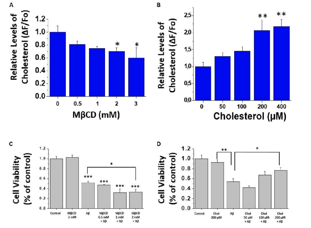
Aβ oligomers decreased neuronal cell viability. Cholesterol depletion worsened Aβ-related toxicity, while increased cholesterol provided protection in the experiment discussed.
Other studies found that cholesterol depletion reduced Aβ aggregation, showing that cholesterol's effects are complex.
Lipid rafts are membrane regions enriched in cholesterol, phospholipids (including sphingolipids), and proteins.
They organize cell signaling, stabilize membranes, and participate in pathogen entry, endotoxicity, exocytosis, and immune responses.
Cholesterol maintains membrane fluidity by preventing excessive rigidity at low temperatures and excessive fluidity at high temperatures.
Cholesterol-rich lipid rafts influence cancer cell migration, invasion, and metastasis, making them potential treatment targets. Altered cholesterol is also implicated in Alzheimer's and Parkinson's disease.
1823: Fats identified as esters of glycerol and fatty acids.
1929: Essential fatty acids recognized as important for infant growth.
1940–1950: Infants with insufficient fatty acids developed severe eczema and other skin disorders.
The simplest lipid class, consisting of a hydrocarbon chain with a hydrophobic methyl end and hydrophilic carboxylic acid end.
Usually has an even number of carbons, although odd numbers exist.
Saturated: no C=C bonds.
Monounsaturated (MUFA): one double bond.
Polyunsaturated (PUFA): two or more double bonds.
Cis: Hydrogens on the same side of the double bond → bent/U-shaped chain; most naturally occurring unsaturated fatty acids are cis.
Trans: Hydrogens on opposite sides → straighter chain; may occur naturally but are commonly produced by partial hydrogenation.

Short-chain: 2–6 carbons.
Medium-chain: 8–10 carbons.
Long-chain: 12–20 carbons.
Very-long-chain: 22 or more carbons.
Fatty acids can be named (ie. palmitate) or assigned a
number representing length and unsaturation (ie. 16:0)
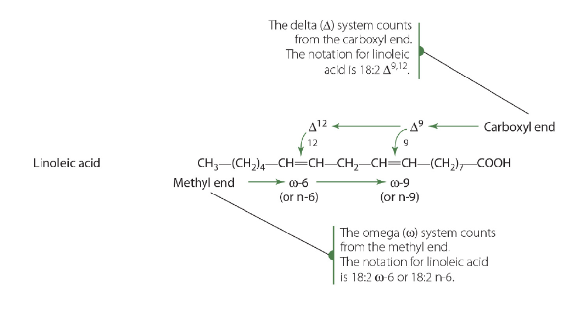
How are fatty acids named using the delta (Δ) and omega (ω) systems?
C:D indicates number of carbons:number of double bonds (e.g., 16:0 = palmitate).
Delta (Δ) counts double-bond positions from the carboxyl (COOH) end.
Ex: Linoleic acid: 18:2 Δ⁹,¹²
18: The fatty acid contains 18 carbons.
2: There are 2 double bonds.
Δ9: One double bond is between carbon 9 and carbon 10.
Δ12: The other double bond is between carbon 12 and carbon 13.
Omega (ω/n) counts from the methyl (CH₃) end.
Ex: Linoleic acid = 18:2 Δ⁹,¹² = 18:2n-6.
It has 18 carbons.
It has 2 double bonds.
The first double bond from the methyl end begins at carbon 6.

Humans cannot synthesize them and must obtain them from food.
Linoleic acid: 18:2n-6 (omega-6; Δ⁹,¹²).
α-Linolenic acid: 18:3n-3 (omega-3; Δ⁹,¹²,¹⁵). Found in plants and aquatic food sources; can be metabolized into other fatty acids.
Acetyl-CoA → acetyl-CoA carboxylase/fatty acid synthase using malonyl-CoA → palmitate (16:0) → elongase → stearate (18:0) → stearoyl-CoA desaturase (Δ9) → oleate (18:1n-9).
Stearate is a precursor for at least 13 other fatty acids in mammals.

Humans cannot introduce double bonds at the required omega-3 and omega-6 positions.
Plants can convert oleate (18:1n-9) into linoleic acid (18:2n-6) and α-linolenic acid (18:3n-3).
Humans must obtain these from food but can further elongate and desaturate them.
Linoleic acid (18:2n-6) → Δ6 desaturase → γ-linolenic acid (18:3) → elongase → 20:3 → Δ5 desaturase → arachidonic acid (20:4).
Arachidonic acid is used to produce eicosanoids.
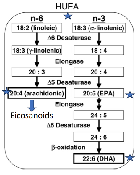
α-Linolenic acid (18:3n-3) → Δ6 desaturase → 18:4 → elongase → 20:4 → Δ5 desaturase → EPA (20:5) → elongase → 24:5 → Δ6 desaturase → 24:6 → β-oxidation → DHA (22:6).
EPA = eicosapentaenoic acid;
DHA = docosahexaenoic acid.

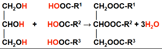
Triglycerides (triacylglycerols) are major storage molecules for fatty acids.
Structure: one glycerol backbone + three esterified fatty acids (R¹, R², R³).
Stored in intracellular lipid droplets and transported through blood in lipoproteins.

Glycerol + 3 fatty acids → triglyceride + 3 H₂O.
This is an esterification/dehydration reaction forming three ester bonds, releasing one water molecule per fatty acid.
In the actual metabolic pathway, fatty acids must first be activated by attachment to CoA → fatty acyl-CoA.
Glycerol or dihydroxyacetone phosphate (DHAP), DHAP comes from glycolysis or gluconeogenesis.
NADH
Activated fatty acids (fatty acyl-CoA)
ATP
Five enzymes
How do triglyceride (TG) and phospholipid synthesis share common steps?
Most glycerol 3-phosphate comes from dihydroxyacetone phosphate (DHAP) from glycolysis / gluconeogenesis.
Some glycerol 3-phosphate is made from glycerol via glycerol kinase – a minor pathway in liver and kidney
Phosphatidic acid is the precursor to TGs and phospholipids.
Advantage of making phosphatidic acid is it can then be made into triacylglycerol OR phospholipid.
Key point: Phosphatidic acid is a common precursor that can be used to synthesize either triglycerides or phospholipids.

Primarily in adipose tissue.
Subcutaneous fat: beneath the skin.
Visceral fat: around internal organs.
Storage provides energy reserves, but excessive accumulation can be harmful.
Epicardial fat is visceral adipose tissue between the heart muscle (myocardium) and visceral pericardium.
Excess epicardial fat is associated with metabolic syndrome, including abdominal obesity, elevated blood pressure, impaired fasting glucose, and dyslipidemia.

When energy is required, stored triglycerides undergo lipolysis → glycerol + free fatty acids.
Glycerol travels to the liver for glucose or pyruvate metabolism.
Fatty acids travel to tissues for β-oxidation → acetyl-CoA.
Acetyl-CoA enters the citric acid cycle for energy or is used for ketone body synthesis in the liver.

Glycerol travels to the liver, where it can enter:
Glycolytic reactions to produce pyruvate
Gluconeogenesis to produce glucose.
This allows part of stored fat to contribute to glucose production.
Free fatty acids enter blood and travel to tissues, including liver, heart, and muscle.
They undergo β-oxidation → acetyl-CoA, which;
Enters the citric acid cycle → CO₂ + H₂O and energy,
Is used for ketone body synthesis in the liver.
1. Fatty acid activation: fatty acid + CoA → fatty acyl-CoA.
2. For long-chain fatty acids, the acyl group transfers from CoA to carnitine for mitochondrial transport.
3. Inside mitochondria, carnitine is exchanged for CoA.
4. Fatty acyl-CoA undergoes β-oxidation → acetyl-CoA.
During prolonged fasting, liver mitochondria use acetyl-CoA to synthesize ketone bodies.
Acetoacetate: formed from HMG-CoA.
β-Hydroxybutyrate: formed by reduction of acetoacetate.
Acetone: formed by decarboxylation of acetoacetate.
How does the triglyceride–fatty acid cycle work between the liver, blood, adipose tissue, and other tissues?
Liver:
Gluconeogenesis: Pyruvate → glucose.
Glyceroneogenesis: Pyruvate → glycerol-3-phosphate (G3P), involving Pck1.
De novo lipogenesis produces fatty acids.
G3P + free fatty acids (FFA) → triglycerides (TG) through re-esterification.
Glucose and TG enter the bloodstream.
2. Adipose tissue (fat storage):
Glucose from blood → G3P through glycolysis.
Pyruvate → G3P through glyceroneogenesis (Pck1).
G3P + FFA → TG through re-esterification (lipid storage).
3. Lipolysis (fat breakdown):
Hormone-sensitive lipase (HSL) promotes TG breakdown → FFA + glycerol.
FFA enter the bloodstream; glycerol is released.
4. Blood transport:
Transports glucose, TG, and FFA between tissues.
FFA can return to the liver or travel to muscle and heart.
5. Muscle and heart:
FFA undergo oxidation to produce energy.

How do CD36 and FABP transport long-chain fatty acids into cells?
Release of fatty acids from albumin
Binding to CD36 which can accommodate up to two fatty acids at a time
Guidance of the fatty acid through the CD36 and be exposed to the plasma membrane surface
The fatty acids leave CD36 and enter the outer leaflet of the membrane. They then move to the inner leaflet through a process called flip-flop of single FA; transmembrane translocation
Binding to the interior of FABP that is anchored by binding to the intracellular part of CD36
Diffusion into the soluble cytoplasm of the fatty acid–FABP complex toward sites of intracellular fatty acid metabolism.

Glycerol-3-phosphate pathway: Begins with glycerol-3-phosphate (G3P).
Monoacylglycerol pathway: Begins with monoacylglycerol (MG).
Both pathways eventually produce diacylglycerol (DAG) and then TAG.
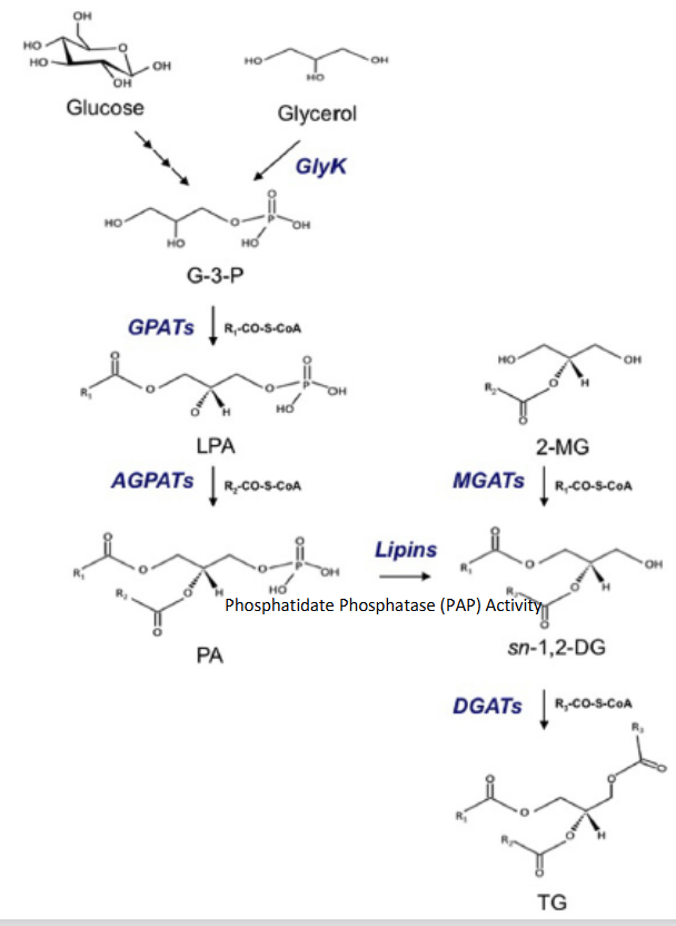
Glucose → glycolysis → G3P (via DHAP).
Glycerol → G3P by glycerol kinase (GlyK). G3P provides the glycerol backbone for TAG synthesis.
1. GPAT (glycerol phosphate acyltransferase) adds fatty acyl-CoA to G3P → lysophosphatidic acid (LPA).
2. AGPAT (acylglycerol phosphate acyltransferase) adds a second fatty acyl-CoA to LPA → phosphatidic acid (PA).
Both reactions release CoA.

1. Lipin (phosphatidate phosphatase/PAP) removes phosphate from PA → sn-1,2-diacylglycerol (DAG).
2. DGAT (diacylglycerol acyltransferase) adds a third fatty acyl-CoA to DAG → TAG. Overall: G3P → LPA → PA → DAG → TAG.

1. Begins with 2-monoacylglycerol (2-MG).
MGAT (monoacylglycerol acyltransferase) adds fatty acyl-CoA → sn-1,2-DAG.
DGAT adds another fatty acyl-CoA → TAG. This bypasses G3P, LPA, and PA.

Lipid droplets are intracellular storage structures containing TAG, cholesteryl esters, and fat-soluble vitamins.
They form at the ER–cytosol interface and are surrounded by a phospholipid monolayer, not a bilayer.
How are lipid droplets synthesized?
Newly synthesized TAG ‘nucleates’ the ER membrane until a critical mass is reached
The critical mass of TAG is extruded into the cytosol in a phosphospholipid (PL) monolayer
Pinched off from the ER to release an LD (that may fuse with other LDs in the cytosol)

Intracellular lipases associate with lipid droplets and hydrolyze TAG, releasing fatty acids.
Studied mainly in white adipose tissue (WAT) and liver, where mechanisms differ. TAG hydrolysis in heart and skeletal muscle is comparable to WAT.
1. ATGL (adipose triglyceride lipase): TAG → DAG + free fatty acid (FFA).
2. HSL (hormone-sensitive lipase): DAG → monoacylglycerol (MAG) + FFA.
3. MGL (monoglyceride lipase): MAG → glycerol + FFA.
Overall: TAG → DAG → MAG → glycerol + 3 FFA.

Insulin and excess intracellular acyl-CoA suppress lipolysis.
Stress hormones stimulate lipolysis.
HSL is hormonally regulated.
The diagram also shows lipid droplet-associated PLIN and CGI-58 alongside ATGL.
Although ATGL, HSL, and MGL are present, most TAG hydrolysis in the liver is attributed to CES1 (carboxylesterase-1), also called triacylglycerol hydrolase (TGH).
CES1 alone can produce 3 free fatty acids + glycerol from TAG.
1. Activated into fatty acyl-CoA for β-oxidation or other metabolic pathways.
2. Used for intracellular signaling and gene regulation.
3. Released into blood bound to albumin, which can carry up to 8 fatty acids.
De novo fatty acid synthesis produces palmitate (16:0), a saturated 16-carbon fatty acid, in the cytosol.
Requires
Acetyl-CoA, ATP, NADPH,
Two multifunctional enzymes:
ACC (acetyl-CoA carboxylase, 2 catalytic activities)
FAS (fatty acid synthase, 6 catalytic activities).
The process is thermodynamically favourable.
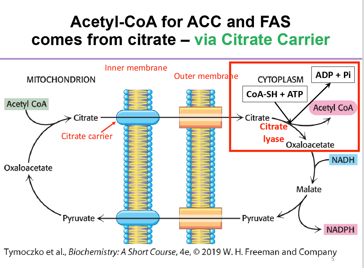
How does mitochondrial acetyl-CoA reach the cytosol for fatty acid synthesis (ACC and FAS) via the citrate shuttle?
Acetyl-CoA cannot cross the inner mitochondrial membrane directly, so it is transported as citrate.
Mitochondria: Acetyl-CoA + oxaloacetate → citrate.
Transport: Citrate crosses the inner mitochondrial membrane via the citrate carrier and enters the cytosol.
Cytosol: ATP-citrate lyase converts citrate + CoA-SH + ATP → acetyl-CoA + oxaloacetate + ADP + Pi.
Oxaloacetate → malate: Uses NADH (produces NAD⁺).
Malate → pyruvate: Produces NADPH, needed for fatty acid synthesis.
Pyruvate returns to the mitochondria and is converted back into oxaloacetate, continuing the shuttle.
Final result: Cytosolic acetyl-CoA is supplied for ACC and FAS, while NADPH provides reducing power for fatty acid synthesis
When energy is abundant, mitochondrial acetyl-CoA and ATP increase → more citrate forms → citrate moves into the cytosol. Citrate supplies acetyl-CoA and allosterically activates ACC (an important site of regulation).
This shifts metabolism from ATP production through the Krebs cycle and ETC toward fatty acid synthesis and energy storage.
ACC catalyzes the rate-limiting step of fatty acid synthesis.
Citrate: allosterically activates ACC.
Palmitate: inhibits ACC through end-product feedback.
Glucagon: inactivates ACC through phosphorylation.
FAS converts excess carbohydrate metabolites into fatty acids in the liver and adipose tissue and is relevant to cancer biology and prognosis.
ACC catalyzes the irreversible carboxylation of acetyl-CoA to malonyl-CoA.
Reaction: Acetyl-CoA + HCO₃⁻ + ATP → malonyl-CoA + ADP + Pi + H₂O.
Requires acetyl-CoA, bicarbonate (CO₂ source), ATP, and biotin.
ACC is a biotin-dependent enzyme complex with 3 functional regions:
1. Biotin carboxylase uses ATP to activate CO₂ from bicarbonate.
2. Transcarboxylase transfers activated CO₂ to acetyl-CoA.
3. Biotin carrier protein holds biotin and moves activated CO₂ between sites.
Biotin with biotin carrier protein attached to Biotin Carboxylase and temporarily carries CO₂ and is not consumed.

What are the steps of malonyl-CoA synthesis by ACC?
Biotin attaches to its carrier protein through a lysine side chain.
Biotin carboxylase uses ATP and HCO₃⁻ to attach activated CO₂ to biotin.
The flexible biotin arm moves CO₂ to transcarboxylase.
Transcarboxylase transfers CO₂ from biotin to acetyl-CoA → malonyl-CoA.
Malonyl-CoA is released, and the biotin carrier is regenerated. ATP supplies energy for this irreversible reaction.
Overall: Acetyl-CoA + HCO3- + ATP → malonyl-CoA + ADP + Pi + H2O

KS (β-ketoacyl-ACP synthase): condensation.
MAT (malonyl/acetyl-CoA-ACP transferase): substrate loading.
KR (β-ketoacyl-ACP reductase): first reduction.
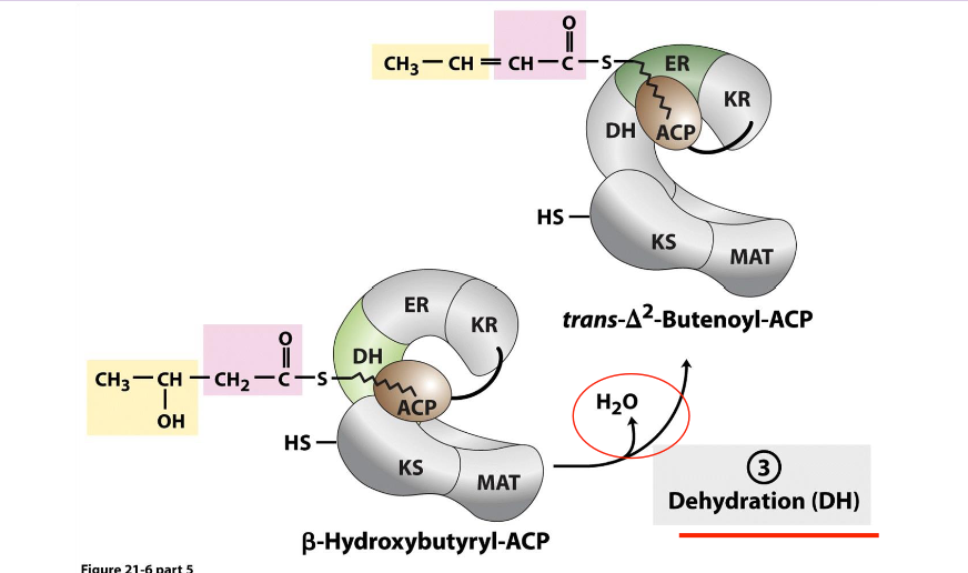
DH (β-hydroxyacyl-ACP dehydratase): dehydration.
ER (enoyl-ACP reductase): second reduction.
TE (thioesterase): releases palmitate.
ACP (acyl carrier protein) is not catalytic; it carries the growing fatty acyl chain.

Domain I: KS and MAT for substrate entry; DH for dehydration.
Domains II and III: KR and ER for reduction, with ACP carrying intermediates between sites.
TE releases the completed palmitate from ACP.
The catalytic sites work together to elongate fatty acid chains.

ACP acts like the biotin carrier protein of acetyl-CoA carboxylase (ACC)
Functions as a moving arm to carry the building fatty acyl chain to one enzyme active site to next enzyme within FAS
The –SH group of the phosphopantetheine esterfies with malonyl-CoA

Acetyl-CoA: initial 2-carbon primer.
Malonyl-CoA: donates 2 carbons per elongation cycle.
NADPH: provides reducing power for the two reduction reactions in each cycle.
FAS repeatedly elongates the chain until it reaches 16 carbons.
How are acetyl-CoA and malonyl-CoA initially loaded onto FAS?
1. MAT transfers the acetyl group from acetyl-CoA to the -SH group of ACP.
2. The acetyl group moves from ACP to the cysteine -SH group of KS.
3. ACP's -SH group becomes available again.
4. MAT transfers the malonyl group from malonyl-CoA to ACP.
Result: acetyl on KS + malonyl on ACP, ready for condensation.
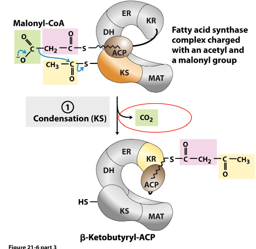
Enzyme: β-ketoacyl-ACP synthase (KS).
The 2-carbon acetyl group combines with the malonyl group on ACP, releasing CO₂ and forming β-ketobutyryl-ACP (4 carbons).
The CO₂ added during the ACC step is removed here, making condensation more exergonic and energetically favourable.
Enzyme: β-ketoacyl-ACP reductase (KR).
β-Ketobutyryl-ACP → β-hydroxybutyryl-ACP.
The β-keto group (C=O) is reduced to a hydroxyl group (-OH).
Uses NADPH + H⁺ → NADP⁺.

Enzyme: β-hydroxyacyl-ACP dehydratase (DH).
β-Hydroxybutyryl-ACP → trans-Δ²-butenoyl-ACP.
Removes H₂O
Forms a carbon-carbon double bond.