apthous stomatitis and hypersensitivity reactions
 Call Kai
 Learn
 Practice Test
 Spaced Repetition
 Match
 Flashcards
 Knowt Play
1/52

Earn XP
Description and Tags
lecture given 10/16/2026
53 Terms
recurrent aptheous stomatitis
most common oral mucosal lesion
cause- different things in different people
T cell mediated immunologic reaction with production of TNF-alpha
etiology of aphthae
still uncertain
different subgroups of pts appear to different causes for the occurance of aphthae
may be multifactorial
immunologic reaction to an oral antigen
allergies, genetic predisposition, hematologic abnormalities, hormonal influences, immmunological factors, infectious agents, nutritional deficiencies, smoking cessation, stress (mental and physical), trauma
factors that increae the frequency of occurrence of aphthae
trauma, nutritional deficiencies, smoking cessation
factors that increae the frequency of occurrence of aphthae
smoking, hormonal changes, attached mucosa
potential antigens of aphthae
sodium lauryl sulphate in tooth paste
systemic medications (NSAIDs, beta blockers)
infectious agents (strep, H. pylori, HSV, VZV, CMV)
foods (cheese, chocolate, coffee, milk, gluten, nuts, strawberries, tomatoes, flavoring agents, preservatives)
recurrent aphthous stomatitis
somtimes associated wtih prior trauma
may be precipitated by psychic stress
may be precipitated by dietary factors
may be associated with hormonal fluctuation
somtimes associated with IBD
uncommon in smokers
clinical variations of apthous stomatitis
minor aphthous ulcerations
major aphthous ulcerations
herpetiform apthous ulcerations
behcet syndrome
minor aphthous ulcerations
children and young adults
fewest recurrences and shortest duration
F>M, frequency declines with age
occur exclusively on non-keratinized mucosa BM, LM, ventral tongue, vestibule, FOM, SP
small, painful, shallow ulcer with red rim
solitary or multiple; <5 at one outbreak
full size within a day; heal within 2 weeks
minor aphthous ulcerations
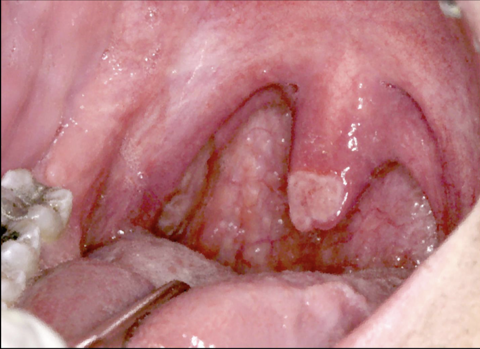
minor aphthous ulcerations

minor aphthous ulcerations

minor aphthous ulcerations

major aphthous ulcerations
larger ulcerations measuring from 1-3cm in diameter
take from 2-6 weeks to heal
may cause scaring with eventual restriction of oral opening
number of lesions varies from 1-10
present for longer duration
deep ulcerations
common locations: labial mucosa, soft palate, tonsillar fauces
major aphthous ulcerations

major aphthous ulcerations

herpetiform aphthous ulcerations
least common of the aphthous stomatitis conditions
adult onset, mostly movable mucosa, female predominance
greatest number of ulcers and most frequent recurrences
small crops of painful ulcers that come and go
resemble herpes lesions except confined to non-keratinized mucosa
heal in 7 to 10 days but recur more frequently
do not respond to antibiotics/antivirals
herpetiform aphthous ulcerations

treatment for aphthous stomatitis
review diet/nutrition/GI symptoms
symptoms control
OCT preparations
topical anesthesia (elixir of benadryl)
corticosteroids (flucocinomide gel 0.05%, dexamethasone solution 0.5mg/5mL)
antimicrobial (tetracycline/chlorhexidine)
laser/chemical cautery
severe cases: TNF alpha inhibitor (thalidomide)
behcet’s syndrome
affects oral, genital, and ocular regions
multisystem disorder caused by systemic vasculitis
abnormal immune process
triggered by infectious or environmental antigen in a genetically predisposed individual
distinct racial predilection for people of middle east descent
young adults; uncommon in blacks; M>F
oral ulcers resembling minor/major aphthae
genital ulcers
ocular involvement
cutaneous involvment
arthritis
CVS, CNS, pulmonary, renal systems can be involved
behcet’s syndrome

behcet’s syndrome

erythema multiforme
blistering ulcerative mucocutaneous disease
uncertain etiopathogenesis
immunologically mediated process
initiating factors: HSV, mycoplasma pneumoniae, medications
acute onset; young adults; F>M
prodromal symptoms are often present
cutaneous lesions: targetoid or bull’s eye lesions
oral lesions: large, shallow erosions and ulcerations
hemorrhagic crusting of the vermillion zone of the lips
self limiting: lesions heal in 2 to 6 weeks
IV rehydration, symptomatic management, identify initiating factor in recurrent lesions
erythema multiforme

erythema multiforme

erythema multiforme

stevens johnson syndrome (SJS)
toxic epidermal necrolysis (TEN)
hypersensitivity reactions like EM but separate clinically and likely separate etiopathogeneticallu
SJS typically associated with medication usage or mycoplasma pneumoniae infection and rarely HSV
SJS considered less severe variant of TEN
flu-like prodromal signs and symptoms
skin lesions always present with predominant involvement of the chest
like EM, both TEN and SJS present with labial crusting, oral bullae/ulcers/erosions
eye and genital involvement in addition to oral lesions
discontinue drug; skin lesions heal in 3 to 5 weeks
stevens johnson syndrome (SJS)
toxic epidermal necrolysis (TEN)

transient lingual papillitis
one to several painful enlarged fungiform papillae
unknown pathogenesis
may represent a hypersensitivity rxn however, the underlying etiology has not been fully elucidated
typically involves the anterior dorsal tongue
red papules with yellow ulcerated cap
no treatment necessary
resolve spontaneously within several hours to several days
transient lingual papillitis

erythema migrans (geographic tongue)
common benign condition predominantly involving the tongue
asymptomatic; occasionally causes mild burning discomfort
etiology unknown; atopic individuals?
multiple well-demarcated erythematous patches; yellow-white scalloped border
filiform papillary atrophy which migrates from one area to the next
often seen in association with fissured tongue
may occasionally occur on other mucosal sites
treatment rarely indicated
erythema migrans (geographic tongue)

erythema migrans (geographic tongue)
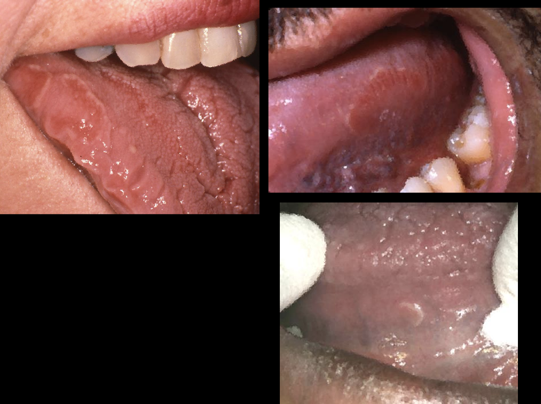
erythema migrans (geographic tongue)

allergic mucosal reaction to systemic medication
stomatitis medicamentosa
several different patterns of oral mucosal lesions- anaphylactic stomatitis, intraoral fixed drug eruptions, lichenoid drug reactions, lupus erythematosus-like eruptions, pemphigus-like drug reactions, pemphigoid like drug reactions, non-specific erosive/ulcerative/aphthous-like lesions
medication associated mucositis
no clinically consistent features- may mimic various other disorders
index of suspicion needed for unusual presentations
may take weeks to improve on withdrawl of stimulus
palliated with topic steroids if the offending medication cannot be discontinued
medication associated mucositis
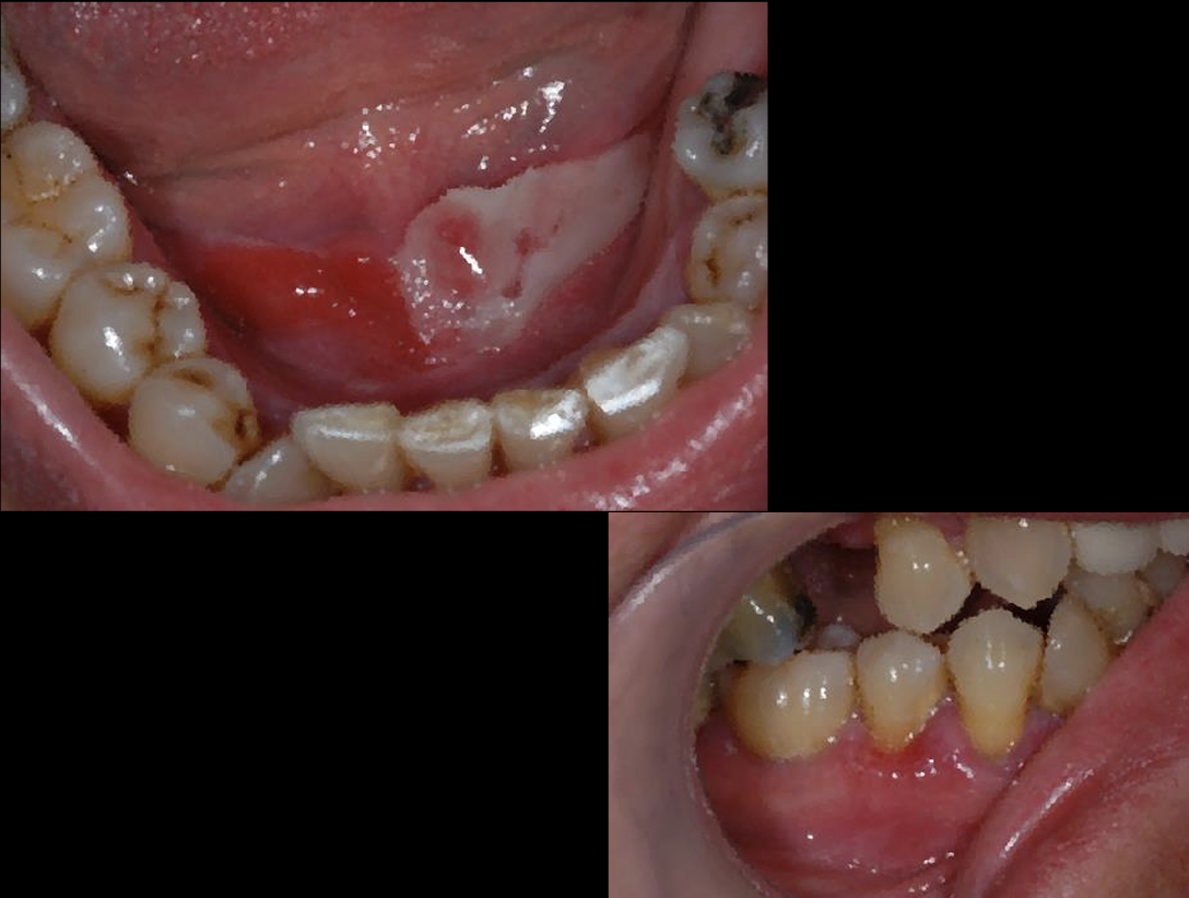
medication associated mucositis

medication associated mucositis

medication associated mucositis

medication associated mucositis
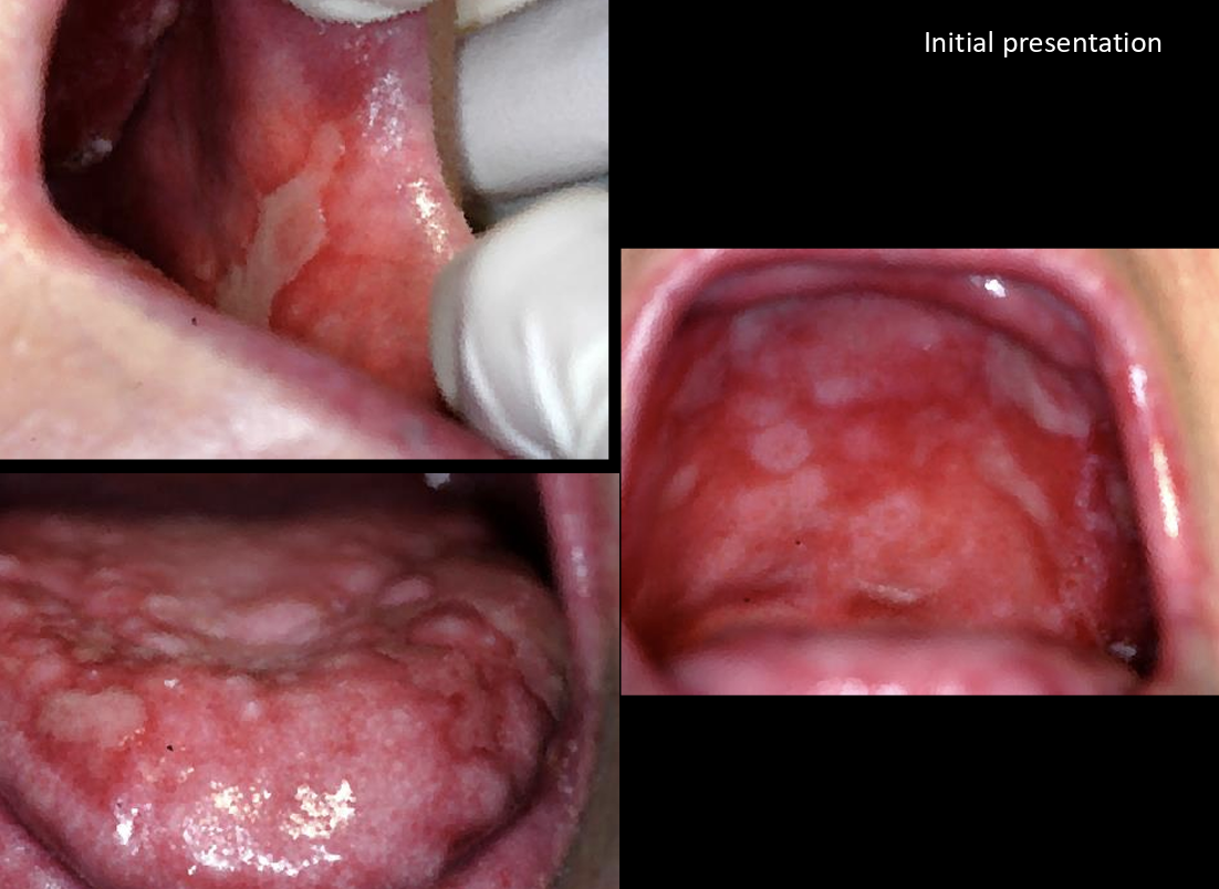
medication associated mucositis

allergic contact stomatitis from dental restorative materials
may involve buccal mucosa, tongue, and gingiva in intimate contact with restoration
typically solitary red/white lesion; may exhibit peripheral striae
cutaneous allergy patch testing may be helpful to identify appropriate choice of material for future restorations
allergic contact stomatitis from dental restorative materials

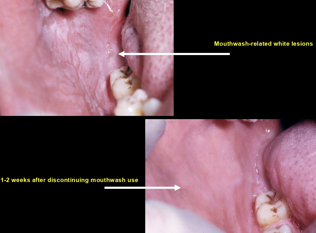
allergic contact stomatitis to oral hygiene products
wispy strips of epithelium lying on surface
no erythema or pain
caused by use of strong dentifrices
treat via discontinuation of inciting agent
allergic contact stomatitis to oral hygiene products

allergic contact stomatitis to oral hygiene products

allergic contact stomatitis to oral hygiene products
allergic contact stomatitis from artificial cinnamon flavoring
cinnamon oil (used as a flavoring agent) is used in concentrations 100x that found in natural spice
reaction typically seen in candy, chewing gum, and toothpaste
toothpaste reactions show changes ranging from diffuse erythema to superficial sloughing; may be associated with exfoliative cheilitis/circumoral dermatitis
candy/gum reactions on buccal mucosa and lateral tongue at level of occlusion; typically white, irregular patches
pain and burning sensation
allergic contact stomatitis from artificial cinnamon flavoring
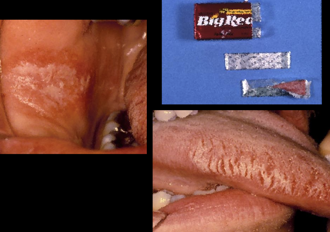
allergic contact stomatitis from artificial cinnamon flavoring
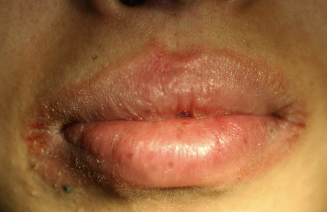
angioedema
mast cell degranulation
IgE- mediated hypersensitivity reactions
allergic contact reaction
physical stimuli
physical stimuli
ACE inhibitiors
activation of the complement pathway: herediatry/acquired
high levels of antigen / antibody complexes
elevated peripheral blood eosinophil counts
angioedema

angioedema
