(1) BOOK 4: Virus
 Call Kai
 Learn
Learn Practice Test
 Spaced Repetition
 Match
Match Flashcards
 Knowt Play
1/28

There's no tags or description
Looks like no tags are added yet.
Name | Mastery | Learn | Test | Matching | Spaced | Call with Kai | Chat |
|---|
No analytics yet
Send a link to your students to track their progress
29 Terms
Reason to study Virus
Sources of enzymes
A number of enzymes used in molecular biology are viral enzymes
Eg: Reverse transcriptases from retroviruses, RNA polymerases from phages
Anti-cancer agents
Genetically modified strains of viruses are being investigated for treatment of cancers
These strains have been modified so that they are able to infect and destroy specific tumour cells, but are unable to infect normal cells
Viruses — Obligate Intracellular Parasites
Viruses are obligate intracellular parasites (not unique to virus)
Obligate - by necessity
Intracellular - within the cell
Parasite - an organism living and feeding on a host
The parasite cannot survive without its living host cell
Viruses lack enzymes for most metabolic processes
Viruses lack amino acids and nucleotides, ribosomes and energy, in the form of adenosine triphosphate (ATP) for protein synthesis
Thus, they are dependent on their living host cells for these substances
Viruses can only reproduce within a living host cell
Virus locations:
In their extracellular states, viruses are termed as virions
The virion is metabolically inert and does not carry out respiratory or biosynthetic functions
The virion is the structure by which the virus genome is carried from the cell in which it has been produced to another cell where the viral nucleic acid can be introduced
Inactive as extracellular virion
In their intracellular states, viral replication occurs
The viral genome and proteins that make up the virus coat are synthesised and assembled to form new viruses
A cell that a virus can infect and reproduce in is called a host cell
Active as intracellular virus state
Virus — Arguments For Being Living Organisms
Viruses can reproduce
Although viruses can reproduce, unlike cells, viruses are only able to reproduce in the intracellular state
The way a virus multiplies is very different from that of cells
In cells, reproduction always forms a new cell directly from a pre-existing cell, but a new virion is never formed directly from pre-existing virion
Replication in viruses involves the synthesis of large number of viral components in host cells, followed by their assembly into virions
Viruses are able to direct metabolic processes
Even though as a virion, a virus does not exhibit most of the metabolic life processes of cells, they can however direct them when existing in a virus state (intracellular)
These life processes include processes like protein synthesis and genome replication
Viral genomes can evolve
Different types of viruses vary greatly in their structural and genetic complexity and no single gene is shared by all viruses/viral lineages
Viral genomes can consist of single- or double- stranded DNA/RNA molecules
This indicates that viruses descended from more than one ancestral lines and do not share a common ancestor
Viruses evolve with their host and acquire their metabolic and translational genes from the host cells
Genetic recombination can result in different / changing viral genomes
Virus — Arguments For Being Non-Living Organisms
Viruses are not cells
Viruses are not cells and do not have protoplasm or organelles
The basic structure of a virus consists of a protein capsid surrounding a nucleic acid core which is composed of DNA or RNA
In some viruses, the capsid is surrounded by a membranous envelope
Viruses lack some of the characteristics of living organisms
Viruses are able to reproduce and respond to stimuli while in the intracellular state (within their host cells)
However, in their extracellular state, they are
Unable to carry out metabolic processes, such as nuclei acid and protein synthesis
Do not require nutrition, such as amino acids and nucleotides
Unable to synthesise their own ATP as an energy source
Unable to respond to stimuli
Neither grow nor excrete
Recall: Cell Theory
All living things are made of cells
Cells are the basic unit of life
All cells come from pre-existing cells
Virus — Challenge Cell Theory
Cells are the smallest unit of life
Viruses lack the necessary molecular machinery to conduct many of the biochemical reactions a normal cell would need
Lack enzymes for most metabolic processes
Lack amino acids and nucleotides, ribosomes and energy, in the form of adenosine triphosphate (ATP) for protein synthesis
However, like cells, they contain the genetic material necessary to form the next generation and are able to evolve in response to the environment
All cells come from pre-existing cells
Viruses are capable of replicating but rely on host cells to provide the energy and materials needed for replicating their genomes and synthesising their proteins
So they cannot replicate unless they have entered a suitable host cell
All living organisms are composed of cells
Viruses are acellular and do not have protoplasm or organelles
They thus are not considered cells since they need a host cell to carry out many functions necessary to reproduce
In the extracellular virion state, it is also metabolically inert and does not carry out respiration or biosynthesis
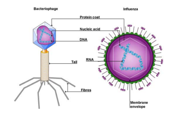
Basic Structure of Virus
Size:
Virus can vary widely in size, shape and genomic content
They are the smallest infectious agents
Most of them range between 20 to 300 nm in size and hence require examination by electron microscopy (EM)
Basic Structures:
Genome → DNA or RNA
Present in all virus
Protein coat (capsid protein)
Comprise of protein subunits, capsomeres
Present in all virus
Envelope
Comprise of phospholipids from host cell
Only animal virus can have envelope
Enzymes
Basic Structure of Virus — Genome
Group I:
Double-stranded DNA viruses
E.g. λ phages
Group V:
(-) Sense single-stranded RNA viruses
Genome must be converted to (+) sense RNA by a RNA-dependent RNA polymerase before translation
E.g. Influenza viruses
Group VI:
Single-stranded RNA-Reverse transcriptase (RT) viruses
Make use of reverse transcriptase (RNA-dependent DNA polymerase), to produce DNA from the initial viral RNA genome
E.g. HIV
All viral genomes would contain genes coding for essential proteins:
Regulatory proteins
Necessary for regulating the action of the host genes
Host cell machinery is used by the virus to replicate the viral genome and synthesise the various components necessary for formation of the virus
Structural proteins
Eg: Viral capsid protein → Essential for assembly of the complete virus
Basic Structure of Virus — Protein Coat / Capsid Protein
All viruses have a protein coat
Protein coat either encloses or surrounds the viral genome
Each protein coat is constructed from identical protein subunits called capsomeres
Depending on how capsomeres are shaped and arranged, this assembly usually results in helical or icosahedral protein coat
Basic Structure of Virus — Envelope
Enveloped viruses:
Envelope is present only in enveloped viruses
Enveloped viruses are mostly viruses that infect animal cells → Animal virus
How (lipid membrane) envelope forms:
The envelope of a virus is derived from host cells
When they are released from the host cell by budding, they take with them the host’s cell surface membrane (phospholipid bilayer) and insert proteins of viral origins into the membrane (envelope)
These proteins include viral glycoproteins
Viral glycoproteins are exposed on the outside of the envelope
They are essential for the attachment of viruses to the next host cell
Purpose of Envelope:
The envelope protects the virion’s nucleic acid from the effects of various enzymes and chemicals
Naked / Non-enveloped Viruses:
Viruses which are not surrounded by the lipid membrane envelope
Basic Structure of Virus — Enzymes in viruses
Lysozyme:
Makes a small hole in the bacterial cell wall that allows the viral nucleic acid to enter
Lysosomes are also produced in large amounts in the later stages of infection, causing lysis of the host cell and release of the virus
Neuraminidase:
Breaks down glycosidic bonds of glycoproteins and glycolipids of the connective tissue of animal cells, aiding in the liberation (freeing) of the virus
Neuraminidase is associated with the viral envelope instead of being located within the capsid like most other viral enzymes
Other enzymes that are involved in nucleic acid replication and / or transcription:
DNA-dependent RNA polymerase
RNA-dependent RNA polymerase / viral replicase
DNA-dependent DNA polymerase
RNA-dependent DNA polymerase / reverse transcriptase
*The "dependent" part of the name indicates the type of nucleic acid being copied
*The "polymerase" part of the name indicates the type of nucleic acid is being synthesised
General Reproductive Cycle of Viruses
Replication:
Each type of virus can replicate in only a very limited number of cell types
Host Range & Host Cells:
Host Range → The suitable types host cells for a particular virus
Viruses identify their host cells by a complementary fit between proteins on the outside of the virus and specific receptor molecules on the surface of the host cells
By host range, viruses can be classified into 3 groups:
Bacterial viruses or bacteriophages
Eg: T4 and lambda phage
Animal viruses
EG: Influenza and HIV
Plant viruses
General Steps in Reproduction of an Enveloped Virus (APSAR):
Adsorption
The virus attaches to its host cell by specific binding of its glycoproteins to host cell receptors
Penetration
The virus is engulfed into a vesicle and its envelope is uncoated, thereby freeing the viral nucleic acid into the cell cytoplasm
Synthesis & Replication: Synthesis of viral proteins and replication of viral nucleic acid
Under the control of viral genes, the cell synthesises the basic components of new viruses: nucleic acid, capsomeres and glycoproteins
Assembly
Viral glycoproteins are inserted into the host cell membrane for the viral envelope
Nucleocapsid / capsid (protein coat) is formed from nucleic acid and capsomeres
Release
Enveloped viruses bud off of the membrane, carrying away an envelope with the glycoproteins
This complete virus or virion is ready to infect another cell
Types of Viruses — Bacteriophages
What are bacteriophages:
Bacteriophages are DNA viruses that infect bacteria
The bacteriophages may undergo lytic and lysogenic cycles, during replication
Bacteriophages can also be involved in the transfer of genetic material between bacteria by transduction
Inheritance of the bacteriophage genome:
Replication of DNA viruses requires that the host cell transcribe genes from the DNA genome of the bacteriophage to produce mRNA
The mRNA is then translated into viral proteins necessary for the replication process
The viral DNA genome is also replicated by the host cell machinery to form multiple copies for use in the assembly of the new bacteriophage
Types of Bacteriophages:
T4 Phage (a virulent phage)
Reproduces only by lytic cycle → virulent
Lambda (λ) Phage (a temperate phage)
Reproduces by lysogenic & lytic cycle → temperate
Types of Viruses — Bacteriophages — T4 Phage (virulent)
Structures of T4 Phage (common):
Genome (nucleic acid)
Linear double-stranded DNA
DNA is transcribed by host cell machinery (nucleic acids, amino acids, ribosomes, ATP) into mRNA for translation of viral proteins
Protein coat / Capsid protein
Capsomeres surrounds the nucleic acid, contained in the head of the phage
Structures of T4 Phage (specific to T4):
Head
Contains the DNA of the virus
A tail
Consisting of a tail sheath, multiple tail fibres and base plate
Tail fibres
Allow the phage to adsorb onto the surface of the bacterial cell by binding to the specific receptors site found on the cell surface
This enables the base plate to come into contact with the surface of the cell
Tail sheath
Tail sheath surrounds a central tube
Tail sheath contracts during penetration to trigger conformational change to thrust the central tube through the host cell wall and membrane
A base plate
Comes into contact with the host cell surface and undergoes a conformational change to allow DNA to be extruded from the head, through the central tube and into the host cell

T4 Phage (virulent) — Reproduction through Lytic Cycle
Lytic Cycle:
It is a phage’s reproductive cycle that finally results in death of the host cell
The term “lytic” refers to the last stage of infection, during which the bacterium lyses and releases the new phages that were produced within the cell
Virulent Phage:
It is a class of phage that reproduces only by a lytic cycle
Eg: T4
Steps:
Adsorption
The multiple tail fibres of the T4 phage attach to specific receptor sites on the surface of a bacterial host cell (eg: E. coli)
The base plate settles down on the host cell surface
Penetration
Conformational changes occur in the tail sheath causing it to contract and its core / tube pierces through the bacterial cell wall and cell membrane
T4 uses lysozyme to hydrolyse peptidoglycan, degrading a portion of the bacterial cell wall for insertion of the tail core
DNA is extruded from the head, through the tail tube into the host cell
The protein coat is left on the outside of the bacterial cell wall
Synthesis and replication
Soon after the phage DNA is injected into the host cell, the synthesis of host DNA, RNA and proteins is halted
Viral Nucleic Acid Synthesis (replication)
Phage DNA is replicated by host DNA polymerase
The host DNA is degraded into nucleotides, providing raw materials for phage DNA replication
Viral Protein Synthesis
Phage mRNAs are synthesised by the host RNA polymerase via transcription
The phage mRNAs are translated by host cell ribosomes, tRNAs and translation factors into viral proteins and enzymes required to take over the host cell and replicate phage nucleic acids
These include enzymes for viral replication and inhibitory factors that stop host cell RNA and protein synthesis
Assembly
Viral proteins are assembled to form phage heads, tails and tail fibres each
The different components are assembled into the complete bacteriophage
Release
The T4 phages lyse the host cell by the action of the enzyme lysozyme, which digests the bacterial cell wall
Water enters the cell by osmosis causing the cell to swell and burst

Types of Viruses — Bacteriophages — Lambda (λ) Phage (temperate)
Structures of Lambda Phage (common):
Genome (nucleic acid)
Linear double-stranded DNA
DNA is transcribed by host cell machinery (nucleic acids, amino acids, ribosomes, ATP) into mRNA for translation of viral proteins
Protein coat / Capsid protein
Capsomeres surrounds the nucleic acid, contained in the head of the phage
Structures of Lambda Phage (specific to Lambda):
Head
Contains the DNA of the virus
(no need know) The 5’-terminus of each DNA strand is a single-stranded tail of 12 nucleotides long which is important in prophage formation
A single tail fibre (non-contractile)
Allows the phage to adsorb onto the surface of the bacterial cell by binding to the specific receptor site found on the cell surface

Lambda (λ) Phage — Reproduce through Lysogenic & Lytic Cycles
Lysogenic Cycle:
Lysogenic cycle involves replication of the phage genome without destroying the host in the initial steps
Temperate Phages:
A class of phages capable of using both the lytic and lysogenic modes of reproduction within a bacterium
Once an environmental trigger (such as the presence of UV light or certain chemicals) activates the virus, it switches from the lysogenic cycle to the lytic cycle
Steps (lysogenic cycle of Lambda phage):
Adsorption
The single tail fibre of lambda (λ) phages attach to specific receptor sites on the surface of a bacterial host cell
The base plate (on tip of the tail) settles down on the host’s cell surface
Penetration
The lambda (λ) phages have tails that are not contractile and serve to deliver the viral DNA to the cell membrane
DNA is extruded from the head, through the tail tube and injected into the host cell passing through both the bacterial cell wall and cell membrane
The protein coat is left on the outside of the bacterial cell wall
Prophage Formation (part 1)
The lambda (λ) phage genome circularises and inserts itself into the prophage insertion site (a specific site on the bacterial chromosome) by genetic recombination
Circularises to protect its genome from degredation
This does not cause any loss of the host DNA
This integrated lambda (λ) phage is known as a prophage
In this integrated state, the viral DNA is replicated along with the chromosome each time the host cell divides, and is passed on to generations of host daughter cells
A single infected cell can soon give rise to a large population of bacteria carrying the viral DNA in prophage form
Prophage Formation (part 2)
When there is an environmental trigger (UV radiation or the presence of certain chemicals), the viruses switches from the lysogenic cycle to the lytic cycle
Lysis genes (lysozymes) which were repressed during lysogeny are activated, allowing the lambda (λ) phage genome to be excised (‘cut out’) from the bacterial chromosome to give rise to new active phages
Upon exiting the lysogenic cycle, steps 3 – 5 of the lytic cycle resumes (see previous flascards)
Types of Viruses — Enveloped Animal Viruses
In general:
Many groups of animal viruses have a membranous envelope surrounding their nucleocapsids
These envelopes are derived from host cell membranes but contain viral membrane proteins and glycoproteins
These viral proteins that are involved in virion binding to host cell receptors often formed on the surface of the envelope
Types of Enveloped Human Viruses:
Influenza Virus
Human Immunodeficiency Virus (HIV)
Enveloped Animal Viruses — Influenza Virus
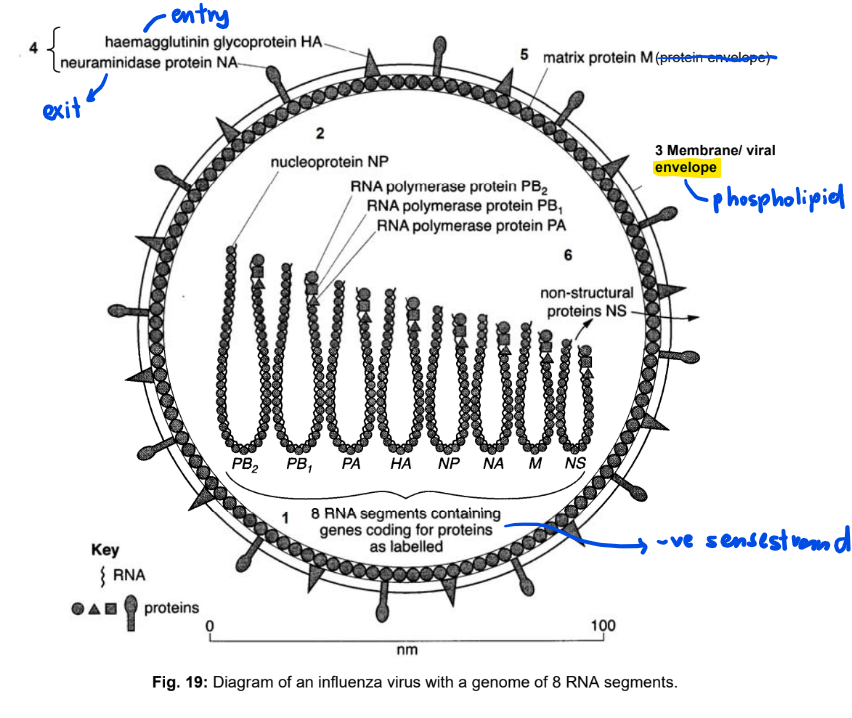
Structure of Influenza Virus (common):
Genome (nucleic acid)
Eight different segments of negative (-) sense single-strand RNA
(-) sense strand RNA must be converted into the complementary (+) sense RNA before it can be used for translation of viral proteins
Protein coat
Nucleoprotein (NP) associate with the viral nucleic acid to form nucleocapsid
Structure of Influenza Virus (unique to influenza virus):
Membrane / Viral Envelope
Phospholipid bilayer obtained from host upon budding
Surface Glycoproteins
Haemagglutinin (HA)
HA binds to sialic acid containing receptors
Attach virus to the receptor on the host cell membrane
Neuraminidase (NA)
Hydrolyses mucus allowing virus to enter cells of the respiratory tract
Facilitate budding by cleaving sialic acid containing receptors
Protein Envelope
It is the matrix protein that forms second layer of envelope, enclosing the nucleocapsid
M1 → Monomers of matrix protein
M2 → Acts as an ion channel to lower or maintain the pH of the endosome in the host cell
Enzymes
PB1, PB2, PA
To form RNA-dependent RNA Polymerase (replicase) responsible for replication and transcription
NS 1
Regulates viral replication mechanisms and cellular signaling pathways
Influenza Virus — Reproductive Cycle
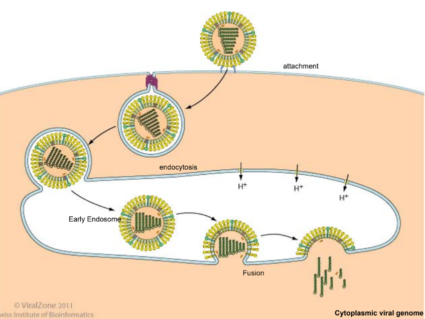
Adsorption
Haemagglutinin (HA) molecules on the viral membrane bind to sialic acid that contains receptors on the membrane of the host cell
Penetration
The virus is then taken in by receptor-mediated endocytosis, forming an endosome (vesicle) within the host cell, with the influenza virus attached to its inner surface
Proton pumps in the endosomal membrane pump H+ ions into the endosome, lowering its pH
This triggers conformational changes in the HA protein, causing the viral envelope to fuse with the endosomal membrane and releasing the viral genome into the host cell cytoplasm
The viral RNAs are then transported into the nucleus
Synthesis & Replication
RNA-dependent RNA polymerase (viral replicase) present in the virion, copies the (-) sense RNA template into complementary (+) sense RNAs
The (+) sense RNA can then be used
Viral Nucleic Acid Synthesis (replication of viral genome)
(+) sense RNAs are used as templates for synthesis of full-length (-) sense strand viral RNAs by viral replicase
These (-) sense viral RNAs can be packaged into new viral particles as their nucleic acid
Viral Protein Synthesis
The (+) sense RNAs are used as mRNA which are translated in the cytoplasm by host amino acids, ribosomes, ATP and tRNAs
Proteins synthesised:
Enzymes
Matrix
Capsomeres proteins
Glycoproteins
Free ribosomes synthesise enzymes, matrix and capsomeres as they are ultimately folded into final conformation in the cytoplasm and packaged into the new virion
rER-bound ribosomes synthesise viral transmembrane surface glycoproteins and transported to Golgi apparatus for glycosylation and then incorporated into the host cell membrane via vesicle which fuses with host cell membrane
Assembly
Assembly of the viral particle is complete when the viral components of eight (-) sense viral RNAs associated with NP and enzymes like viral replicase (PB1, PB2 and PA) are packaged
Acquisition of the glycoprotein studded membrane envelope occurs during the release of the virus
Release
The virus is finally released from the host cell by budding, acquiring with it the host cell’s lipid bilayer as the virus’s envelope
The host membrane containing HA, NA and a third protein, M2, buds off from the host cell with the virion components
With the presence of HA on the viral envelope and sialic-acid containing cellular receptors on the host cell’s membrane, budding invariably bring both together and results in the new viral particle remaining attached to the host cell
Neuraminidase (NA) then aids to release the virus by cleaving sialic acid residues on the cellular receptor that bind the newly formed virions to the cell
This releases the virions, allowing infection to continue
Enveloped Animal Viruses — Human Immunodeficiency Virus (HIV)
Retrovirus:
HIV is a retrovirus that causes AIDS (Acquired Immunodeficiency Syndrome)
Retroviruses are enveloped RNA viruses which replicate by means of a DNA intermediate synthesised by the enzyme reverse transcriptase
Structure of HIV (common):
Genome (nucleic acid)
Two identical single-stranded RNA
The single-stranded RNA is converted to DNA for integration into the host genome
The DNA is then used for transcription of viral mRNA which is translated into viral proteins and for use as the viral genome in the progeny virions
Protein Coat
Protein coat surrounds the nucleic acid
Structure of HIV (unique to HIV):
Viral Envelope
Phospholipid bilayer obtained from host upon budding
Surface glycoproteins
gp120
Binds to CD4 receptors on white blood cells like macrophages and T helper cells
gp41
Aids in the fusion of the HIV envelope and the host cell membrane
Protein Coat
Matrix protein forms second layer of protein envelope, enclosing the protein coat
Enzymes
Reverse transcriptase
2 molecules, each associated with 1 RNA molecule
To reverse transcribe viral RNA into DNA
Integrase
Facilitates incorporation of double-stranded DNA into host cell’s genome
Protease
Cleaves viral polypeptide into functional proteins during viral maturation
Human Immunodeficiency Virus (HIV) — Reproductive Cycle
Adsorption
Glycoprotein gp120 on the surface of the HIV binds to the CD4 receptor, a cell-surface receptor found on T helper cells and macrophages of the host immune system
Penetration
Upon binding to CD4, gp120 undergoes a conformational change, allowing it to bind to a co-receptor (CXCR4) on the surface of T helper cells and (CCR5) on macrophages
Binding of gp120 to the co-receptor causes a further conformational change, exposing gp41, which pulls the viral envelope closer to the host cell membrane
The HIV envelope fuses with the host cell membrane, releasing the viral contents consisting of viral nucleic acid and enzymes into the host cell
Synthesis & Replication
Reverse transcriptase first reverse transcribes the viral RNA into a complementary DNA strand
The RNA strand of the DNA-RNA is broken down by the ribonuclease H component of the reverse transcriptase
The newly synthesised DNA strand is used as a template for synthesis of the other complementary DNA strand, forming a double-stranded DNA molecule
This DNA molecule then passes through the nuclear pore and enters into the host nucleus
The enzyme integrase catalyses the integration of the viral DNA into the genetic material of the host
This newly integrated viral DNA is called a provirus
May remain in a latent (inactive) state for several years, producing few or no copies of HIV
The latent phase ends when the host cell is stimulated in an immune response
Viral Nucleic Acid Synthesis (replication):
When the host cell receives a signal, the proviral DNA is transcribed by the host RNA polymerase into RNA which serves as nucleic acid for new virions
Viral Protein Synthesis
The proviral DNA is also transcribed into viral mRNA, which is then translated to produce a single long chain of HIV proteins which is later cleaved
Viral proteins synthesised → Enzymes, matrix and capsomeres proteins and glycoproteins
Viral surface glycoproteins are synthesised by rER-bound ribosomes and transported to Golgi apparatus for glycosylation and then incorporated into the host cell membrane via vesicles which fuses with host cell membrane
Assembly
Copies of HIV proteins and HIV's RNA genome assemble near the host cell membrane to form a new virus particle
Assembly of the viral components occur when the viral components of 2 single-stranded RNA molecules associated with reverse transcriptase and enzymes like integrase and protease are surrounded by assembled capsid
Release
Acquisition of the glycoprotein studded membrane envelope occurs during the release of the virus
The newly assembled immature HIV buds off from the host cell, surrounded by host membrane
Outside of host cell, viral maturation occurs when the HIV protease cleaves the single long chains of HIV proteins into smaller functional proteins, forming a mature HIV particle (but still a virion that is mature)
Variation in Viral Genomes — Mechanism
Mutation
Occurs in HIV / Influenza virus
As RNA is typically only a messenger molecule in eukaryotic cells, there are no proofreading mechanisms in the host cell
Hence, RNA viruses do not undergo rigorous proofreading during replication of genome and will experience much higher rates of mutations since errors are not corrected
Certain viral enzymes such as reverse transcriptase in HIV have very low fidelity (‘not exact’) and regularly introduce errors into the genome
This results in errors in every replication cycle and is responsible for antigenic drift
Recombination
Occur in HIV
Viruses may also undergo recombination with the genome of another strain, resulting in these genomes exchanging genetic information and resulting in genomes with new combinations of alleles
Viral progeny formed from such a process contain some genes from both the original viral strains
Reassortment
Occurs in Influenza virus
A host cell may be infected with 2 viral strains which introduce two sets of genetic material into the host cell
During formation of the viral progeny, different segments of the viral genome may be packaged into the progeny virus
This would result in a sudden and drastic change in the viral genome and leading to an antigenic shift

Variation in Viral Genomes — Antigenic Terminologies
Antigen
An antigen is any substance that can be recognized by the immune system
It is a molecule that binds to an antibody or a T-cell receptor (TCR) which elicits a B cell or T cell response respectively
Major classes of antigens include proteins, carbohydrates, lipids and nucleic acids
They are usually foreign and are typically large molecules
Location of antigens:
Protrude from the surface of foreign cells or viruses
Other antigens, such as toxins secreted by bacteria, are released into the extracellular fluid
Antigenicity
The capacity of an antigen to induce an immune response in a host
Antibody
A type of glycoprotein molecule, immunoglobulin (Ig), produced by B lymphocytes, that binds antigens, often with a high degree of specificity and high affinity
Variation in Viral Genomes — Antigenic Shift (reassortment)
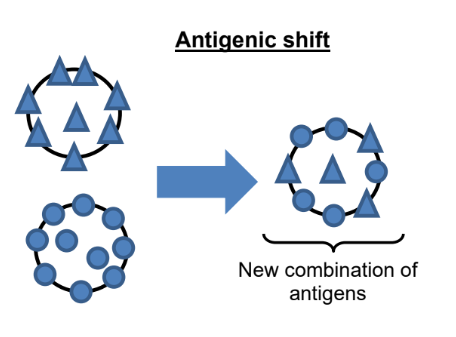
Antigenic shift (definition):
It is a sudden change in the antigenicity of a virus owing to reassortment of the segmented virus genome with another genome of a different antigenic type
In the influenza virus, antigenic shift results in new hemagglutinin and/or new hemagglutinin and neuraminidase proteins in the viruses that infect humans
Antigenic shift results in a new influenza A subtype or a virus that has emerged from an animal population that is so different from the same subtype in humans that most people do not have immunity to the new virus
Influenza Virus:
An antigenic shift arises when different influenza A strains infect one host and subsequently form progeny viruses whose genome is a new combination of RNA from the different strains
The influenza virus, which contains 8 single- stranded RNA, is susceptible to such reassortment since it can easily reshuffle it’s genome during packaging of the new virion
Different viruses of different origins may infect a pig, which provides the opportunity for the viruses to reassort, resulting in formation of a new virus which contains different surfaces antigens from both the avian and human influenza strains
This would result in the formation of viruses with new combinations of hemagglutinin and neuraminidase
As the new strain would not have been circulating in the population, there would not likely be any immunity against this novel strain and resulting in the virus being easily
Variation in Viral Genomes — Antigenic Shift — Inter-Species Transmission
Antigenic Shift 1:
A bird passes a strain of avian influenza A to an intermediate host (chicken or pig)
Concurrently, a human passes a human strain of influenza A to the same host, allowing the genes from the bird strain mix with genes from the human strain to yield a new strain which can spread to humans
Antigenic Shift 2:
Without undergoing genetic change, a bird strain of influenza A can jump directly from a duck or other aquatic bird to humans
Antigenic Shift 3:
Without undergoing genetic change, a bird strain of influenza A can jump directly from a duck or other aquatic bird to an intermediate animal host and then to humans
Variation in Viral Genomes — Antigenic Drift (mutation)
Antigenic drift (definition):
It is the gradual accumulation (slow process) of minor mutations in the genes of influenza viruses that results in altered antigenicity
It happens continually over time in the genes of viruses like the influenza virus as the virus replicates
These result in small changes which produce viruses that are closely related to each other and usually share the same antigenic properties
Influenza Virus:
Antigenic drift arises when viruses undergo continuous, subtle antigenic changes due to accumulation of mutations to the hemagglutinin and/or neuraminidase genes
Factors which influence the rate of mutation of viral genomes:
As the genome of influenza consists of 8 single- strand RNA strands which lack a complementary strand, polymerases cannot perform proofreading during replication
Viral polymerases are also prone to errors and will introduce mutations during the course of DNA replications
Retroviruses, such as HIV, average of one point mutation for every replication cycle as a result of viral reverse transcriptase being unable to correct nucleotide misincorporation errors
These mutations result in the production of surface proteins with different 3D conformations, resulting antibodies no longer being complementary to them and thus will not recognise and bind to them
Variation in Viral Genomes — Effect of Antigenic Shift and Drift
Negative effects:
Due to antigenic shift or antigenic drift, viruses are able to mutate and change the antigens presented on their surfaces
This prevents antibodies, which are proteins generated by the host immune system to help identify and remove foreign antigens on viruses, from recognising them and allowing the viruses to evade detection by the host immune system
Vaccines would become ineffective since the antibodies generated would not be able to bind specifically to the new surface antigen due its new and different 3D conformation
Virus can evade the host’s immune system and continue its reproductive cycle
Effect on Humans:
The magnitude of impact on the human population would be different due to the different mechanisms for change in surface proteins
The major change in surface proteins during an antigenic shift causes in a large number of individuals being susceptible to the new virus, thus resulting in a pandemic which could spread across the globe
Relatively gradual and minor change in surface proteins during an antigenic drift allows some people to be unaffected due to pre-existing immunity, thus resulting in only a seasonal epidemic
Variation in Viral Genomes — Antigenic Shift VS Antigen Drift
