Balance Week 7
 Call Kai
 Learn
 Practice Test
 Spaced Repetition
 Match
 Flashcards
 Knowt Play
1/260

Earn XP
Description and Tags
Acid-Base and Respiratory Control
261 Terms
B7LG1.1. State the Henderson–Hasselbalch equation and identify what lungs and kidneys regulate.
Answer: pH = 6.1 + log10([HCO3−]/[0.03 × PaCO2]). The kidney changes the numerator by controlling bicarbonate, while the lung changes the denominator by eliminating CO2.
Extra Information:
- The ratio of base to dissolved carbon dioxide, not either number in isolation, determines pH.
Source: B7LG1 slide 5 | First Aid 2025 p. 609

B7LG1.1. Why does increased bicarbonate raise pH but increased PaCO2 lower pH?
Answer: Rising HCO3− enlarges the numerator and increases the ratio, so pH rises. Rising PaCO2 enlarges the denominator and decreases the ratio, so pH falls. CO2 combines with H2O to produce carbonic acid, H+ and HCO3−.
Extra Information:
- The same pH can conceal two opposing primary disorders if both variables change markedly.
Source: B7LG1 slide 5 | First Aid 2025 p. 609
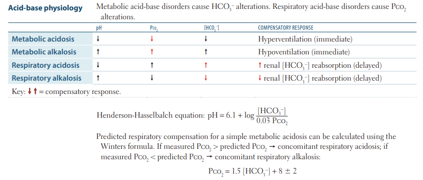
B7LG1.1. Explain how normal HCO3− = 24 and PaCO2 = 40 yield pH approximately 7.40.
Answer: 0.03 × 40 = 1.2, and 24/1.2 = 20. log10(20) ≈ 1.30, so pH = 6.1 + 1.30 = 7.40.
Extra Information:
- A 20:1 bicarbonate-to-dissolved-CO2 ratio is the normal physiologic benchmark.
Source: B7LG1 slide 5 | First Aid 2025 p. 609

B7LG1.1. Distinguish acidemia, alkalemia, acidosis, and alkalosis.
Answer: Acidemia means measured arterial pH below 7.35, and alkalemia means pH above 7.45. Acidosis and alkalosis are underlying processes that push pH down or up, and can coexist even when measured pH is near normal.
Extra Information:
- A normal pH does not rule out mixed acid–base disease.
Source: B7LG1 slides 9, 25–27 | First Aid 2025 p. 610

B7LG1.1. Why do pH disturbances affect cellular function and protein activity?
Answer: Changes in hydrogen ion concentration alter protonation and charge of proteins, influencing enzyme activity, ion binding, membrane excitability and cellular metabolism. Alkalemia can reduce ionized calcium despite unchanged total calcium.
Extra Information:
- Carpopedal spasm with hyperventilation reflects increased albumin binding of Ca2+ as pH rises.
Source: B7LG3 slides 8–9 | B7LG2 slide 3

B7LG1.1. Trace the carbonic-acid equilibrium and name the catalyst that couples CO2 transport to acid–base buffering.
Answer: CO2 + H2O ⇌ H2CO3 ⇌ H+ + HCO3−. Carbonic anhydrase accelerates hydration and dehydration of CO2, particularly in erythrocytes and renal tubular cells. Removing CO2 through ventilation pulls the equilibrium toward consuming H+.
Extra Information:
- The open bicarbonate buffer system works because the lungs can excrete CO2 and kidneys can replace HCO3−.
Source: B7LG1 slide 5 | First Aid 2025 pp. 608–609

B7LG1.2. Compare the speed, sensed variable, and effector of respiratory versus renal compensation.
Answer: Respiratory compensation starts within minutes by altering alveolar ventilation and PaCO2. Renal compensation develops over hours to days through H+ secretion, bicarbonate reabsorption and new bicarbonate generation.
Extra Information:
- Lungs primarily compensate for metabolic disorders, whereas kidneys compensate for respiratory disorders.
Source: B7LG1 slides 5, 7, 12 | B7LG2 slides 3–4

B7LG1.2. Why does metabolic acidosis trigger hyperventilation, and does that correct the underlying metabolic problem?
Answer: Reduced HCO3− lowers pH, stimulating respiratory drive and increasing alveolar ventilation. More CO2 is exhaled, PaCO2 falls, and the Henderson–Hasselbalch ratio rises toward normal. This is compensation, not elimination of the metabolic acid source.
Extra Information:
- Deep rapid Kussmaul respirations are seen in severe metabolic acidosis such as DKA.
Source: B7LG1 slides 5, 12–14 | B7LG2 slide 36

B7LG1.2. Predict compensatory HCO3− shifts in acute versus chronic respiratory acidosis.
Answer: For each 10-mmHg rise in PaCO2 above 40, HCO3− rises approximately 1 mEq/L acutely due to buffering, versus 3–4 mEq/L chronically as kidneys retain and generate bicarbonate.
Extra Information:
- A large bicarbonate elevation with high CO2 favors chronic hypercapnia or an additional metabolic alkalosis.
Source: B7LG1 slides 7, 12 | B7LG3 slide 25

B7LG1.2. Predict bicarbonate changes during acute versus chronic respiratory alkalosis.
Answer: For each 10-mmHg decline in PaCO2 below 40, bicarbonate decreases by about 2 mEq/L acutely through buffering and 4–5 mEq/L chronically through decreased renal bicarbonate retention.
Extra Information:
- Renal compensation is larger after sustained hypocapnia because kidneys require time to change transporter activity.
Source: B7LG1 slide 12 | First Aid 2025 p. 609

B7LG1.2. What are the expected respiratory compensation formulas for metabolic acidosis and metabolic alkalosis?
Answer: Metabolic acidosis: expected PaCO2 = 1.5 × HCO3− + 8 ± 2 (Winter). Metabolic alkalosis: expected PaCO2 ≈ 40 + 0.7 × (HCO3− − 24) ± 5.
Extra Information:
- Compensation moves pH toward normal but cannot create overshoot alkalemia or acidemia by itself.
Source: B7LG1 slides 12–14 | First Aid 2025 p. 609
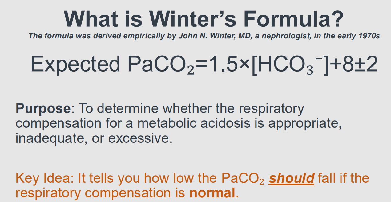
B7LG1.2. Why is a PaCO2 change called compensation rather than a second primary disorder only when it matches the predicted response?
Answer: The appropriate response follows a predictable physiologic relationship between the primary variable and the compensatory variable. PaCO2 higher than the expected range indicates additional respiratory acidosis and PaCO2 lower than expected indicates additional respiratory alkalosis.
Extra Information:
- An independent second disorder is diagnosed by the discrepancy, not simply by PaCO2 being abnormal.
Source: B7LG1 slides 12–14 | B7LG3 slides 6, 23–25

B7LG1.3. Recite the exact six steps of the core ABG interpretation algorithm.
Answer:
1 Determine acidemia versus alkalemia.
2 Identify primary metabolic versus respiratory disturbance.
3 Assess expected compensation.
4 If metabolic acidosis, calculate and albumin-correct the anion gap.
5 If the gap is elevated, perform delta-delta analysis.
6 Integrate all abnormalities with clinical context.
Extra Information:
Use the same full sequence on every ABG to avoid overlooking mixed disorders.
Source: B7LG1 slide 8 | B7LG3 slides 5–10
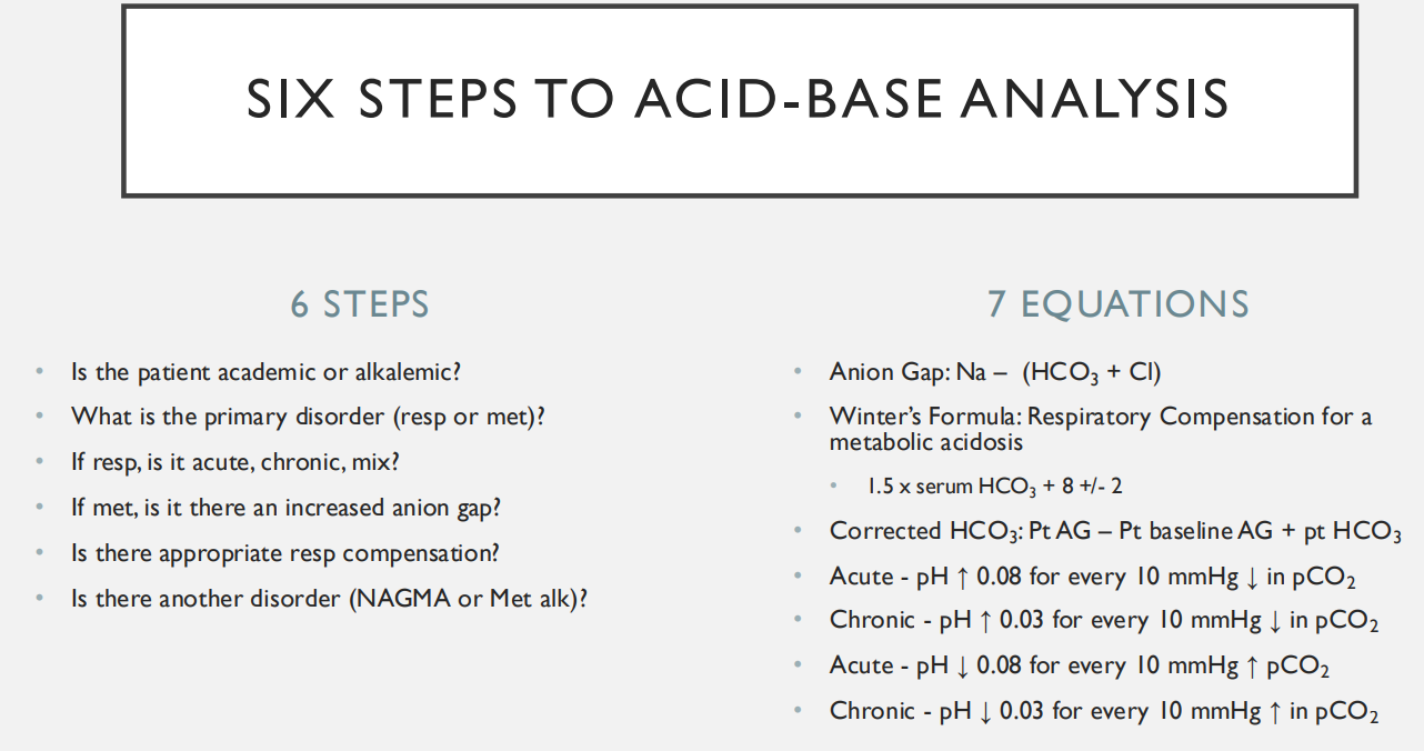
B7LG1.3. How do pH, HCO3− and PaCO2 identify the four primary acid–base disturbances?
Answer:
Acidemia plus low HCO3− = primary metabolic acidosis.
Acidemia plus high PaCO2 = primary respiratory acidosis.
Alkalemia plus high HCO3− = primary metabolic alkalosis.
Alkalemia plus low PaCO2 = primary respiratory alkalosis.
Extra Information:
HCO3− and PaCO2 frequently move together from compensation, so identify the variable that explains the pH direction.
Source: B7LG1 slides 9–11 | First Aid 2025 p. 609

B7LG1.3. In metabolic acidosis with HCO3− 12 and PaCO2 28, assess respiratory compensation.
Answer: Winter: 1.5 × 12 + 8 = 26 ± 2, so expected PaCO2 is 24–28. The measured 28 lies within range, meaning appropriate respiratory compensation, without a proven second respiratory disturbance.
Extra Information:
- This calculation does not distinguish gap versus nongap causes.
Source: B7LG1 slides 13–15, 32

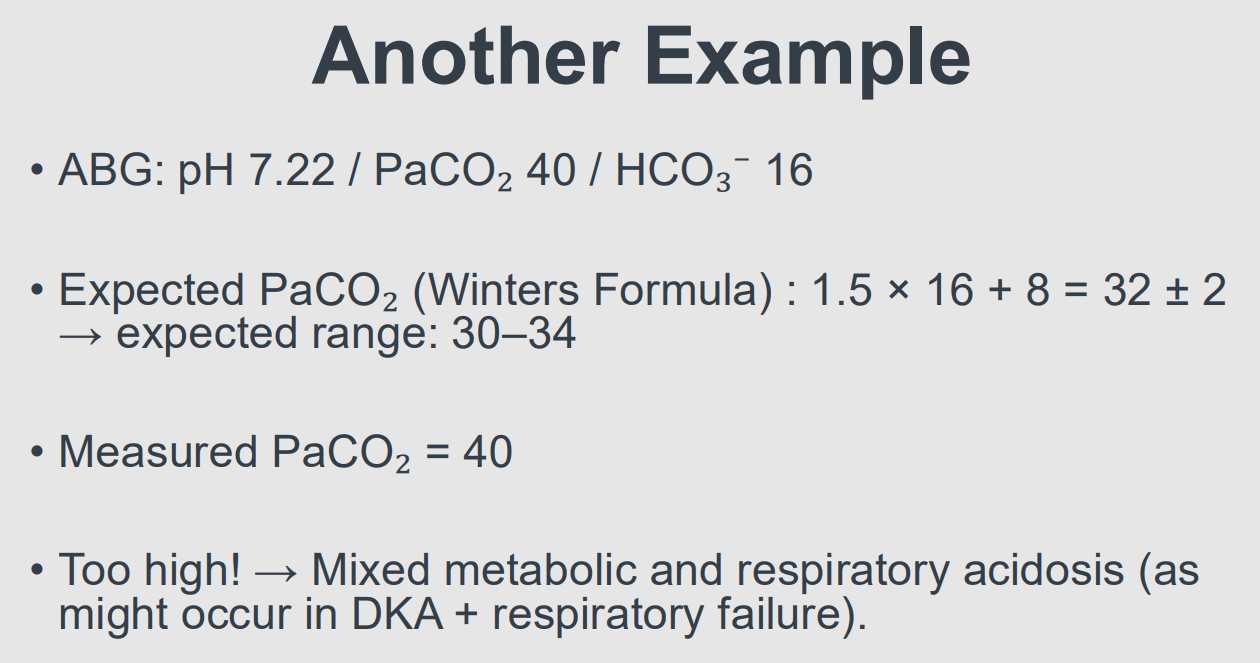
B7LG1.3. In metabolic acidosis with HCO3− 16 and measured PaCO2 40, diagnose the mixed component.
Answer: Winter: expected PaCO2 = 1.5 × 16 + 8 = 32 ± 2, or 30–34. Measured 40 is above this range, proving concomitant respiratory acidosis due to insufficient CO2 elimination.
Extra Information:
- Consider simultaneous metabolic acid accumulation and hypoventilation or respiratory failure.
Source: B7LG1 slides 14, 16
B7LG1.3. Compute anion gap and corrected gap if Na 140, Cl 102, HCO3− 13 and albumin 2.
Answer: AG = 140 − (102 + 13) = 25. Corrected AG = 25 + 2.5 × (4 − 2) = 30 mEq/L. The albumin correction reveals the full severity of unmeasured anion accumulation.
Extra Information:
Corrected AG=AG+2.5(4−albumin)
Albumin is a major unmeasured anion, so hypoalbuminemia deceptively lowers the observed AG.
Source: B7LG1 slides 3, 18 | B7LG3 slide 7
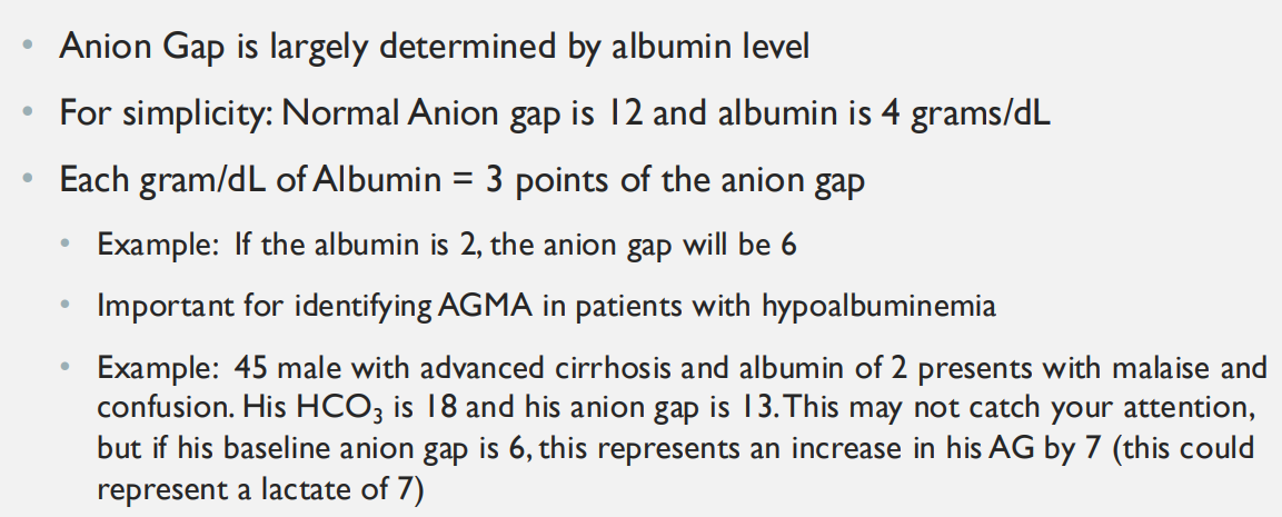
B7LG1.3. Use delta ratio to interpret AG 28 and bicarbonate 12.
Answer: Delta ratio = (28 − 12)/(24 − 12) = 16/12 = 1.33. This is compatible with predominantly high-anion-gap metabolic acidosis, without a clear second metabolic disturbance by the lecture thresholds.
Extra Information:
- The delta-ratio convention and Dr. Leveno’s corrected-HCO3 method test the same question by different arithmetic.
Source: B7LG1 slides 19, 26

B7LG1.3. Work through Dr. Rice’s Patient A: pH 7.29, PaCO2 28, HCO3− 13, Na 140, Cl 102, albumin 2.
Answer: Acidemia with primary metabolic acidosis. AG = 140 − (102 + 13) = 25, and corrected AG ≈25 + 2.5(4 − 2) = 30. Winter = 1.5(13)+8 = 27.5 ±2, range 25.5–29.5, so PaCO2 28 is appropriate. With corrected AG, ΔAG = 18 and ΔHCO3 = 11, ratio ≈1.64: high-AG metabolic acidosis without a demonstrated additional metabolic process.
Extra Information:
- Using albumin-corrected AG prevents underestimating a high-gap acidosis when albumin is low.
Source: B7LG1 slides 3, 8, 13–19

B7LG1.3. Why can a low bicarbonate and low PaCO2 indicate either compensation or two simultaneous primary disorders?
Answer: Bicarbonate loss lowers pH, causing compensatory hyperventilation and PaCO2 reduction. But independently increased respiratory drive, as in salicylate poisoning, can push PaCO2 even lower than Winter’s predicted range. Always calculate expected compensation to distinguish these.
Extra Information:
- Look for a measured PaCO2 below the lower endpoint of Winter’s interval.
Source: B7LG1 slides 13–16, 25–27

B7LG1.3. Apply Dr. Leveno’s corrected-bicarbonate method to AG 25, measured HCO3− 22 and baseline AG 12.
Answer: Corrected HCO3− = measured HCO3− + (patient AG − baseline AG) = 22 + (25 − 12) = 35 mEq/L. This exceeds normal HCO3− ≈24, revealing an additional metabolic alkalosis despite measured bicarbonate of 22.
Extra Information:
- The method conceptually adds back bicarbonate consumed by high-gap acid, then examines the remaining bicarbonate level.
Source: B7LG3 slides 4, 6, 13

B7LG1.4. Explain why a normal-gap metabolic acidosis is usually hyperchloremic.
Answer: Bicarbonate loss lowers one major plasma anion. Chloride rises or becomes relatively more abundant to preserve electroneutrality, so Na − (Cl + HCO3−) remains near normal despite lower pH.
Extra Information:
- Typical mechanisms include diarrhea, renal tubular acidosis and chloride-rich fluid administration.
Source: B7LG1 slides 21–24 | First Aid 2025 p. 610

B7LG1.4. Contrast anion-gap versus non-anion-gap metabolic acidosis mechanistically.
Answer: High-gap acidosis adds unmeasured organic anions such as lactate, ketoanions or toxin metabolites as bicarbonate buffers H+. Normal-gap acidosis primarily loses bicarbonate or impairs renal acid excretion, with an accompanying chloride increase.
Extra Information:
- An elevated AG can be missed if hypoalbuminemia is not corrected.
Source: B7LG1 slides 18, 21–24 | First Aid 2025 p. 610

B7LG1.4. What clinical processes cause the four primary acid–base disorders?
Answer:
Metabolic acidosis: ketoacidosis, lactic acidosis, toxins, diarrhea or RTA.
Metabolic alkalosis: vomiting, diuretics or mineralocorticoid excess.
Respiratory acidosis: hypoventilation from COPD, sedatives or muscle weakness.
Respiratory alkalosis: hyperventilation from pulmonary embolism, hypoxemia, anxiety or salicylates.
Extra Information:
Connect each diagnosis to its mechanism rather than using pH pattern alone.
Source: B7LG1 slides 10–11, 18, 21–24 | First Aid 2025 p. 610

B7LG1.4. Explain the ABG pattern for acute and chronic CO2 retention.
Answer: Both cause elevated PaCO2 and respiratory acidification. Acutely buffering raises HCO3− only slightly, leaving marked acidemia. Chronically renal H+ excretion and bicarbonate retention raise HCO3− substantially, bringing pH closer to normal.
Extra Information:
- Compare observed bicarbonate against the 1 versus 3–4 mEq/L per 10-mmHg compensation rules.
Source: B7LG1 slides 7, 12 | B7LG3 slides 17–19
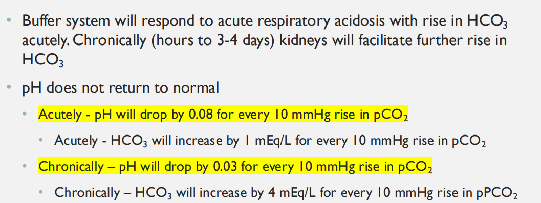
B7LG1.4. Why can a patient have pH 7.40 despite an important metabolic acidosis?
Answer: Two opposing acid–base processes can offset one another, producing a near-normal net pH. A high AG, depleted HCO3−, abnormal PaCO2 or abnormal delta ratio reveals the disease despite normal pH.
Extra Information:
- Do not call a blood gas normal until all major variables and compensation have been checked.
Source: B7LG3 slide 13 | B7LG1 slides 25–27

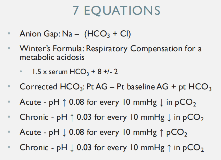
B7LG1.4. Contrast acute and chronic respiratory acidosis using Dr. Leveno’s approximate arterial pH changes.
Answer: Each acute 10-mmHg rise of PaCO2 decreases pH by approximately 0.08. Each chronic 10-mmHg rise lowers pH by approximately 0.03 because kidney bicarbonate retention has had time to develop.
Extra Information:
- For respiratory alkalosis, pH rises approximately 0.08 acutely and 0.03 chronically per 10-mmHg PaCO2 decline.
Source: B7LG3 slides 6, 17–19
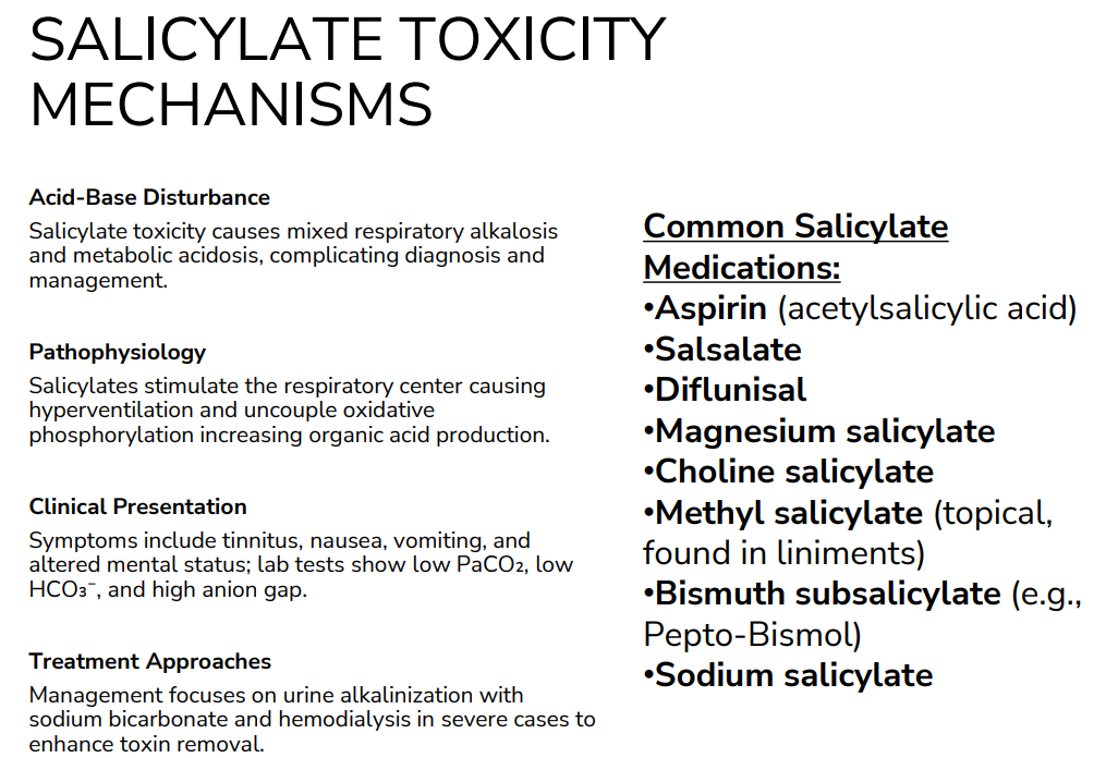
B7LG1.5. Explain the two distinct primary acid–base effects of salicylate poisoning.
Answer:
Salicylates stimulate the medullary respiratory center, causing hyperventilation, CO2 loss and primary respiratory alkalosis.
They also uncouple oxidative phosphorylation and promote accumulation of organic acids, using HCO3− and producing high-anion-gap metabolic acidosis.
Extra Information:
The opposed primary processes can create a deceptively normal pH.
Source: B7LG1 slides 25–28 | B7LG2 slides 13, 47

B7LG1.5. Fully calculate the acid–base disturbance for salicylate case pH 7.38, PaCO2 20, HCO3− 12, Na 140, Cl 100.
Answer: AG = 140 − (100 + 12) = 28. Winter predicts 1.5 × 12 + 8 = 26 ± 2, so expected PaCO2 24–28. Actual PaCO2 20 is below range. The diagnosis is high-gap metabolic acidosis PLUS primary respiratory alkalosis.
Extra Information:
- A pH of 7.38 is not reassuring because the two primary disorders oppose each other.
Source: B7LG1 slides 25–27
B7LG1.5. Interpret the delta ratio and typical symptoms in the salicylate case.
Answer: Delta ratio = (28 − 12)/(24 − 12) = 1.33, consistent with a high-gap process. Clinical clues include tinnitus, nausea, vomiting, tachypnea, hyperthermia and altered mental status.
Extra Information:
- Toxic salicylates can come from aspirin or bismuth subsalicylate, not just obvious aspirin overdose.
Source: B7LG1 slide 26 | B7LG2 slides 44–48
B7LG1.5. Why does salicylate poisoning increase temperature and oxygen demand?
Answer:
Salicylates act as mitochondrial protonophores, dissipating the proton gradient across the inner mitochondrial membrane.
Electron transport consumes oxygen but ATP synthesis becomes inefficient, so more energy is released as heat.
Extra Information:
Organic acid formation worsens metabolic acidosis while respiratory-center stimulation lowers PaCO2.
Source: B7LG2 slide 47 | First Aid 2025 p. 76

B7LG1.5. Interpret Dr. Pearson’s salicylate case with pH 7.48, PaCO2 25, HCO3− 14, AG 26, salicylate 55 mg/dL and normal osmolar gap.
Answer: The high AG and HCO3− 14 indicate high-gap metabolic acidosis. Winter predicts 1.5(14)+8 = 29 ± 2, or 27–31 mmHg. PaCO2 25 is below the interval, consistent with an additional respiratory alkalosis. However, the slide’s pH 7.48 is inconsistent with its paired HCO3− 14 and PaCO2 25 (which predict pH ≈7.37), so treat that pH as a teaching-case transcription discrepancy rather than a physiologically consistent ABG.
Extra Information:
- Salicylate simultaneously stimulates ventilation and disrupts mitochondrial ATP production.
Source: B7LG2 slides 44–47
B7LG1.5. Why does a normal osmolar gap not rule out salicylate poisoning?
Answer: The osmolar gap screens for abundant unmeasured dissolved particles such as unmetabolized toxic alcohols. Salicylate poisoning chiefly produces mixed acid–base effects and high-gap acid accumulation rather than the characteristic high parent-alcohol osmolar load.
Extra Information:
- Dr. Pearson’s salicylate patient has a normal measured osmolar gap despite significant toxicity.
Source: B7LG2 slides 45–47

B7LG2.1. Describe the integrated immediate, respiratory and renal defenses against acid accumulation.
Answer: Chemical buffers immediately bind H+, ventilation increases within minutes to lower PaCO2, and kidneys subsequently reclaim filtered HCO3− while excreting H+ as titratable acid and NH4+ to generate new bicarbonate.
Extra Information:
- These coordinated defenses resist pH change at different time scales, but cannot replace treatment of the cause.
Source: B7LG2 slides 3–4, 53 | First Aid 2025 p. 609

B7LG2.1. Why does compensation fail to normalize pH fully when the primary process persists?
Answer: Compensating organs change the opposite component of the HCO3−/CO2 ratio but are physiologically limited and do not remove the initiating disease. Kidneys require days to maximally adapt to CO2 retention and lungs cannot eliminate fixed nonvolatile acids.
Extra Information:
- Compensation by itself does not cross the normal pH to create an opposite disturbance.
Source: B7LG1 slide 12 | B7LG2 slides 3–4
B7LG2.1. Differentiate high PaCO2 from low HCO3− in a tachypneic, acidemic patient.
Answer: High PaCO2 indicates inadequate effective alveolar ventilation and primary respiratory acidosis. Low HCO3− indicates a metabolic acid load or loss of base and often provokes compensatory tachypnea to reduce PaCO2. If measured PaCO2 exceeds Winter’s expected range, both processes coexist.
Extra Information:
- Visible rapid breathing does not prove adequate alveolar ventilation if tidal volume is small or dead space increases.
Source: B7LG1 slides 10–14 | B7LG2 slides 4, 38–40
B7LG2.1. Explain how an opioid overdose can create simultaneous respiratory and metabolic acidosis.
Answer:
Opioid-induced depressed respiratory drive causes hypoventilation, CO2 retention and respiratory acidosis.
Hypoxemia or impaired tissue perfusion raises anaerobic lactate production, consuming HCO3− and causing a second high-gap metabolic acidosis.
Extra Information:
Faculty case: PaCO2 60, HCO3− 18, AG 22, lactate 6 and pinpoint pupils.
Source: B7LG2 slides 38–40
B7LG2.1. Why can Kussmaul breathing accompany severe diabetic ketoacidosis?
Answer:
Insulin deficiency increases lipolysis and hepatic ketogenesis, accumulating β-hydroxybutyrate and acetoacetate.
H+ consumes HCO3−, decreasing pH, and chemoreceptor stimulation produces deep rapid ventilation to lower PaCO2.
Extra Information:
Kussmaul breathing is a respiratory compensation to metabolic acidosis rather than primary lung disease.
Source: B7LG2 slides 41–43 | First Aid 2025 p. 610
B7LG2.1. Explain why an elevated respiratory rate is not equivalent to adequate CO2 clearance.
Answer:
Alveolar ventilation equals respiratory frequency times tidal volume minus dead-space volume.
A patient taking rapid but very shallow breaths may ventilate mostly dead space, retain CO2 and develop respiratory acidosis despite visible tachypnea.
Extra Information:
Work of breathing and effective tidal volume matter as much as respiratory rate.
Alveolar ventilation= (VT-VD) x RR
Source: B7LG2 slides 3–4 | First Aid 2025 p. 685

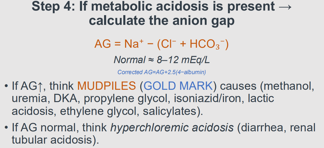
B7LG2.2. Calculate anion gap and explain its biochemical meaning.
Answer: AG = Na+ − (Cl− + HCO3−), typically about 8–12 mEq/L in the lectures. It estimates unmeasured anions after accounting for the principal measured ions. Elevation suggests lactate, ketones, renal acids or toxic metabolites.
Extra Information:
- Albumin is an important negatively charged unmeasured anion, requiring correction when low.
Source: B7LG2 slides 9–10, 15 | B7LG1 slide 18
B7LG2.2. Calculate serum osmolar gap, including adjustment if ethanol is present.
Answer:
Calculated osmolality in mOsm/kg ≈ 2Na + glucose/18 + BUN/2.8 using conventional units.
Osmolar gap = directly measured osmolality − calculated osmolality.
If ethanol is present, include its osmotic contribution, conventionally ethanol mg/dL divided by 4.6.
Extra Information:
An unexplained gap greater than about 10 raises concern for unmeasured osmoles, but is not toxin-specific.
Source: B7LG3 slides 27–30 | B7LG2 slide 10

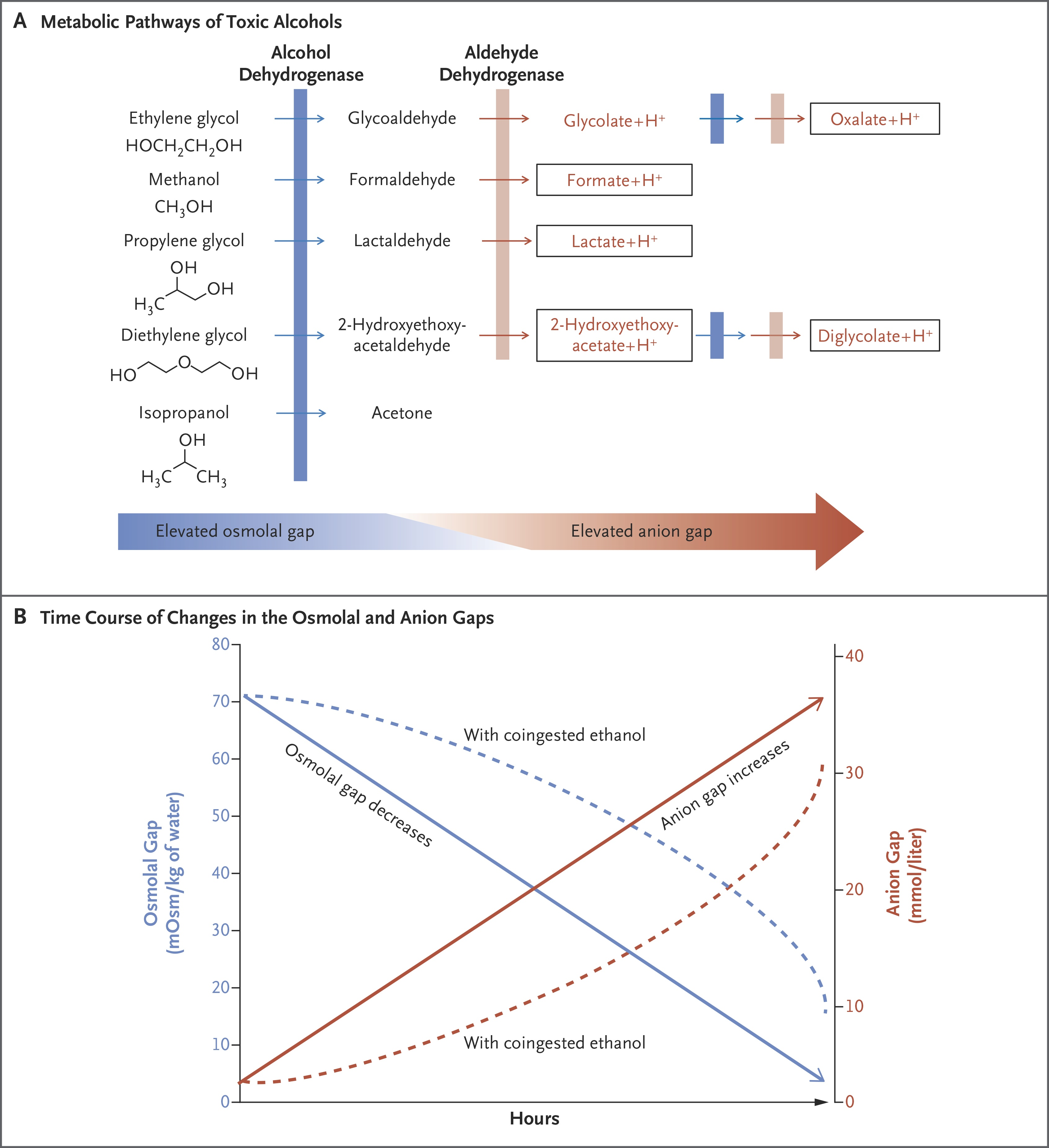
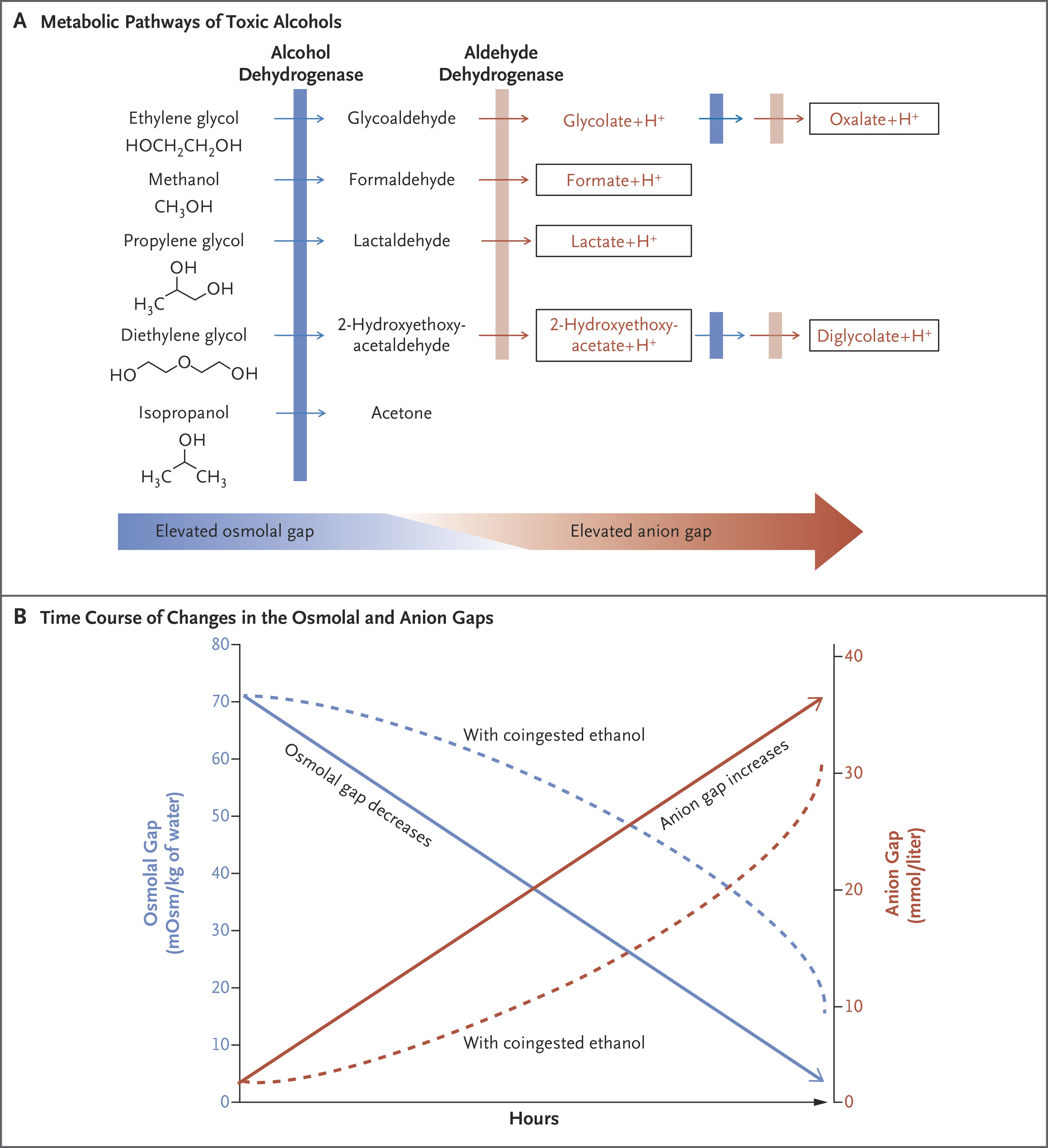
B7LG2.2. Why can toxic alcohol poisoning show an early osmolar gap and a later anion gap?
Answer: Initially unmetabolized parent alcohol is osmotically active but uncharged, increasing the osmolar gap without necessarily increasing AG. Alcohol dehydrogenase then produces organic acid metabolites, raising AG as parent alcohol concentration and osmolar gap may fall.
Extra Information:
- A normal osmolar gap late in poisoning does not exclude methanol or ethylene glycol toxicity.
Source: B7LG2 slides 12, 14, 29–30 | B7LG3 slides 27–30
B7LG2.2. Differentiate the characteristic clinical-laboratory patterns of methanol, ethylene glycol and salicylates.
Answer:
Methanol: visual injury, elevated AG and often osmolar gap.
Ethylene glycol: AKI, flank pain, calcium oxalate crystals, elevated AG and often osmolar gap.
Salicylates: tinnitus and tachypnea with mixed respiratory alkalosis plus high-gap metabolic acidosis, commonly without an elevated osmolar gap.
Extra Information:
All three may produce severe acid–base disturbances, but their organ toxicities differ.
Source: B7LG2 slides 13–14, 21–22, 33–36, 44–46
B7LG2.2. Apply both gaps to the DKA teaching case: Na 132, Cl 96, HCO3− 10, glucose 620, BUN 28, measured osm 320.
Answer: AG = 132 − (96 + 10) = 26. Calculated osm = 2(132) + 620/18 + 28/2.8 = 264 + 34.4 + 10 = 308.4. Osmolar gap ≈ 320 − 308.4 = 11.6 mOsm/kg.
Extra Information:
- DKA explains the high AG through ketoanions, while a mildly raised osmolar gap is not specific for toxic alcohol.
Source: B7LG2 slides 41–43
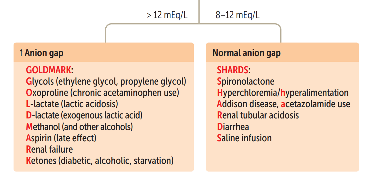
B7LG2.2. Recall the GOLD MARK differential for high-anion-gap metabolic acidosis.
Answer: Glycols, Oxoproline, L-lactate, D-lactate, Methanol, Aspirin/salicylates, Renal failure, Ketoacidosis. These raise circulating unmeasured anions as bicarbonate buffers the associated acids.
Extra Information:
- Distinguish this from common normal-gap causes such as diarrhea and renal tubular acidosis.
Source: B7LG2 slide 19 | First Aid 2025 p. 610

B7LG2.2. Solve Dr. Pearson’s ethylene glycol case: pH 7.10, PaCO2 20, HCO3− 8, Na 140, Cl 100 and calcium oxalate crystals.
Answer:
AG = 140 − (100 + 8) = 32, establishing high-AG metabolic acidosis.
Winter predicts 1.5(8)+8 = 20 ± 2, or 18–22, so PaCO2 20 indicates appropriate respiratory compensation.
Calcium oxalate crystals, flank pain and creatinine 2.0 point toward ethylene glycol renal injury.
Extra Information:
Glycolic acid contributes substantially to acidosis and oxalate contributes to crystal-associated injury.
Source: B7LG2 slides 33–37
B7LG2.3. State Winter's formula and classify measured PaCO2 relative to its expected range.
Answer:
Expected PaCO2 = 1.5 × HCO3− + 8 ± 2 during primary metabolic acidosis.
A measured value within the interval represents expected compensation, above means additional respiratory acidosis and below means additional respiratory alkalosis.
Extra Information:
Do not use Winter’s formula as the diagnostic compensation rule for primary metabolic alkalosis.
Source: B7LG1 slides 13–17 | B7LG2 slide 25

B7LG2.3. In methanol case with HCO3− 10 and PaCO2 25, is there evidence of extra respiratory acidosis?
Answer: Winter predicts 1.5 × 10 + 8 = 23 ± 2, a range of 21–25 mmHg. Actual PaCO2 = 25 is at the upper boundary, so it is within expected compensation, not above range.
Extra Information:
- PaCO2 at the exact endpoint of the predicted interval remains within that interval.
Source: B7LG2 slides 22, 25–26 | B7LG1 slide 14
B7LG2.3. Derive and interpret the delta ratio in a high-gap metabolic acidosis.
Answer: Delta ratio = (albumin-adjusted AG − baseline AG)/(baseline HCO3− − measured HCO3−). For the common baseline AG 12 and HCO3− 24, this is (AG − 12)/(24 − HCO3−). A ratio below roughly 1 suggests an added normal-gap acidosis, around 1–2 is typical of an isolated high-gap process, and above 2 raises concern for concurrent metabolic alkalosis or a high baseline bicarbonate.
Extra Information:
- Interpret cutoffs as approximations and use Dr. Leveno’s corrected-HCO3 method when his lecture specifically asks for it.
Source: B7LG3 slide 4 | B7LG1 slides 18–19

B7LG2.3. Why does a small delta ratio suggest added normal-gap acidosis?
Answer:
The denominator, 24 − HCO3−, is disproportionately large compared with the rise in AG.
Thus more bicarbonate has been lost than can be explained by the accumulating unmeasured anions alone, suggesting an added hyperchloremic bicarbonate-loss process.
Extra Information:
Example: DKA plus diarrhea can combine high-gap ketoacids with normal-gap stool bicarbonate loss.
Source: B7LG1 slides 19, 21–22

B7LG2.3. Why does a delta ratio above 2 suggest a concurrent metabolic alkalosis?
Answer:
The anion gap has risen markedly, but the observed bicarbonate has fallen less than expected for the acid load.
An independent alkalinizing process, such as vomiting with HCl loss, keeps bicarbonate higher and makes the denominator smaller.
Extra Information:
A high baseline bicarbonate in chronic respiratory acidosis can also raise the ratio.
Source: B7LG1 slide 19 | B7LG3 slides 12–14

B7LG2.3. Why must mixed disorders be sought even when pH or PaCO2 appears nearly normal?
Answer: Two independent processes may offset in pH while individual parameters remain abnormal. Evaluate primary direction, compensation, albumin-corrected AG and delta ratio systematically to find all processes before assigning a clinical diagnosis.
Extra Information:
- Faculty cases include salicylate toxicity and opioid-induced mixed respiratory and metabolic acidosis.
Source: B7LG1 slides 8, 25–27 | B7LG2 slides 38–40
B7LG2.3. Work Dr. Leveno’s Case 1: pH 7.31, PaCO2 10, Na 123, Cl 99, HCO3− 5 and albumin 4.
Answer:
AG = 123 − (99 + 5) = 19. Winter = 1.5(5)+8 = 15.5 ±2, so expected PaCO2 13.5–17.5 and observed 10 is additional respiratory alkalosis.
Corrected HCO3− = 5 +(19−12)=12, much below 24, proving additional normal-gap metabolic acidosis.
Extra Information:
Three simultaneous processes: HAGMA, NAGMA and respiratory alkalosis.
Source: B7LG3 slide 12
B7LG2.3. Work Dr. Leveno’s Case 2: pH 7.40, PaCO2 41, Na 137, Cl 90, HCO3− 22 and albumin 4.
Answer: AG = 137 − (90 + 22) = 25. Winter predicts 1.5(22)+8 = 41 ±2, so 41 is appropriate. Corrected HCO3− = 22+(25−12)=35, showing a concurrent metabolic alkalosis even though pH is 7.40.
Extra Information:
- A normal pH does not establish a normal acid–base state when opposing metabolic processes coexist.
Source: B7LG3 slide 13
B7LG2.3. Work Dr. Leveno’s Case 3: pH 7.20, PaCO2 25, Na 130, Cl 80 and HCO3− 10.
Answer: AG = 130 − (80 + 10) = 40. Winter predicts 1.5(10)+8 = 23 ±2, interval 21–25, so PaCO2 25 is appropriate. Corrected HCO3− = 10+(40−12)=38, strongly indicating concurrent metabolic alkalosis.
Extra Information:
- The corrected-bicarbonate approach detects hidden vomiting/diuretic-type alkalosis alongside high-gap acidosis.
Source: B7LG3 slide 14
B7LG2.3. Work Dr. Leveno’s Case 4: pH 7.07, PaCO2 28, Na 125, Cl 100 and HCO3− 8.
Answer: AG = 125−(100+8)=17. Winter predicts 1.5(8)+8=20±2, but PaCO2 28 exceeds 18–22, indicating added respiratory acidosis. Corrected HCO3− = 8+(17−12)=13, below 24, indicating added NAGMA.
Extra Information:
- This is a triple disorder: high-gap metabolic acidosis, normal-gap metabolic acidosis and respiratory acidosis.
Source: B7LG3 slide 16
B7LG2.4. Trace methanol metabolism from ingestion to high-gap acidosis and visual injury.
Answer: Methanol is converted by alcohol dehydrogenase to formaldehyde, then by aldehyde dehydrogenase to formic acid/formate. Formate inhibits mitochondrial cytochrome c oxidase, impairing oxidative phosphorylation, causing cellular energy failure, organic acid accumulation and retinal/optic nerve injury.
Extra Information:
- Think blurred vision or visual loss with unexplained AG and osmolar gaps.
Source: B7LG2 slides 14, 21–22, 27–31 | First Aid 2025 p. 247

B7LG2.4. Trace ethylene glycol metabolism and explain kidney injury plus hypocalcemia.
Answer: Alcohol dehydrogenase initiates metabolism of ethylene glycol, yielding glycolaldehyde, glycolate, glyoxylate and oxalate. Glycolate contributes strongly to high-gap acidosis, while oxalate binds calcium and forms calcium oxalate crystals that injure renal tubules.
Extra Information:
- Look for flank pain, acute kidney injury, crystals and possible low calcium.
Source: B7LG2 slides 14, 33–37 | First Aid 2025 p. 247

B7LG2.4. Connect methanol and ethylene glycol clinical findings to specific organ-level mechanisms.
Answer:
Methanol-derived formate injures energy-dependent optic structures and brain, causing blurred vision, confusion and acidosis.
Ethylene glycol metabolites damage renal tubules through acid injury and calcium oxalate deposition, causing rising creatinine, flank pain and urinary crystals.
Extra Information:
An elevated osmolar gap identifies unmeasured osmoles but not which alcohol is present.
Source: B7LG2 slides 21–22, 33–36

B7LG2.4. Why does salicylate toxicity cause tinnitus, fever and a mixed acid–base disturbance?
Answer:
Salicylates stimulate medullary ventilation to lower CO2 and cause respiratory alkalosis.
Uncoupling of mitochondrial oxidative phosphorylation increases oxygen demand, heat release and organic-acid accumulation, lowering bicarbonate and producing metabolic acidosis.
Extra Information:
Tinnitus, vomiting, tachypnea, fever and mental-status changes help distinguish the syndrome.
Source: B7LG2 slides 13, 44–48 | First Aid 2025 p. 610
B7LG2.4. Explain how opioid toxicity produces a misleadingly high PaCO2 even when lactate is elevated.
Answer:
Respiratory depression lowers effective ventilation and traps CO2, causing respiratory acidosis.
Concurrent hypoxia or poor perfusion generates lactic acid and lowers bicarbonate.
Thus a high measured PaCO2 rather than expected compensatory hypocapnia reveals a second respiratory disorder.
Extra Information:
The faculty patient has pinpoint pupils, AG 22 and lactate 6 mmol/L.
Source: B7LG2 slides 38–40
B7LG2.4. Explain the time-dependent reversal of osmolar-gap and anion-gap prominence in toxic alcohol poisoning.
Answer: Early after ingestion, much of the parent alcohol remains: it adds unmeasured osmoles and raises the osmolar gap. As alcohol dehydrogenase converts it into acidic metabolites, the parent osmolar contribution decreases while the anion gap rises.
Extra Information:
- Neither a normal osmolar gap nor the absence of a massive gap safely excludes a late presentation.
Source: B7LG2 slides 10, 12, 14, 30, 32
B7LG2.5. Explain fomepizole's mechanism and why it helps in toxic alcohol ingestion.
Answer: Fomepizole competitively inhibits alcohol dehydrogenase, preventing methanol conversion to toxic formate and ethylene glycol conversion to glycolate/oxalate-generating intermediates. It halts additional toxic metabolite production but does not rapidly remove metabolites already formed.
Extra Information:
- Start it promptly when methanol or ethylene glycol poisoning is strongly suspected.
Source: B7LG2 slides 31, 37 | First Aid 2025 p. 247

B7LG2.5. Construct a mechanism-based initial treatment plan for suspected methanol poisoning.
Answer: Stabilize airway, breathing and circulation, obtain serial ABGs, electrolytes, AG, osmolality and renal studies, begin fomepizole to block bioactivation, correct dangerous acidemia with indicated bicarbonate, and obtain toxicology support for potential hemodialysis.
Extra Information:
Dialysis eliminates methanol and formate and corrects severe acid–base abnormalities.
NBME memory trick
Ethanol = Distracts ADH.
Fomepizole = Blocks ADH.
Source: B7LG2 slides 21–22, 31 | B7LG3 slide 31

B7LG2.5. Construct a treatment plan for ethylene glycol poisoning and explain monitoring priorities.
Answer: Give supportive resuscitation, fomepizole to block toxic metabolite formation, and bicarbonate when indicated for severe acidemia. Assess electrolytes, ionized calcium, kidney function and urine findings, and use hemodialysis in severe poisoning or organ dysfunction.
Extra Information:
- Dialysis removes parent toxin/metabolites and can address marked acidosis or renal failure.
Source: B7LG2 slides 33–37
B7LG2.5. Why is urinary alkalinization useful in salicylate poisoning?
Answer: Sodium bicarbonate increases urinary pH, favoring ionized salicylate that crosses tubular membranes poorly. This reduces passive tubular reabsorption and increases urinary elimination while simultaneously treating systemic acidemia.
Extra Information:
Potassium depletion can make urine alkalinization difficult, so monitor and correct electrolytes.
Because when potassium is low (hypokalemia), the kidneys try to conserve K⁺, but this causes them to secrete more H⁺ into the urine.
That makes the urine more acidic, which is the opposite of what we want when treating aspirin (salicylate) poisoning!
Source: B7LG2 slides 13, 48 | First Aid 2025 p. 247

B7LG2.5. When should hemodialysis be considered in severe toxic acid–base disturbances?
Answer: Consider it when severe or refractory acidemia, renal failure, serious neurologic or visual toxicity, clinical deterioration, or a substantial toxic burden makes elimination and correction urgent. Dialysis decisions require toxin-specific criteria and consultation.
Extra Information:
- The supplied toxicology lectures discuss dialysis indications qualitatively but do not provide verified toxin-specific numerical thresholds.
Source: B7LG2 slides 14, 31, 37, 48

B7LG2.5. Why can emergent airway management be particularly challenging in severe metabolic acidosis?
Answer:
A patient may rely on marked hyperventilation to keep PaCO2 low.
Sedation or apnea removes this compensation, so PaCO2 rises quickly and pH can precipitously fall.
Airway support must preserve appropriate ventilation while definitive therapy addresses the acid source.
Extra Information:
Faculty emphasizes initial ABC stabilization and protection of the airway when obtunded.
Apnea is a medical condition defined as the temporary cessation of breathing, lasting for at least 10 seconds, which can happen voluntarily or involuntarily
Source: B7LG3 slide 31 | B7LG1 slides 13–14
B7LG2.5. Why should acidemia and serum potassium be monitored during salicylate urine alkalinization?
Answer: Systemic acidemia increases nonionized salicylic acid distribution into tissues, while raising urinary pH keeps salicylate ionized and less reabsorbable. Hypokalemia promotes renal H+ secretion and impairs urinary alkalinization, so potassium assessment and replacement matter.
Extra Information:
- Bicarbonate therapy is a targeted elimination strategy, not simply an attempt to normalize a laboratory pH.
Source: B7LG2 slides 13, 48 | First Aid 2025 p. 247
B7LG3.1. Define metabolic acidosis and explain why pH and bicarbonate alone are not enough to subtype it.
Answer: A primary decrease in serum HCO3− reduces the HCO3−/PaCO2 ratio and generally produces acidemia. To distinguish high- versus normal-gap forms, calculate AG = Na − (Cl + HCO3−), account for albumin, and assess measured PaCO2 against expected compensation.
Extra Information:
- A mixed alkalosis can mask acidemia despite clinically important metabolic acidosis.
Source: B7LG3 slides 5–10, 20–23 | B7LG1 slide 18
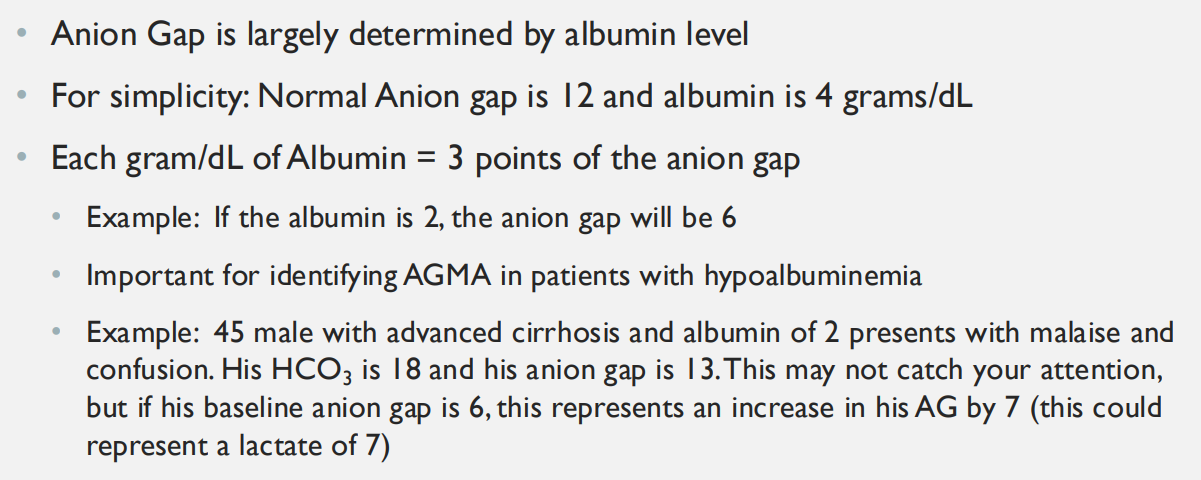
B7LG3.1. Why must anion gap be corrected for a low albumin level?
Answer: Albumin carries substantial negative charge and accounts for much of the normal unmeasured-anion pool. Low albumin lowers baseline measured AG, potentially hiding new organic acids. Corrected AG ≈ observed AG + 2.5 × (4 − albumin in g/dL).
Extra Information:
- The faculty alternative approximates 3 mEq/L of AG per 1 g/dL of albumin, while Dr. Rice uses 2.5 for correction.
Source: B7LG3 slides 7–8 | B7LG1 slide 18
B7LG3.1. Distinguish a normal-gap metabolic acidosis from high-gap metabolic acidosis by the changes in plasma anions.
Answer: With GI or renal bicarbonate loss, HCO3− declines while chloride rises, leaving AG near normal. With lactate, ketones or toxic acids, HCO3− declines while an unmeasured conjugate anion accumulates, increasing AG.
Extra Information:
- Electroneutrality is maintained in both scenarios, but the accompanying negative charge differs.
Source: B7LG1 slides 18, 21–24 | First Aid 2025 p. 610
B7LG3.1. How do potassium and urine pH help classify renal tubular acidoses?
Answer:
Types 1 and 2 are usually hypokalemic, whereas type 4 is hyperkalemic.
Type 1 distal RTA cannot excrete H+ and typically maintains urine pH above 5.5 despite acidemia.
Type 2 has bicarbonate wasting and may acidify urine below 5.5 after plasma bicarbonate falls.
Type 4 impairs NH4+ excretion.
Extra Information:
RTA commonly produces normal-gap hyperchloremic metabolic acidosis.
Source: B7LG1 slide 24 | First Aid 2025 p. 611

B7LG3.1. What does Winter's formula test when the primary disorder is metabolic acidosis?
Answer: It tests whether ventilation has lowered PaCO2 to the expected level. Calculate 1.5 × HCO3− + 8 ± 2, then compare the observed PaCO2. Values above suggest added respiratory acidosis, below added respiratory alkalosis, and within the range appropriate compensation.
Extra Information:
- It does not independently establish whether AG is elevated or identify the acidosis etiology.
Source: B7LG1 slides 13–17 | B7LG3 slide 23
B7LG3.1. How does hypoalbuminemia alter the baseline anion gap in Dr. Leveno’s teaching convention?
Answer: Albumin supplies much of the normal unmeasured negative charge. Dr. Leveno uses approximately 3 mEq/L of AG per 1 g/dL albumin: albumin 2 g/dL implies baseline AG around 6 rather than 12.
Extra Information:
- His cirrhosis example has observed AG 13 but elevation of about 7 above its albumin-adjusted baseline.
Source: B7LG3 slide 7
B7LG3.2. Give the high-anion-gap metabolic acidosis differential using GOLD MARK and explain the shared mechanism.
Answer: Glycols, Oxoproline, L-lactate, D-lactate, Methanol, Aspirin, Renal failure and Ketoacidosis. Each adds circulating unmeasured anions as H+ consumes bicarbonate, raising AG.
Extra Information:
- History, renal function, glucose, ketones, lactate, osmolar gap and toxicology clues differentiate these causes.
Source: B7LG3 slide 20 | B7LG2 slide 19 | First Aid 2025 p. 610
B7LG3.2. Give a mechanism-based differential for non-anion-gap metabolic acidosis.
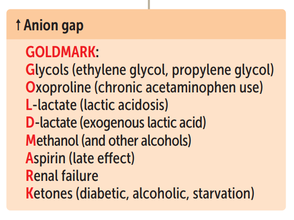
Answer: GI bicarbonate loss from diarrhea or pancreatic/biliary drainage, renal bicarbonate wasting or reduced distal H+ excretion from RTA, carbonic-anhydrase inhibitors such as acetazolamide, ileal urinary diversion, and chloride-rich saline loading.
Extra Information:
- Cl− increases relative to HCO3−, so AG stays close to baseline.
Source: B7LG3 slide 22 | B7LG1 slides 21–24 | First Aid 2025 p. 610

B7LG3.2. Contrast type 1, type 2 and type 4 RTA by defect, potassium and urine pH.
Answer: Type 1: alpha-intercalated cell distal H+ secretion defect, low K+, urine pH usually above 5.5, calcium phosphate stones. Type 2: proximal HCO3− reabsorption defect, low K+, variable urine pH that can fall below 5.5. Type 4: low aldosterone effect plus reduced ammoniagenesis, high K+, variable urine pH.
Extra Information:
- Type 4 often occurs with diabetes, ACE inhibitors, ARBs or potassium-sparing drugs.
Source: B7LG1 slide 24 | First Aid 2025 p. 611
B7LG3.2. How do respiratory acidosis and respiratory alkalosis differentials differ?
Answer: Respiratory acidosis reflects alveolar hypoventilation from obstructive lung disease, sedatives/opioids, obesity hypoventilation, neuromuscular weakness or CNS depression. Respiratory alkalosis reflects excessive effective ventilation from hypoxemia, pulmonary embolism, anxiety, pregnancy or early salicylate toxicity.
Extra Information:
- Determine whether PaCO2 change explains the pH rather than assuming dyspnea equals respiratory alkalosis.
Source: B7LG3 slides 20–21 | First Aid 2025 p. 610
B7LG3.2. A patient with DKA also has diarrhea: what mixed pattern can occur?
Answer: Ketones add unmeasured anions and raise AG while stool bicarbonate loss lowers HCO3− with compensating chloride rise. The bicarbonate deficit can therefore exceed the AG rise and delta ratio falls below approximately 0.8.
Extra Information:
- Use the anion-gap calculation, Winter’s formula and delta ratio rather than naming only DKA.
Source: B7LG1 slides 18–22 | B7LG3 slide 4
B7LG3.2. Why do diarrhea and distal renal tubular acidosis both cause normal-gap metabolic acidosis but yield different urine ammonium responses?
Answer: Diarrhea causes extrarenal bicarbonate loss, so healthy kidneys increase NH4+ excretion. Distal renal acidification failure reduces effective NH4+ excretion. A urine anion gap can indirectly help distinguish those responses in selected cases.
Extra Information:
- Urine indices have important limitations, including unmeasured urinary anions and low urine sodium.
Source: First Aid 2025 pp. 610–611
B7LG3.3. Identify metabolic alkalosis and explain the initial role of loss of gastric HCl.
Answer: A primary HCO3− increase produces alkalemia and compensatory CO2 retention. Vomiting or nasogastric suction removes gastric H+ and Cl−, leaves bicarbonate in extracellular fluid, and causes volume and chloride depletion that impair renal bicarbonate excretion.
Extra Information:
- Vomiting removes acid, whereas lower GI diarrhea usually removes bicarbonate.
Source: B7LG3 slide 24 | B7LG1 slides 11–12, 22 | First Aid 2025 p. 610
B7LG3.3. Distinguish chloride-responsive versus chloride-resistant metabolic alkalosis using urine chloride.
Answer: Urine Cl− below about 20 mEq/L usually indicates chloride-responsive alkalosis from vomiting or remote diuretic effects. Urine Cl− above about 20 mEq/L favors ongoing diuretic exposure or chloride-resistant causes such as mineralocorticoid excess, Bartter syndrome or Gitelman syndrome.
Extra Information:
- Urine Cl− often predicts response to saline and chloride replacement better than urine Na+.
Source: First Aid 2025 p. 610 | B7LG3 slide 24
B7LG3.3. Explain why volume depletion maintains metabolic alkalosis.
Answer: Low effective circulating volume activates RAAS and proximal NaHCO3 reabsorption, reducing bicarbonate excretion. Aldosterone promotes distal Na+ absorption coupled with K+ and H+ secretion, maintaining hypokalemia and alkalosis until volume/chloride deficits are corrected.
Extra Information:
- This is contraction and maintenance physiology, not simply a one-time gastric acid loss.
Source: B7LG1 slides 5, 11–12 | First Aid 2025 pp. 608, 610
B7LG3.3. How does hypokalemia perpetuate metabolic alkalosis?
Answer: K+ depletion shifts K+ out of cells and H+ into cells, increases renal bicarbonate reabsorption and promotes renal H+ secretion and ammoniagenesis. The resulting renal processes maintain elevated serum HCO3− even after the initiating vomiting or diuretic exposure.
Extra Information:
- Correcting potassium is often necessary for successful reversal of alkalosis.
Source: First Aid 2025 pp. 608–610 | B7LG3 slide 24
B7LG3.3. Predict compensation for HCO3− 34 in primary metabolic alkalosis and suggest its differential.
Answer: Expected PaCO2 ≈ 40 + 0.7(34 − 24) = 47 mmHg, usually within ±5. If actual PaCO2 is much greater there is added respiratory acidosis, and if much lower there is added respiratory alkalosis. Consider vomiting, diuretics and mineralocorticoid excess, guided by urine Cl−.
Extra Information:
- Compensatory hypoventilation is limited by the need to maintain oxygenation.
Source: B7LG1 slide 12 | B7LG3 slide 24 | First Aid 2025 p. 610
B7LG3.3. Why does ongoing loop- or thiazide-diuretic use often produce urine chloride above 20 mEq/L despite volume contraction?
Answer: Active diuretics block tubular NaCl reabsorption, increasing chloride loss into urine despite low effective circulating volume. When the drug effect wears off, kidneys may avidly conserve chloride and urinary chloride can fall.
Extra Information:
- Timing of the urine sample relative to a diuretic dose changes its interpretation.
Source: B7LG3 slide 22 | First Aid 2025 p. 610
B7LG4.1. Explain panacinar emphysema pathogenesis from SERPINA1 abnormality through dyspnea.
Answer: SERPINA1 mutations can lower functional alpha-1 antitrypsin (AAT), a liver-made inhibitor of neutrophil elastase. Unopposed elastase degrades alveolar elastin throughout the acinus, destroying alveolar walls, reducing elastic recoil, promoting expiratory collapse and air trapping, and reducing gas-exchange area.
Extra Information:
- Lower-lobe-predominant emphysema in a young never-smoker is classic.
Source: B7LG4 slides 8–11 | First Aid 2025 pp. 692–693
B7LG4.1. Why can alpha-1 antitrypsin deficiency injure both lungs and liver by different mechanisms?
Answer: Lung damage is loss of antiprotease function, permitting elastase injury. Z-variant AAT misfolds and polymerizes in hepatocyte endoplasmic reticulum, causing toxic intracellular retention, ER stress, fibrosis/cirrhosis and increased hepatocellular carcinoma risk.
Extra Information:
- PAS-positive, diastase-resistant hepatocyte globules indicate retained abnormal protein.
Source: B7LG4 slide 9 | First Aid 2025 p. 400
B7LG4.1. Compare panacinar, centriacinar and paraseptal emphysema by acinar distribution.
Answer: Panacinar destroys the entire acinus and favors bases with AAT deficiency. Centriacinar primarily injures proximal respiratory bronchioles with initial distal sparing, favoring apices in smokers. Paraseptal affects distal acinus near pleura and may lead to spontaneous pneumothorax.
Extra Information:
- An acinus extends from respiratory bronchiole through alveolar ducts and alveoli.
Source: B7LG4 slides 3, 11–12 | First Aid 2025 pp. 692–693
B7LG4.1. What workup confirms suspected AAT deficiency and why is a single serum level insufficient?
Answer: Screen with serum AAT concentration, then confirm with protease inhibitor (Pi) phenotype and/or SERPINA1 genotype. AAT is an acute-phase reactant, so inflammation can elevate serum levels and mask deficiency.
Extra Information:
- Faculty recommends testing patients with COPD for AAT deficiency at least once.
Source: B7LG4 slide 10
B7LG4.1. Why does smoking worsen AAT-associated emphysema even without changing a patient's genotype?
Answer: Smoke recruits inflammatory neutrophils that release elastase, increases oxidative stress and can oxidatively inactivate protective AAT. More protease activity combined with less antiprotease protection accelerates alveolar elastin destruction.
Extra Information:
- Lung injury can occur in a never-smoker because the inherited inhibitor deficit exists independently.
Source: B7LG4 slides 9, 11 | First Aid 2025 pp. 692–693
B7LG4.1. Identify the classic Dr. Givler patient pattern that should trigger suspicion for AAT deficiency.
Answer: A relatively young never-smoker with obstruction (for example FEV1/FVC 0.58), basilar-predominant panacinar emphysema, mildly elevated liver enzymes and a family history of early liver disease strongly suggests SERPINA1-related AAT deficiency.
Extra Information:
- This links the same genetic disorder to lung antiprotease loss and hepatic misfolded-protein accumulation.
Source: B7LG4 slides 4–10
B7LG4.2. Identify silicosis exposure, chest imaging, and histology.
Answer: Occupational crystalline silica exposure occurs with sandblasting, mining and engineered-stone cutting. Imaging shows upper-lobe rounded nodules and possible eggshell hilar lymph-node calcification. Histology shows concentric or whorled dense collagen nodules with dust-laden macrophages.
Extra Information:
- Silicosis can progress to severe fibrotic lung disease.
Source: B7LG4 slides 14–15, 18–19 | First Aid 2025 p. 696
B7LG4.2. Why does silica exposure predispose to tuberculosis?
Answer: Inhaled silica accumulates in macrophages, damaging their phagolysosomal function and impairing intracellular killing. Mycobacterium tuberculosis survives more easily, increasing TB susceptibility and reactivation risk.
Extra Information:
- Silicosis also increases lung-carcinoma risk.
Source: B7LG4 slides 18–19 | First Aid 2025 p. 696
B7LG4.2. Describe the exposure, imaging distribution and histology of asbestosis.
Answer: Exposure occurs with shipyards, insulation, construction and brake/clutch work, with long latency. CT commonly shows lower-lobe reticular interstitial fibrosis, traction bronchiectasis or honeycombing, and pleural plaques that may calcify. Histology shows golden-brown beaded iron-protein-coated ferruginous bodies, Prussian-blue positive.
Extra Information:
- Pleural plaques document previous exposure but do not automatically establish severe parenchymal fibrosis.
Source: B7LG4 slides 20–22 | First Aid 2025 p. 696
B7LG4.2. Compare asbestos-associated bronchogenic carcinoma and mesothelioma, including effect of smoking.
Answer: Bronchogenic carcinoma is the more common asbestos-associated cancer, and cigarette smoking greatly amplifies its risk. Malignant mesothelioma is less common but more specifically linked to asbestos exposure and arises from pleura. Smoking does not similarly increase mesothelioma risk.
Extra Information:
- Think pleural thickening or recurrent effusion with mesothelioma, not simply lower-lobe fibrosis.
Source: B7LG4 slide 23 | First Aid 2025 p. 696
B7LG4.2. Differentiate silicosis, asbestosis, berylliosis and coal workers pneumoconiosis.
Answer: Silica: upper-lobe nodules, eggshell nodes, whorled collagen, TB risk. Asbestos: lower-lobe fibrosis, pleural plaques, ferruginous bodies, lung cancer/mesothelioma. Beryllium: aerospace exposure, granulomatous disease mimicking sarcoidosis. Coal dust: coal miners and carbon-laden macrophages, potentially progressive massive fibrosis.
Extra Information:
- Use exposure occupation plus radiographic distribution to distinguish them.
Source: B7LG4 slides 14–23 | First Aid 2025 p. 696
B7LG4.2. What histologic distinction separates silicotic nodules from asbestos bodies?
Answer: Silicotic nodules are dense concentrically whorled collagen with nearby dust-laden macrophages. Asbestos bodies are elongated beaded rods with a golden-brown iron-protein coating, stainable with Prussian blue.
Extra Information:
- Both reflect inhaled particulate injury, but their morphology and typical lobe distributions differ.
Source: B7LG4 slides 18–22
B7LG4.2. For a former shipyard worker with calcified pleural plaques and new pleural thickening/effusion, what two neoplasms must be contrasted?
Answer: Asbestos increases bronchogenic carcinoma and malignant pleural mesothelioma risk. Bronchogenic carcinoma is more common and risk is greatly amplified by smoking. Mesothelioma is more specific to asbestos and presents with diffuse pleural involvement or effusion, without the same smoking interaction.
Extra Information:
- Pleural plaques mark exposure and by themselves are not malignant mesothelioma.
Source: B7LG4 slides 20–23