Exam #3
 Call Kai
 Learn
 Practice Test
 Spaced Repetition
 Match
 Flashcards
 Knowt Play
1/39

Earn XP
Description and Tags
brain #2, ANS, Blood, Heart
40 Terms
Effectors

Autonomic NS and innervation
Sympathetic
Parasympathetic
Preganglionic Neurons – Cell bodies originate in the CNS
Postganglionic Neurons – Cell bodies found at ganglia
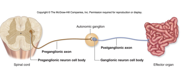
Parasympathetic Division
maintain homeostasis (stable internal enviro)
fx: Slows heart rate, increases digestive fxs, controls sphincters of digestive,
urinary, and reproductive systems. Constricts pupils
Preganglionic neurons:
Long (axons)
Secrete acetylcholine (ACh) as a neurotransmitter (cholinergic)
Arise from cranial and sacral regions
A.K.A. “Cranio-sacral division”
• Cranial = CN III, V, VII, IX, X; Sacral = S2-S4
• The Vagus nerve (CN X) – one of the most important psym nerves (innervates heart, lungs,
stomach, intestines, other viscera)
Postganglionic neurons:
Short (axons)
Also secrete ACh

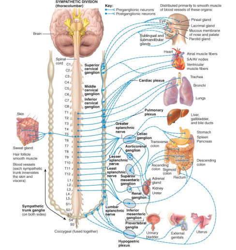
Sympathetic Division
response to emergencies or stress
fx: Increases heart rate, shunts blood toward skeletal mms, increases respiration rate. Dilates pupils
Preganglionic neurons
Short (axons)
Secrete acetylcholine (ACh) as a neurotransmitter.
Arise from thoracic/lumbar regions
A.K.A. “sympathetic trunk”
T1-L2
Synapse with postganglionics at Sympathetic Chain Ganglia
Postganglionic neurons:
Long (axons)
Secrete norepinephrine/noradrenaline (adrenergic).
also innervates the adrenal medulla which releases epinephrine (adrenaline)
blood Fx
Transportation - oxygen and carbon dioxide as well as nutrients and waste products
Regulation - body temperature, pH, and fluid volume
Protection - by mounting an immune response and the production of antibodies
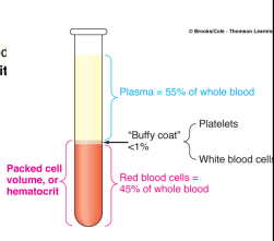
composition of blood
erythocytes: 44% = hematocrties
Buffy Coat: 1% (WBC and Platelets)
Plasma: 55%
Blood Plasma Composition
water 92%
plasma proteins 7%
albumin 58%
globulins 37%
fibrinogen 4%
regulatory proteins 1%
other 1%
nutrients, respiratory gases, wastes
Hematocrit
% of the volume of all formed elements in ones blood (primarily RBC)
female: 38-45%
males: 40-50%
Erythocytes
not true cells, no organelles, no nucleus
7.5 um. biconcave to increase SA
rouleau - line up like a stack of coins in capillaries
Hemoglbin in erythocytes
280 per rbc
reversible binding to 4 O2 or 4 CO2
heme: ringed moleucle with iron ion

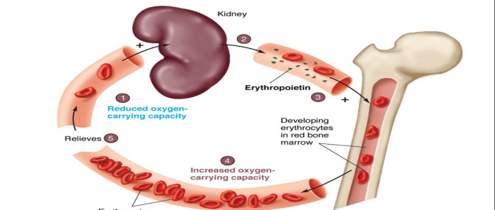
Erythopoiesis
creation of RBCs/erythrocytes @ red marrow
120 day cycle
erythropoietin (EPO) = hormone produced by kidneys low o2. stimulates erythopoiesis
3 mill a sec. 200 billion a day. 25 trillion total
recycling aged or damaged erythocytes
aged erythocytes gets phagocytized in liver and spleen
-recycled heme (without Fe) turnin into bile from liver
-Fe is transported ad stored in liver
-proteins are borken down into amino acids which can make new erythocytes
Doped blood
rbc from donor or previously removed
or the homrone EPO is injected
muscles work longer and harder wout cramping
Leukocytes
true cells: nucleus and organelles
“buffy coat”
initiate immune response
1.5 to 3 times larger than erythocytes
diapedesis: leaving bv to enter tissues
chemotaxis: attraction to site of infection/damage by chemicals

Classification of Leukocytes
granulocytes (visible granules)
Neutrophils 50-70% (baceterial fungal)
Eosinophils: 1-4% (allergens, parasites)
Basophils: 0.5-1% (release histamine in inflammation)
Agranulocytes
lymphocytes: 20-40% (activate b and t cells against viruses, tumors, adaptive, immune responses return to normal fx)
monocytes: 2-8% (migrate from blood, form mmacrophages, target bacteria, tumor cells)

platelets (thrombocytes)
fragments of large cells = megakaryoctyes (15x)
¼ size of erythocytes
clotting of blood when exposed to collagen
fibrin provides meshwork of clot
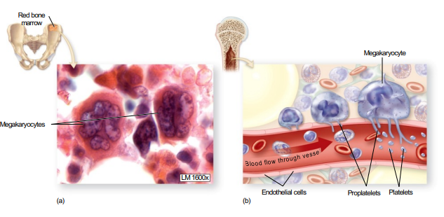
Blood Clot

hemopoiesis
formed elements (rbcs, platelets) short life span
occurs in red marrow
hemocytoblasts = stem cells (pluripotent)
two lines of dev
myeloid = erythocytes, (erythopoiesis) megakaryocyes (thrombopoiesis), all leukocytes (leukopoiesis)
lymphoid = lymphocytes exception (b and t lymphoblast)
Pulmonary vs Systemic Circuit
one pump directs blood to lungs (right)
one pumps directs blood to most body tissues (left)
blood pressure
force of the blood on walls of arteries
normal 120/80mmHg
Pericaridum
within mediastinum
fibruos pericardium: outer covering (dense irrct)serous pericardium
parietal and visceral (epicardium)
pericardial cavity and pericardial fluid

pericarditis vs pericardial effusion
-inflammation of the pericardium
-fluid accumulates around heart
heart wall
epicardium - areolar ct
myocardium - thickest
endocardium - internal surface of heart chambers and external of heart valves
coronary sulcus

Right Atrioventricular (tricuspid)
forced closed when ventricle begins to contract (increases pressure)
then after ventricle into: pulmonary semilunar valve
coronary circulation
right and left coronary arteries are only branches off ascending aorta
right
marginal (r border)
posterior interventricular artery
left
anterior interventircular arteris
circumflex artery: left atrium and vent
Coronary veins
great cardiac vein: anterior interventircular a
middle cardiac vein: posterior interventricular A.
small cardiac vein: to R marginal a.
gap junctions
comprised as intercalated discs
work as single unit
funcitonal syncytium = mechanical/chemical linkage
Heart Sounds
lub: AV valves (into ventricle)
dub: closing of the semilunar valves (out of heart)
autorythmicity
heart capable of initiaing its own heartbeat
SA (sinoatrial): roof of R atrium (sinus)
AV (antrioventricular) node: floor of R atrium
AV (antrioventricular bundles) = bundles of his
right and left bundles @ superior septum
purkinje fibers: located @ walls of vents
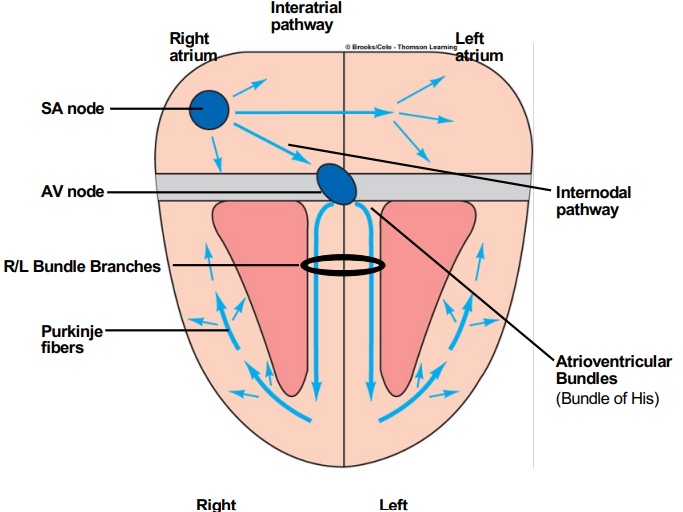
Coordinated sequence of heart chamber contractions
SA node generates impulse
atria contract (sys), ventricle relax (dia)
impulse goes to av node then ventricles (via conducting fibers)
ventricles contract (sys), while atria relaces (dia)
BBB
tight junctions bewtween capillary and endothelial cells
perivascular feet
Brainstem
autonomic and reflex centers
mesencephalon (midbrain, maintain posture)
superior coliculus: visual reflexes
inferior colliculus: auditory reflexes
reticular formation: consciousness
pons
medulla oblongata

cranial nerves
trochlear: moves eye muscles
trigeminal: sensory and motor information of head and neck
abducens: moves eye muscles
glossopharyngeal: moves neck and tongue muscles
hypoglossal: neck muscles
