Non-Echocardiographic Assessment of CAD/MI U2 L1
 Call Kai
 Learn
Learn Practice Test
 Spaced Repetition
 Match
Match Flashcards
 Knowt Play
1/87

There's no tags or description
Looks like no tags are added yet.
Name | Mastery | Learn | Test | Matching | Spaced | Call with Kai | Chat |
|---|
No analytics yet
Send a link to your students to track their progress
88 Terms
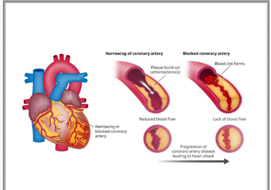
What is a myocardial infarction? & What does it cause?
A blockage prevents (O2) oxygen-rich blood from reaching the heart muscle, causing tissue death.
List the 3 ways a patient with an MI may present.
Chest pain
Discomfort/pain in other areas, such as the neck, jaw, or back
Shortness of breath
What are serum cardiac markers?
Molecules released into the bloodstream because of compromise of a cell membrane
List the 4 serum cardiac markers.
Troponin*****
Creatine kinase
Myoglobin
Lactate dehydrogenase
What is troponin, and why is it important for detecting myocardial injury?
Troponin is a protein that regulates the interaction between actin and myosin filaments in cardiac and skeletal muscle.
It is the gold standard for detecting heart-muscle injury.
When does troponin rise and peak after an injury?
Rises: 2–4 hours
Peaks: 12–24 hours
What are the normal values for Troponin I and Troponin T?
Troponin I: <0.04 ng/mL
Troponin T: <0.01 ng/mL
(ng = nanogram)
What does creatine kinase do, and how specific is it compared with troponin?
Creatine kinase is an enzyme that helps convert ATP to ADP so muscles can produce energy.
It is less specific than troponin, but is still used.
(ATP (Adenosine Triphosphate) ADP (Adenosine Diphosphate)
What is Creatine Kinase helpful in detecting? & Why?
Helpful in detecting Reinfarction because it returns to normal faster
Reinfarction = a second heart attack occurring shortly after the first one.
After the first MI:
CK-MB goes up
Then it comes back down to normal fairly quickly
So if CK-MB has already returned toward normal and then rises again, doctors can suspect that the patient has had another MI.
Heart attack happens → CK-MB rises → then drops back to normal sooner
That is why it helps with reinfarction. If CK-MB already went back to normal, and then it goes up again, that suggests a new heart attack happened.
What is the normal Creatine Kinase/ CK-MB value?
<5 ng/mL.
What is myoglobin, and why is it not specific to the heart?
Myoglobin is a protein released into the bloodstream with myocardial injury.
It’s a Very early marker but not specific to the heart because it can also increase with skeletal-muscle damage.
When can myoglobin be detected after the onset of an MI?
Detected within the first few hours after the onset of an MI
Within the first 1–4 hours.
What are the normal myoglobin values for men and women?
Men: 20–90 ng/mL
Women: 12–75 ng/mL
What does lactate dehydrogenase do?
It is an enzyme that assists in the conversion of pyruvate and lactate for ATP during anaerobic cellular respiration.
Lactate dehydrogenase (LDH) is an enzyme that helps your cells make energy when there isn’t enough oxygen.
It helps change:
pyruvate ↔ lactate
so the cell can keep making a little bit of ATP = energy during anaerobic respiration.
So the easiest way to remember it:
LDH helps cells make energy when oxygen is low.
naerobic cellular respiration means:
Cells make energy without using oxygen.
Aerobic = with oxygen
Anaerobic = without oxygen
So if a cell does not have enough oxygen, it can still make a small amount of ATP (energy) using anaerobic respiration.
For your slide, think:
Low oxygen → anaerobic respiration → cells still make some energy
When is lactate dehydrogenase helpful for detecting an MI? & When does it appear?
It helps with late detection and appears about 1–2 days after the onset of MI.
What does an electrocardiogram detect?
The heart's electrical activity using electrodes placed on the surface of the skin.
List the 2 types of information an EKG can provide.
Electrical activity
Heart structure and function
What does electrical depolarization lead to mechanically?
Mechanical contraction.
What does the P wave represent, and what occurs afterward?
The P wave represents atrial stimulation/depolarization.
Mechanical contraction of the atria occurs afterward.
What does the PR interval represent?
Atrial depolarization and the delay through the AV node.
It means the PR interval shows two things:
The atria are electrically activated → atrial depolarization
Then the electrical signal slows down briefly at the AV node before it goes to the ventricles
So simply:
PR interval = atria depolarize + brief delay at the AV node before the ventricles depolarize.
Why is there a brief pause during the PR interval?
Depolarization slows through the AV node, causing a brief pause or plateau.
How is the PR interval measured?
From the start of the P wave to the start of the QRS complex.
What does the QRS complex represent, and what occurs afterward?
The QRS represents ventricular stimulation/depolarization.
Mechanical contraction of the ventricles occurs afterward
What does electrical repolarization lead to mechanically?
Mechanical relaxation.
What does the ST segment represent?
The period between ventricular depolarization and repolarization.
What does the T wave represent?
Rapid repolarization.
List the 2 EKG findings that may be seen with ischemia.
Inverted T wave
ST depression

What EKG finding is associated with myocardial injury?
ST elevation.
What does ST elevation indicate on the myocardial injury slide?
Acute MI.

List the 2 conditions that may have an EKG appearance similar to myocardial injury.
Ventricular aneurysm
Pericarditis
What other ST-segment change may occur with myocardial injury?
ST depression.
List the 2 criteria for a significant Q wave associated with what.
Significant Q wave = Infarct
At least one small square wide, or 0.04 seconds
At least 1/3 of the QRS amplitude
Those bullets are describing when a Q wave is considered significant/pathologic, which can suggest an old infarct.
Super simple:
At least one small square wide (0.04 sec) = the Q wave has to be wide enough
1/3 of QRS amplitude = the Q wave has to be deep enough, about one-third the height of the whole QRS
So think:
Abnormal Q wave = wide + deep
On your slide, that means a Q wave suggesting infarction is at least 0.04 seconds wide and about 1/3 of the QRS amplitude.

What EKG finding may occur with a subendocardial infarct?
ST depression

What is an NSTEMI?
A Non-ST-Elevation Myocardial Infarction caused by a partial or transient blockage of a coronary artery, or blockage of a smaller branch artery.
What EKG findings may occur with an NSTEMI? List 3
ST-segment depression
T-wave inversions
The EKG may also appear normal
What is a STEMI?
An ST-Elevation Myocardial Infarction is a severe heart attack resulting from a complete blockage of a major coronary artery.
What EKG finding is associated with a STEMI? & What does it indicate?
ST-segment elevation, indicating extensive heart-muscle injury.
What does cardiac catheterization involve?
Insertion of hollow tubes or catheters under fluoroscopic guidance into a peripheral artery or vein.
What is the Injection in the Cardiac cath?
Injection of radiographic contrast agents
What does cardiac catheterization help determine? & Helps decide what for the pt?
Helps distinguish severity or extent of pathology
Helps decide appropriate plan for medical, surgical, or interventional
purposes
List the 5 purposes of cardiac catheterization.
Define coronary anatomy and identify blockage or spasm in a coronary artery or bypass graft
Measure pressures within the chambers and great vessels
Evaluate valve and ventricular function
Provide access to coronary arteries for PTCA, streptokinase, laser therapy, or stents
Provide access to valves for percutaneous valvuloplasty
A bypass graft is a new route for blood to go around a blocked coronary artery.
Basically:
Blocked artery → graft creates a detour → blood reaches the heart muscle past the blockage
The graft is usually made from a blood vessel taken from somewhere else in the body.
Streptokinase is a clot-busting medicine.
It helps break down blood clots so blood can flow again.
For a heart attack:
Blood clot blocks coronary artery → streptokinase helps dissolve the clot → blood flow improves
During cardiac cath, it means it may be used to help open a blocked coronary artery.
They can put a laser catheter inside the coronary artery and use laser energy to help break down or remove plaque/clot material that is blocking the artery.
List the 3 arteries that may be used for left-heart catheterization access.
Femoral artery
Brachial artery
Radial artery
How is the catheter advanced during left-heart catheterization?
Catheter is advanced retrograde across AV into LV
It means the catheter is moved backward against the normal direction of blood flow through the aorta, across the aortic valve, and into the left ventricle.
So:
artery → aorta → across aortic valve → LV
Retrograde = going opposite the normal blood-flow direction.
List the 2 main assessments performed during left-heart catheterization.
Obtain pressures from the aorta and left-sided chambers
Perform angiography of the coronary arteries, aortic root, and LV
That means they inject contrast dye and take X-ray images to look at those structures.
Coronary arteries → look for blockages
Aortic root → look at the beginning of the aorta
LV → look at the left ventricle, including its size, function, and wall motion
So simply:
Angiography = contrast dye + X-ray imaging to see the coronary arteries, aortic root, and left ventricle.
How is LV ventriculography performed?
Radiographic contrast is injected directly into the LV cavity to define the LV silhouette.

At what 2 points in the cardiac cycle is the LV evaluated during ventriculography?
End-diastole
End-systole
List the 2 things LV ventriculography can assess.
Ejection fraction
Regional wall-motion abnormalities
Where is the catheter tip guided during coronary angioplasty?
Into the right or left coronary artery under fluoroscopic guidance.
Why is radiopaque contrast injected during coronary angioplasty?
To visualize the coronary vessels and detect coronary artery stenosis >50%.
List the 4 additional things that may be assessed during coronary angioplasty.
Congenital anomalies
Rapidity of coronary flow
Patency of bypass grafts
Collaterals
It means how fast the blood/contrast moves through the coronary arteries.
So during coronary angiography, they can watch the dye travel through the vessels.
Normal rapid flow = blood is moving well
Slow flow = may suggest narrowing or another problem affecting blood flow
List the 7 RELATIVE CONTRAINDICATIONS for cardiac catheterization.
(same for Right and Left Heart Catheterization)
Uncontrolled ventricular irritability
Uncontrolled hypertension
Febrile illness
Decompensated heart failure with pulmonary edema
Severe allergy to contrast
Severe renal insufficiency — dialysis should be planned
Anticoagulated state
Febrile illness simply means an illness that causes a fever.
So:
Febrile = fever
Examples would be infections like the flu or pneumonia.
How is the catheter advanced during right-heart catheterization?
From a suitable vein to the SVC.
“Suitable vein” just means a vein that is safe and large enough to insert the catheter into.
List the 4 venous access sites for right-heart catheterization.
Femoral
Brachial
Subclavian
Internal jugular vein
Measurements that may be calculated:
List the 3 main assessments performed during right-heart catheterization.
RA, RV, PA, and pulmonary wedge pressures
Oximetry to locate shunts
Cardiac-output calculation
Catheter goes into a small pulmonary artery branch → it “wedges” there → the pressure measured reflects pressure backing up from the left atrium.
So think:
Pulmonary wedge pressure ≈ left atrial pressure
If it is high, it can suggest increased left-sided filling pressure, such as with left-sided heart failure.
Oximetry to locate shunts means they measure the oxygen level of blood in different chambers and vessels of the heart during the right heart cath.
They are looking for a sudden change in oxygen level.
For example:
RA oxygen lower → RV oxygen suddenly higher
That can mean oxygen-rich blood is entering the RV through a left-to-right shunt, such as a VSD.
So simply:
Oximetry = measure oxygen levels in different heart locations to help find where a shunt is occurring.
List the 6 complications of cardiac catheterization.
Death
Stroke or MI — 1/1000
Dysrhythmias, including bradycardia or VF — 1/500
Severe reaction to contrast agents
Perforation leading to tamponade
Dissection
List the 3 main purposes of nuclear cardiac imaging.
Evaluate cardiac physiology and function
Evaluate myocardial perfusion
Evaluate resting regional and global function
When discussing the heart, physiology simply means how the heart works or how it functions to keep you alive.
List the 3 conditions under which myocardial perfusion may be evaluated.
Rest
Exercise
Pharmacologic stress
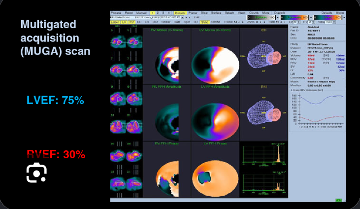
What type of imaging is MUGA? MUGA = Multiple Gated Acquisition scan
Radionuclide imaging. (Evaluation of resting regional and global function)
MUGA is a type of radionuclide imaging, meaning it uses a small amount of radioactive tracer to take pictures of the heart.
MUGA = Multiple Gated Acquisition scan
Very simply:
radioactive tracer → camera tracks it in the blood → pictures show how the ventricles pump
What is administered during nuclear imaging, and what does it contain?
A radiopharmaceutical containing a radioisotope.
How is the radioisotope detected during nuclear imaging?
The radioisotope emits gamma rays, which are detected by a gamma camera.
How does the gamma camera acquire images?
It rotates around the patient and obtains a series of images gated to the EKG.
It means the camera times each picture to the patient’s heartbeat using the EKG.
So instead of taking random pictures, it takes images at the same point in each cardiac cycle.
For example:
EKG detects the R wave → camera knows when systole starts → images are matched to that part of the heartbeat
So simply:
“Gated to the EKG” = the camera is synchronized with the EKG so it can take pictures at specific times during each heartbeat.
What do the reconstructed nuclear images reflect?
They show the location of the isotope, which reflects blood flow to the myocardium
Projections mathematically reconstructed showing the location of the isotope
It means the computer takes all the pictures the camera collected from different angles and combines them into a clearer image.
Then it shows where the radioactive tracer is located in the heart.
So simply:
Many camera pictures → computer combines them → final image shows where the isotope/tracer went.
List the 2 main types of nuclear cardiac imaging.
Myocardial perfusion imaging (stress)
Radionuclide angiography — MUGA
List the 2 types of myocardial perfusion imaging.
PET — Positron Emission Tomography
SPECT — Single Photon Emission Computed Tomography
What pharmacologic agent is used with myocardial perfusion imaging?
Regadenoson (Lexiscan).
Myocardial Prefusion Imaging can be utilized with what imaging, and what is use to create the images?
Can be utilized with stress imaging
• Gamma rays to create images
What does myocardial perfusion imaging identify during rest and stress?
The location and extent of myocardial ischemia during rest and stress.
What is a “cold spot” on myocardial perfusion imaging?
The imaging agent concentrates in normal tissue, but a cold spot occurs in infarcted or ischemic tissue where blood flow is relatively reduced.
How does a Radionuclide Angiography (MUGA) scan image the heart?
It uses gamma rays and radioactive tracers to image the heart cavities over time.
List the 3 measurements or assessments MUGA can provide.
Ejection fraction
Wall motion
Cardiac output
What is Radionuclide Angiography (MUGA) the Gold standard for?
Gold standard for EF*
List the 2 main purposes of exercise stress testing.
Evaluate the cardiovascular response to physical stress
Determine adequacy of coronary blood supply
Adequacy means whether something is enough or sufficient.
So when your slide says:
“Determine adequacy of coronary blood supply”
it means:
Check whether the coronary arteries are supplying enough blood and oxygen to the heart muscle, especially during exercise.
What 2 findings can help determine adequacy of coronary blood supply during exercise stress testing?
Symptoms
EKG changes
List the 6 things exercise stress testing can evaluate.
Chest pain
Exercise-induced symptoms
Exercise-induced dysrhythmias
Effectiveness of drug therapy
Cardiovascular fitness
Exercise tolerance and functional capacity
List the 3 advantages of exercise stress testing.
Provides indicators of the patient's cardiovascular status by EKG
Assesses functional capacity
Assesses presence of CAD
List the 4 disadvantages or pitfalls of exercise stress testing.
High rate of false-positive EKGs
LBBB
Digoxin therapy invalidates EKG changes
Hormone therapy
List the 4 things a chest X-ray can assess.
Cardiac size and location
Assess pulmonary changes:
Pulmonary edema
Pulmonary hypertension
List the 3 advantages of a chest X-ray.
Inexpensive
Portable
Easily repeated and compared
What is the disadvantage of chest X-ray listed in the slides?
Repeated radiation exposure.
What type of images does Computed Tomography (CT) obtain of the heart?
Obtain cross - sectional images of the heart

List the 4 conditions or findings CT can evaluate. Which one is the Gold standard?
Aortic dissection — gold standard
Pericardial effusion
Masses or anatomical abnormalities
Coronary artery calcification
What is Electron Beam Computed Tomography (EBCT) used to image, and how sensitive is it? And what its Technique?
It images coronary artery calcification associated with atherosclerosis and is highly sensitive for detecting coronary calcification.
(Highly sensitive = good at detecting it when it is there)
Radiographic density is recorded and given a score
List the 4 radiographic-density score ranges used with EBCT.
130–200 = 1
201–300 = 2
301–400 = 3
>401 = 4
What relationship exists between coronary artery ? for EBCT
There is a significant relationship between the amount of calcium deposited and the severity of atherosclerosis.
How does Magnetic Resonance Imaging (MRI) create images of the heart?
It uses a magnetic field to reconstruct images of the heart.
List the 4 things cardiac MRI can assess.
Aortic and pericardial disease
Regional and global ventricular function
Tissue/myocardium
Perfusion and viability
List the 3 advantages of MRI.
Can see through bone
Can detect flow in the heart
High spatial resolution
List the 5 disadvantages or limitations of MRI.
Lengthy — 30–90 minutes
Not portable
No metal
Anxiety
Weight restrictions