Toe, foot, ankle, Tib/Fib Exam
 Call Kai
 Learn
 Practice Test
 Spaced Repetition
 Match
 Flashcards
 Knowt Play
1/86

There's no tags or description
Looks like no tags are added yet.
87 Terms
Base of 1st and 2nd metatarsals are separated, which position has been performed?
AP Foot
Sinus Tarsi appears open, tuberosity of the 5th metatarsal is in profile (stick out like nose on a face, tarsal joint spaces are opened nicely. What position has been performed?
Oblique foot
Laterally rolled Oblique shows what anatomy in the foot?
Medial Anatomy (Navicular, Talus)
Medially rolled Oblique shows what anatomy in the foot?
Lateral anatomy (Cuboid, part of calcaneous)
Heads of metatarsals superimposed, 5 metatarsal in profile, Distal fibula superimposed over posterior tibia. What position has been performed?
Lateral Foot
Do you ever roll medially for a lateral foot or ankle?
No, always laterally
Sustentaculum Tali in profile, foot is dorsiflexed, joint space between calcaneous and other tarsals is visable. What position has been performed?
Platodorsal calcaneous
Entire calcaneous is visable, distal fibula covers posterior head of tibia. What position has been performed?
Lateral calcaneous
Distal 1/3 of lower leg included, both malleoli visable, lateral mortise joint closed, superior and medial mortise joint appears open. What position has been performed?
AP Ankle
Leg and foot rotated 15 degrees, Distal 1/3 of lower leg included, both malleoli visable, lateral mortise joint OPEN. What position has been performed?
Mortise ankle
Can you do a 45 degree medial or external oblique for an ankle instead of a mortise ankle to show the mortise joint space opened?
Yes but it would not be considered mortise
Distal, posterior ½ of the tibia covers the fibula, tibiotalar joint apprears open
Lateral Ankle
Knee joint down to the malleoli are included, no rotation. What position has been performed?
AP Tib/Fib
Part of fibular head is superimposed by tibia, distal fibula covers posterior half of tibia. What position has been performed?
Lateral Tib/Fib

A
Calcaneal Sulcus

B
Peroneal Trochlea (Trochlear Process)

C
Posterior articular process

D
Lateral Process

E
Tuberosity

F
Medial Process

G
Sustentaculum Tali

H
Middle articular surface

I
Anterior Articular Surface

A
Fibula
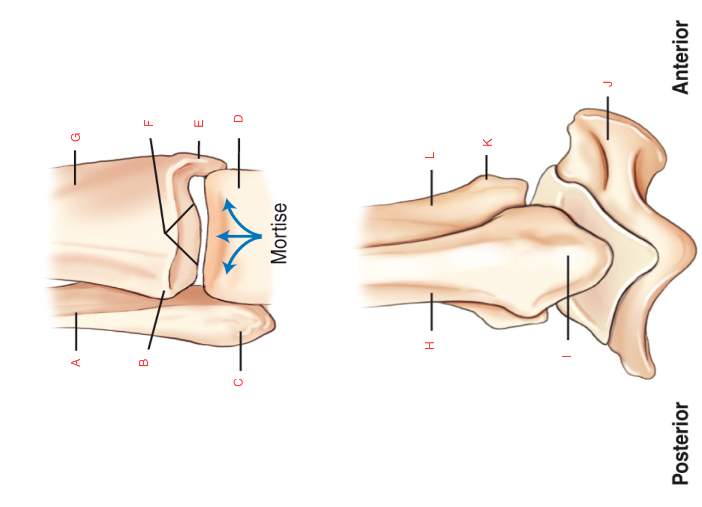
B
Anterior Tubercle
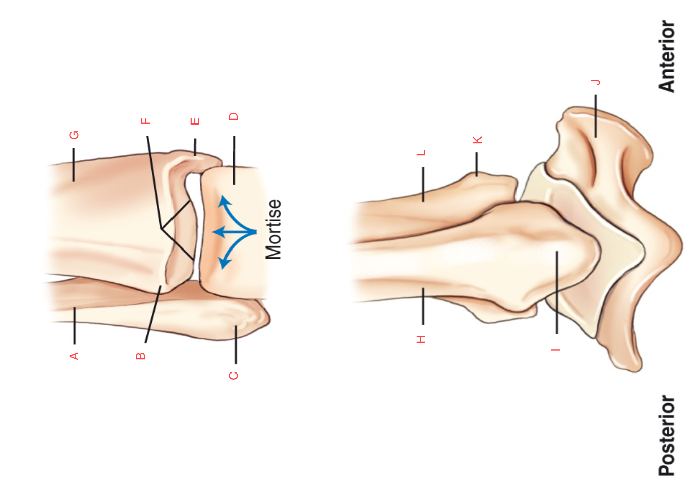
C
Lateral Malleolus
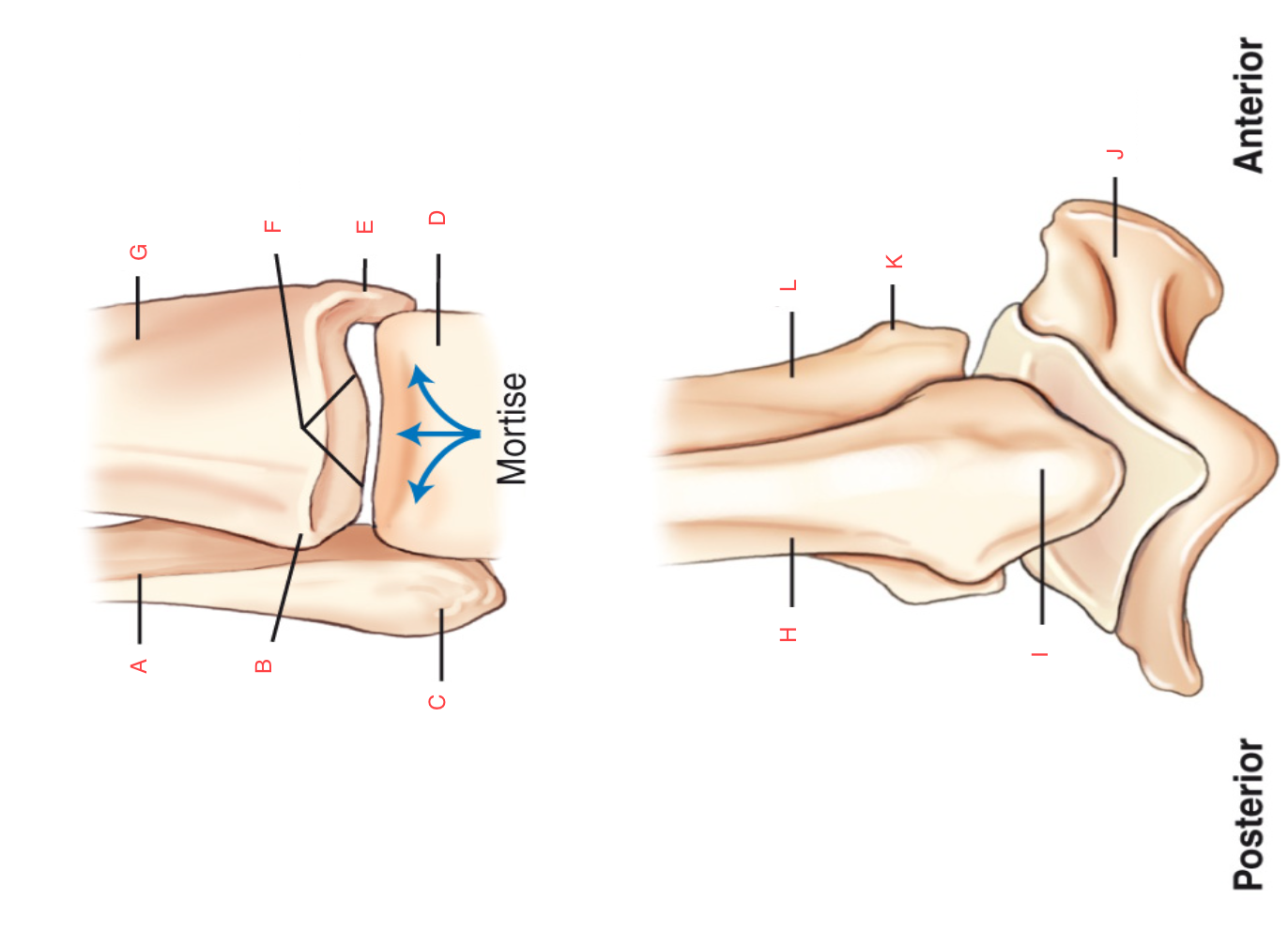
D
Talus

E
Medial Malleolus

F
Tibial plafond
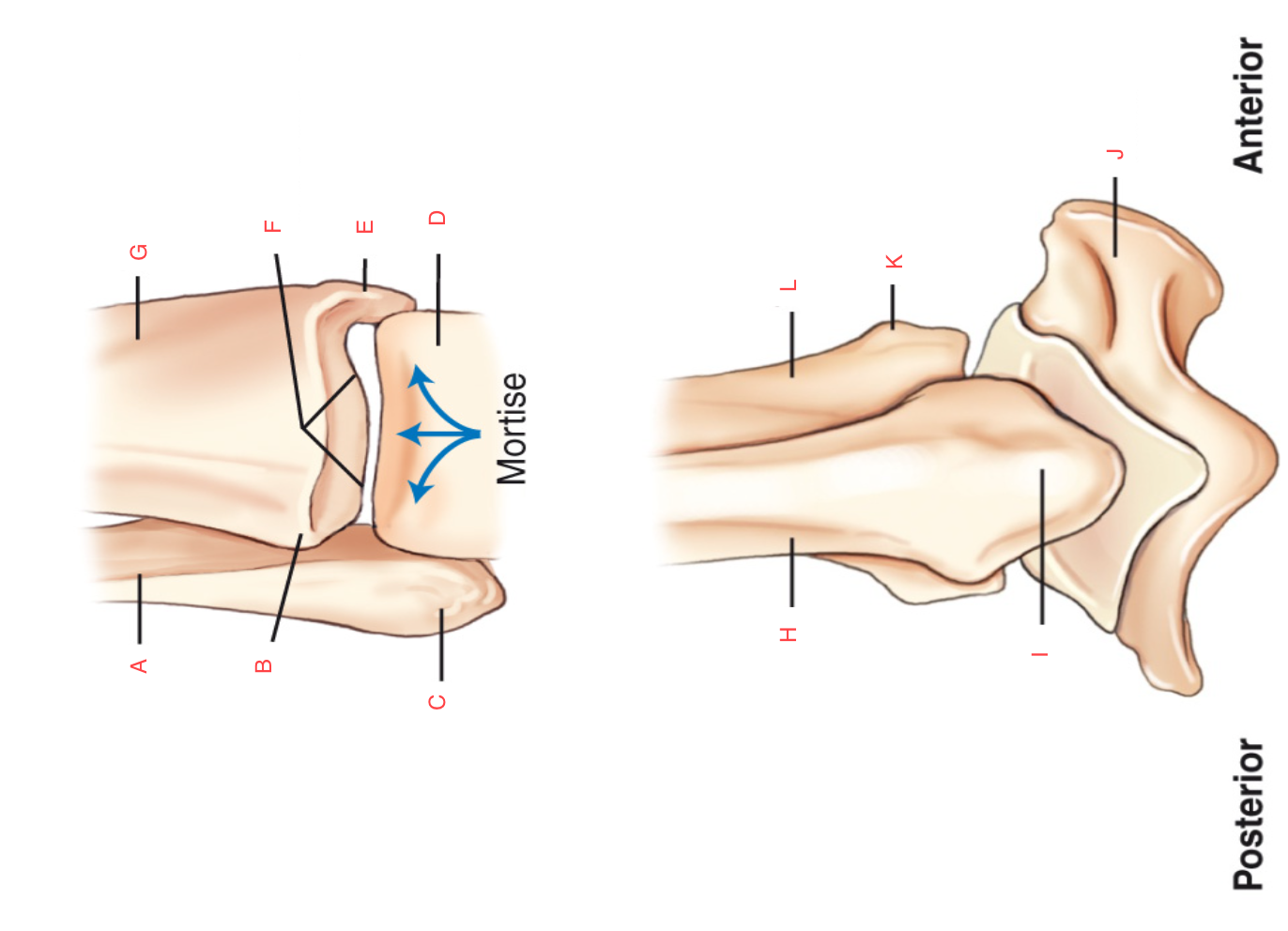
G
Tibia

H
Fibula

I
Lateral Malleolus
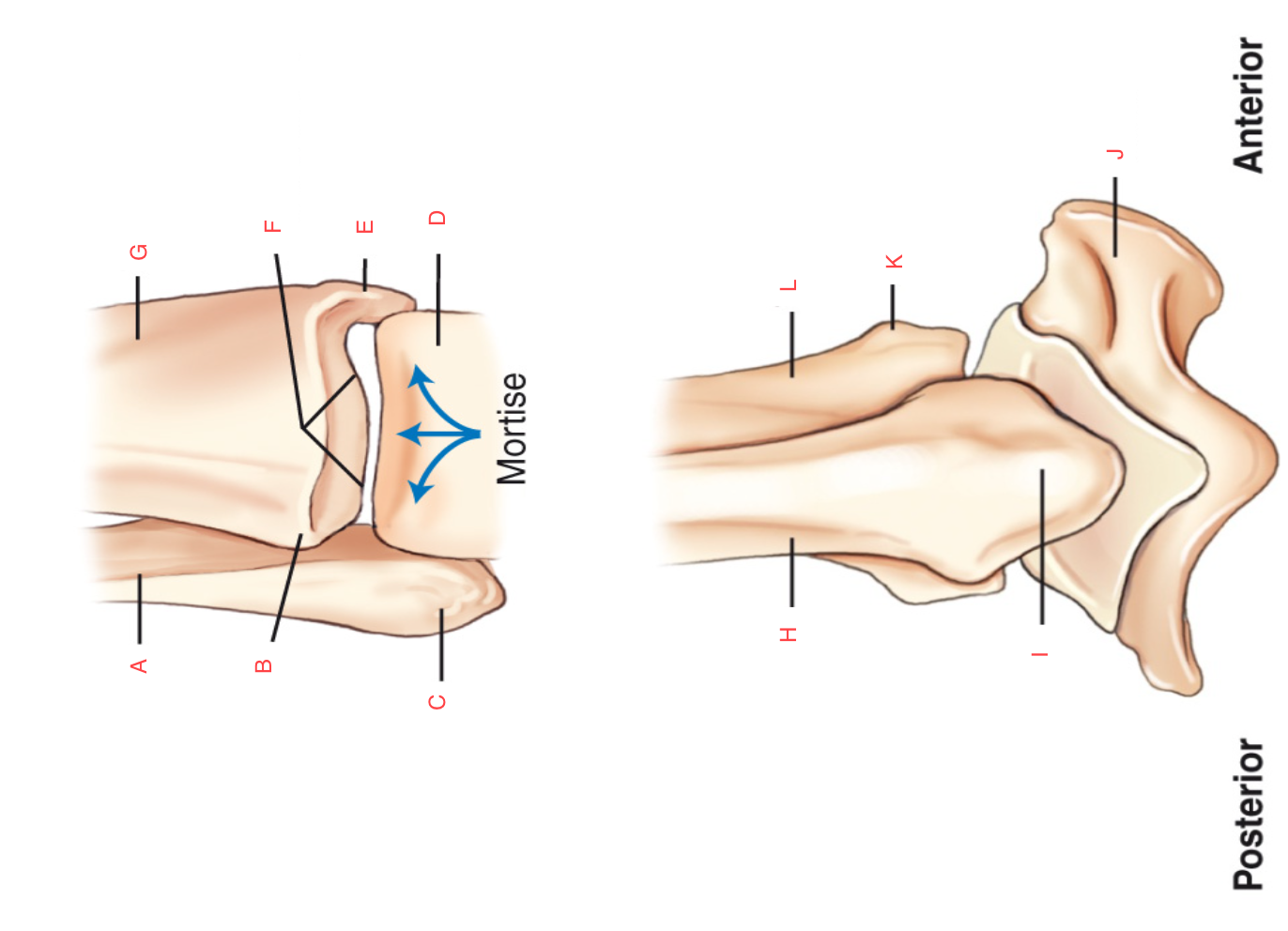
J
Talus

K
Anterior Tubercle

L
Tibia

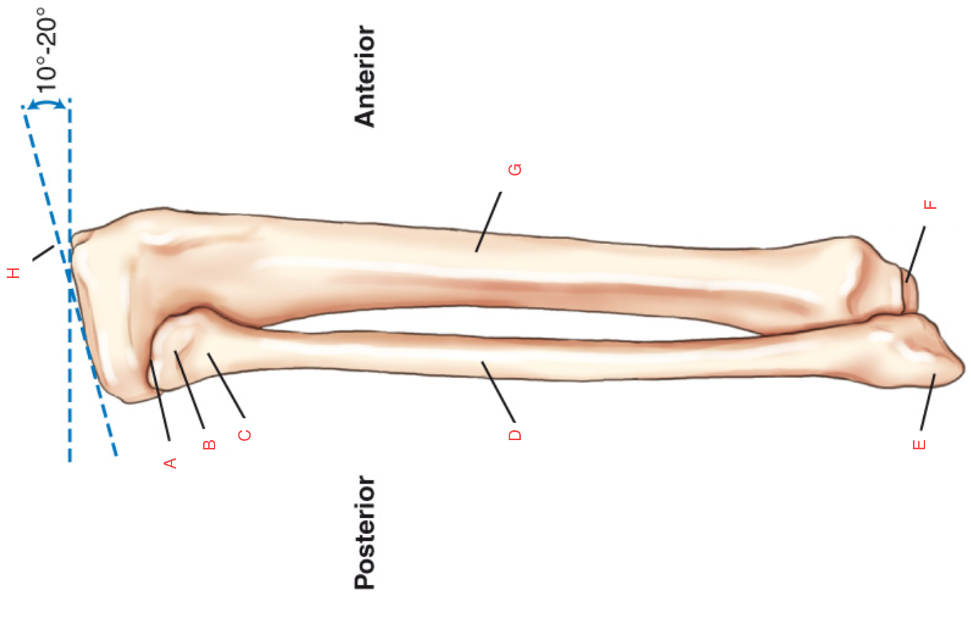
A
Lateral Condyle

B
Articular Facets (tibial plateau)

C
Proximal tibiofibular joint

D
Distal Tibiofibular joint

E
Fibular notch of the tibia

F
Medial Malleolus

G
Distal Extremity

H
Body (shaft) of the tibia

I
Anterior Crest

J
Proximal Extremity

K
Medial Condyle

L
Intercondyloid eminence (medial and lateral intercondyloid tubercles)
A
Apex

B
Head

C
Neck

D
Body (shaft) of Fibula

E
Lateral Malleolus

F
Medial Malleolus

G
Body (shaft) of Tibia
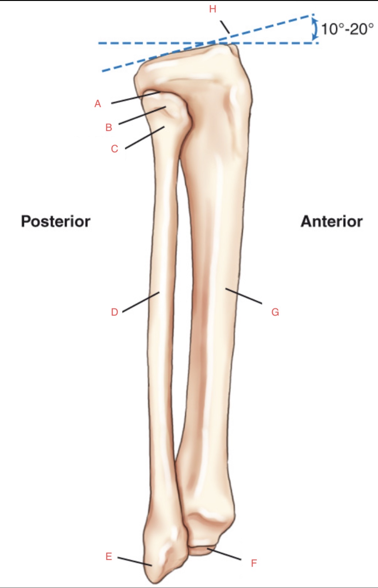
H
Tibial Plateau

A
Distal 1st Phalanx

B
Proximal 1st Phalanx

C
1st MTP joint

D
Medial Cuneiform

E
Intermediate Cuneiform

F
Navicular

G
Talus

H
Calcaneus

I
Cuboid

J
Base/Tuberosity of 5th
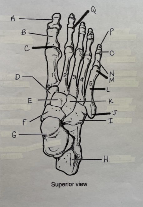
K
Lateral Cuneiform

L
Body of 5th Metatarsal
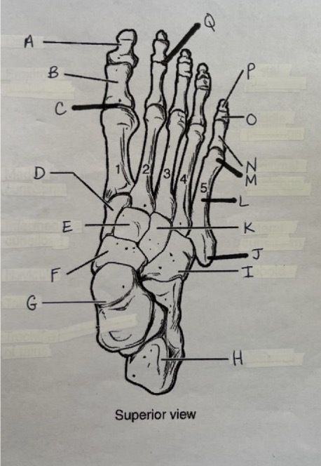
M
Head of 5th Metatarsal

N
Proximal 5th Phalanx

O
Middle 5th Phalanx

P
Distal 5th Phalanx

Q
2nd PIP joint

R
1st Metatarsal

S
Intermediate Cuneiform

T
Navicular

U
Talus

V
Sustentaculum Tali
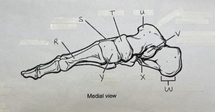
W
Tuberosity

X
Calcaneous

Y
Medial Cuneiform

Z
Navicular

AA
Intermediate Cuneiform

BB
Lateral Cuneiform

CC
Body of 5th Metatarsal

DD
Cuboid

EE
Calcaneous
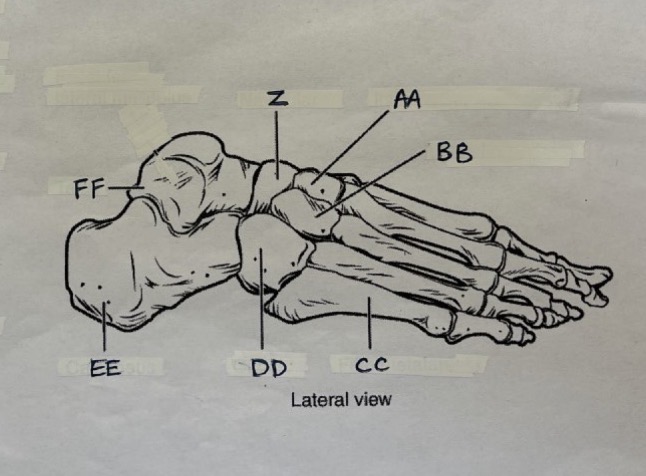
FF
Talus