heart, great vessels, & fetal circulation
 Call Kai
 Learn
 Practice Test
 Spaced Repetition
 Match
 Flashcards
 Knowt Play
1/59

Earn XP
Description and Tags
cwru biol346
60 Terms

highlighted blue
base of the heart

apex of the heart
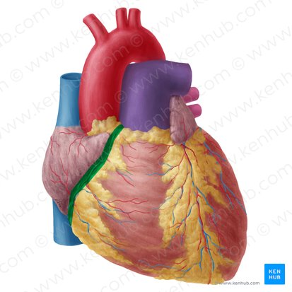
divides atra from ventricles, coronary arteries and coronary sinus run here
coronary sulcus
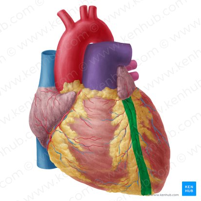
anterior interventricular groove

posterior interventricular groove

right atrium

left atrium

right ventricle

left ventricle

epicardium

myocardium

endocardium

pectinate muscles

what structure divides the atria?
interatrial septum

what is this structure called when it is open?
fossa ovalis

what is this structure called when it is closed?
foramen ovale

point to the tricuspid valve
(bottom right)

point to the mitral valve
(bottom left)

trabeculae carneae

papillary muscles

interventricular septum

point to the pulmonary semilunar valve
(topmost valve)

point to the aortic semilunar valve
(second topmost valve)

superior vena cava

inferior vena cava

pulmonary trunk

point to the pulmonary arteries
(splits from the pulmonary trunk, purple vessel)

pulmonary veins

aorta (specifically, arch of the aorta)
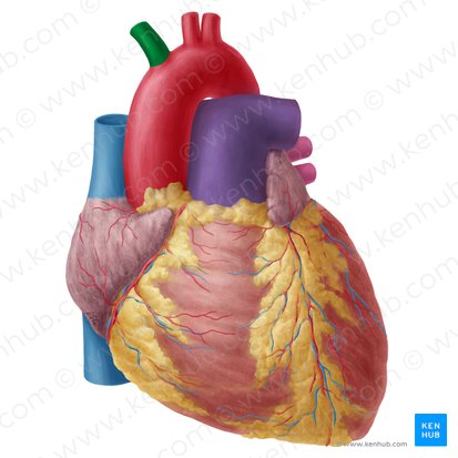
brachiocephalic trunk

ascending aorta

descending aorta

left common carotid artery

left subclavian artery

internal jugular vein

external jugular vein

subclavian arteries

subclavian veins

vertebral arteries

vertebral veins
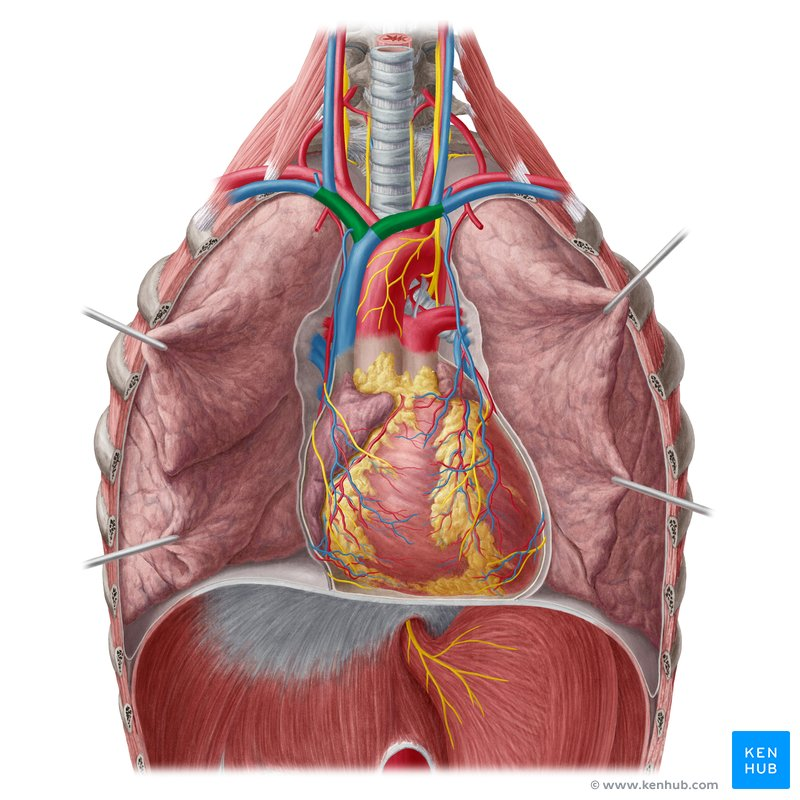
brachiocephalic vein

point to the left coronary artery
(see image labels)
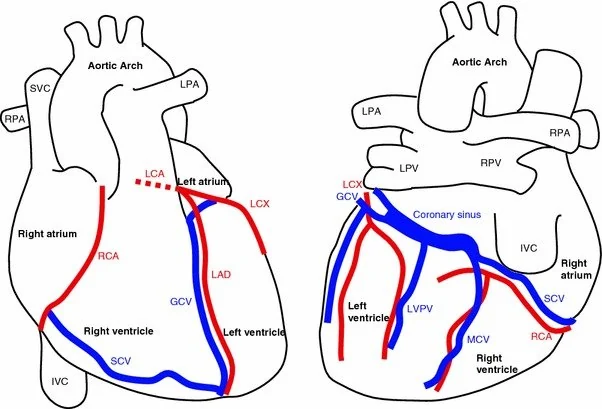
point to the right coronary artery
(see image labels)

point to the circumflex artery
(see image labels) (branches from the left coronary artery)

point to the left anterior descending artery
(see image labels) (branches from the left coronary artery)

point to the posterior descending artery
(see image labels) (branches from the right coronary artery)

point to the right marginal artery
(see image labels) (branches from the right coronary artery)
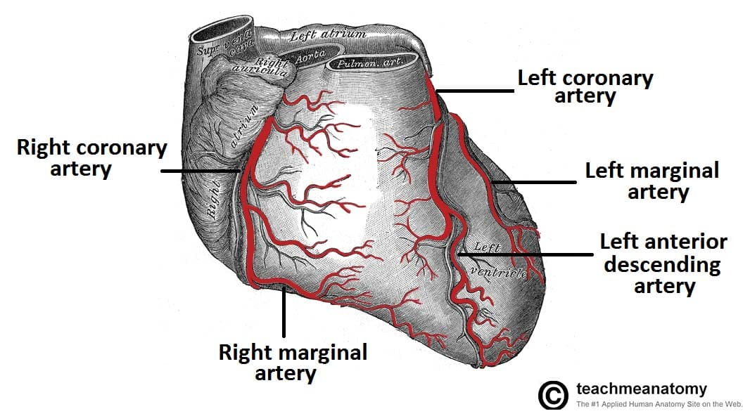
point to the left marginal artery
(see image labels) (branches from the circumflex artery)

point to the coronary sinus
(see image labels) (drains into the right atrium, follows the path of the circumflex loop on the posterior surface of the heart in the coronary sulcus)

point to the great cardiac vein
(see image labels) (parallels the anterior descending artery)

point to the small cardiac vein
(see image labels) (may or may not be present in some people)

point to the middle cardiac vein
(see image labels) (parallels the posterior descending artery)

anterior cardiac veins

what is the inner layer of this sac called?
visceral pericardium

what is the outer layer of this sac called?
parietal pericardium

what is this called when it is open?
ductus arteriosus

what is this called when it is closed?
ligamentum arteriosum

(the closed remnant of the umbilical vein)
ligamentum teres hepatis

the closed remnant of the distal portion of the umbilical artery
medial umbilical ligaments
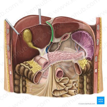
remnant of the ductus venosus
ligamentum venosum