10. Scleral Lenses - Advanced Contact Lenses Fall 2026
 Call Kai
 Learn
 Practice Test
 Spaced Repetition
 Match
 Flashcards
 Knowt Play
1/176

There's no tags or description
Looks like no tags are added yet.
177 Terms

What was the first material used for molded scleral lenses?
PMMA

What are the 3 zones of scleral lenses?
-Landing (haptic) zone
-Optic Zone
-Transition zone

Where does the landing (haptic) zone rest?
Lens rests on sclera

Where does the optic zone rest?
Vaults over the cornea
What are the indications for scleral lenses?
-Correction of corneal induced refractive error
-Regular corneal astigmatism
-Irregular corneal astigmatism
-Corneal Ectasia
What are the reasons for irregular corneal astigmatism that are indications for a scleral lens fit?
-Corneal scars
-Post penetrating keratoplasty
-Post surgical cornea
-Post refractive surgery
What are the corneal ectasias that are indications for a scleral lens fit?
-Keratoconus
-Pellucid Marginal Degeneration
-Keratoglobus
What are the advantages of a scleral lens vs a corneal lens?
-Improved patient comfort
-More stable vision
-Less chance of lens dislodge
-Easier fit
Why do scleral lenses have improved patient comfort over corneal lenses?
-Lens does not touch the cornea
-Sclera is tough with few vessels and nerves
Why do scleral lenses have more stable vision that patients with corneal lenses?
Minimal movement of the lens
Why are fitting sclerals an easier fit than a corneal lens?
Fitting sclera instead of irregular cornea
What are the disadvantages of a scleral lens vs a corneal lens?
-Large lens
-Need for non-preserved saline
-Fitting the sclera
-Requires a fit set
-Cost of lens more
What is the disadvantage to scleral lenses being larger lenses than corneal lenses?
Difficult insertion and removal methods
Because you need to fill sclerals with Non-Preserved saline, is there an advantage for dry eye patients?
Yes

Is the sclera spherical?
No -- and not symmetrical
Are sclerals easily emperically fit?
No -- requires a fit set and diagnostically fit
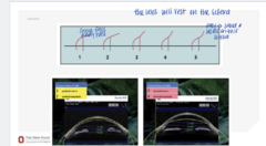
Curvature of the Sclera (Pic)
Curvature of the Sclera (Pic)
Traditional GP fitting is to match ______ of the lens to the curvature of the cornea
base curve
Where does a corneal GP rest?
on the cornea
How are corneal GP lenses fit?
Fit to achieve even distribution of lens resting on the cornea
Where do scleral lenses rest and what do they vault?
Scleral lenses rest on the sclera and completely vault the limbus and cornea
The base curve of a scleral lens should match what?
relatively match the curvature of the cornea

Scleral Lens on An Irregular Cornea (Pic)
Scleral Lens on An Irregular Cornea (Pic)

Scleral Lens on Cornea (Pic)
**vaulting over the cornea
Scleral Lens on Cornea (Pic)

Scleral Lens Shape (Pic)
Scleral Lens Shape (Pic)

With a larger chord length, you will get a (bigger/smaller) sag depth
bigger

Avg Sag Depth @ 15mm Chord (Pic)
**Largest = temporal
Avg Sag Depth @ 15mm Chord (Pic)

Scleral Angle by Eye and Location (Pic)
**Steepest = temporal
Scleral Angle by Eye and Location (Pic)

Corneal-Scleral Junction by Eye and Location (Pic)
**Steepest = temporal
Corneal-Scleral Junction by Eye and Location (Pic)

What is the function of the scleral topographer?
Image the cornea and the sclera

sMap 3D and Eaglet topographer will need ____ in order to image scleral lenses on the eye
NaFl

Does Pentacam need NaFl in order to image the cornea and sclera?
No

If this was the printout you recieved from the corneal topographer. That is the total sag depth that you should initially fit the patient in?
-2176.8um at 10mm chord
-Add 2000um for a 15mm chord
-Add 300um for vault
-Final sag depth is 4476.8um

How much will a scleral lens settle over a few hours?
100-120um

A 10um lens is 1652um on corneal topographer. You up the lens to 15mm. What is the new sag depth that you should select?
-Want to add 2000um of vault
-Approx 3600um for a 15mm chord

What is the goal of fitting a scleral lens?
-To fit the sclera to minimize conj blanching
-Central optics vault over the cornea
-Fitting height of the cornea from the conj

Where will a scleral lens typically decenter?
Temporally and inf
How should you select an initial scleral lens?
It depend -- usually based on depth/height of central cornea to sclera
You can use ____, ____ and/or ___ of the cornea to determine an initial lens selection of a scleral lens
-HVID
-K readings
-Sag depth

____% of the population has a spherical sclera
6

____% of the population has a regular toric sclera
20

____% of the population has a irregular toric sclera
64

What are the scleral imaging technologies that are frequently used?
-Eaglet
-Pentacam CSP
-Smap

What are the base curve options for scleral lenses?
-Spherical Curves
-Toric landing zone
-Zenlens Bioelevation (2 different sags)

What are the free-form scleral lenses that are used?
-Zenlens Echo
-Gaudi
-BostonSight Smart 360

What are free form scleral lenses? What is the benefit of free-form scleral lenses?
-Highly customized CLs designed using digital 3D surface mapping of the eye rather than relying on fitting sets.
-Can specify quad specific landing zone curves for irregular corneas
What are the changes to scleral lenses that can be made to better fit an irregular eye?
-Changes in mid peripheral curves to fit the cornea and limbus
-Changes in edge lift to achieve alignment on the sclera
-Toric edge curves available
-Fenestrations available
Are fenestrations generally recommended to be included in scleral lenses? What is the benefit of these fenestrations?
-Not recommended
-May make removal easier but risk bubbles
How to determine the lens power needed for a scleral lens fit?
With diagnostic lens on after the lens settles, perform a spherical OR (or SCOR if BCVA is reduced with sphere only)
True or False:
Amount of central vault and change in mid-periphery shape, tear reservoir (tear lens) can contribute significant power to a scleral lens system
true
Is the power of a lens needed difficult to calculate without an over refraction?
Yes -- there are compensation tools that can help
Why are sclerals able to be much less power than expected?
Able to vault over the cornea and have a lower base curve

How can you evaulate a scleral lens fit?
-View NaFl pattern with cobalt light and Wratten filter
-View with slit lamp optic section to observe lens surface, tear reservoir layer, and corneal thickness

With optic section looking at the lens surface, tear reservoir later, and corneal thickness, what are you able to judge and measure?
tear reservoir thickness

Scleral Lens NaFl Pattern (Pic)
Scleral Lens NaFl Pattern (Pic)

Evaluating the Fit of a Scleral Lens (Pic)
**Take an optic section and then estimate the tear reservoir in microns related to thickness of the scleral lens
Evaluating the Fit of a Scleral Lens (Pic)
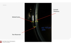
Corneal Thickness, Scleral Lens, Tear Reservoir (Pic)
Corneal Thickness, Scleral Lens, Tear Reservoir (Pic)
What should you observe and evaluate when looking at a scleral lens with optic section?
-Observe the central and limbal vault
-Evaluate conj impingement and blanching
-Observe the conj vasculature
Before you observe the tear meniscus or other conj vasculature, how long must the scleral lens settle on the eye?
10-15 minutes
Why must we wait 10-15min before evaluating a scleral lens?
-Central vault can decrease over time
-Conj blanching can increase over time

Scleral Lens Fit Scales (Pic)
Scleral Lens Fit Scales (Pic)

Tear Reservoir Thickness Over Time (Pic)
Tear Reservoir Thickness Over Time (Pic)
What is the perfect ideal vault for a scleral lens fit?
50um vault from limbus to apex to limbus
We usually select an initial lens with ______ of vault knowing it will settle to about ____um
300; 200
Limbal vault is usually around ____ initially and will settle to _____
150; 50
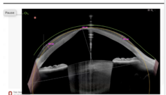
Significant Cone - Scleral Crashing into the Cornea and Excessive Vault in Periphery (Pic)
Significant Cone - Scleral Crashing into the Cornea and Excessive Vault in Periphery (Pic)

How would you try to improve the fit of this scleral lens?
-Increase the sag depth
-Steepen the BC to decrease vault in the mid periphery

Impingement (Pic)
Impingement (Pic)

Conj Compression and Vessel Impingement (Pic)
Conj Compression and Vessel Impingement (Pic)

Impingement Scale (Pic)
Impingement Scale (Pic)

How to prepare a scleral lens for insertion into the eye?
-Use large DMV plunger as a lens stand; cut off end so there is no suction
-Fill the bowl of the lens with PF saline

Will there be any tear exchange behind a scleral lens?
No

How much do you fill the scleral before inserting?
Fill to almost overflow to create the tear meniscus

For fitting purposes, what can also be put into the scleral lens bowl?
NaFl

Filling Scleral Lens (Pic)
Filling Scleral Lens (Pic)

How to insert a scleral lens?
-Place a mirror over counter top or horizontal surface
-Patient bends over and stands with face horizontal
-Hold lids open with one hand
-Look straight down at the center of the lens
-Bring filled lens straight up and place on eye with the other hand
-Move the plunger away

_____ will keep a scleral lens on the eye
Capillary forces

Inserting the Lens (Pic)
Inserting the Lens (Pic)

Inserting the Lens (Pic)
Inserting the Lens (Pic)
With a proper fitting lens, bubbles are most likely d/t what?
improper lens insertion
If a bubble shows up in the scleral lens bowl, what should the patient do?
Remove the lens, refill with saline, and reinsert into the eye
Bubbles can cause what?
immediate discomfort or mild irritation

Scleral Lens Care and Handling (Pic)
Scleral Lens Care and Handling (Pic)

How to insert a scleral lens?
Lens application; DMV lens inserter

How to remove a scleral lens?
-Use the SMALL suction cup
-Place suction cup at the edge of the lens
-Lift off the edge of the lens to break lens suction to the eye
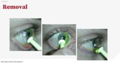
Should you put the small DMV in the center of the scleral to remove the lens from the eye?
No -- put in at the edge of the lens
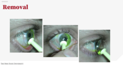
Removal of Scleral Lens (Pic)
Removal of Scleral Lens (Pic)

What are the PF options for saline that can fill a scleral lens bowl?
-ScleralFil B + L
-LacriPure Menicon
-Nutrifill Contamac
-Purilens
-Addipak

iPrint Pro (Pic)
iPrint Pro (Pic)

No Limbal Vault with NaFl (Pic)
No Limbal Vault with NaFl (Pic)

Limbal Vault with NaFl (Pic)
Limbal Vault with NaFl (Pic)

You will need to get a _____ or use a ______ for vault depth
OCT; optic section

Landing Zone Aligned (Pic)
Landing Zone Aligned (Pic)

Heel Down Compression (Pic)
Heel Down Compression (Pic)
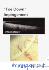
Toe Down Impingment (Pic)
Toe Down Impingment (Pic)

Heel Down Compression (Pic)
Heel Down Compression (Pic)

Toe Down Impingment (Pic)
Toe Down Impingment (Pic)

Landing Zone Aligned (Pic)
Landing Zone Aligned (Pic)

You should assess landing zone in ____ gaze
primary

Why not assess landing zone in downgaze?
Looking down encourages the lens to move in opposite direction and can give false positive of impingement