Dentistry Midterm Study Guide
 Call Kai
 Learn
Learn Practice Test
 Spaced Repetition
 Match
Match Flashcards
 Knowt Play
1/87

There's no tags or description
Looks like no tags are added yet.
Name | Mastery | Learn | Test | Matching | Spaced | Call with Kai | Chat |
|---|
No analytics yet
Send a link to your students to track their progress
88 Terms

Which head shape is this?
dolichocephalic

Which head shape is this?
mesocephalic

Which head shape is this?
brachycephalic

name the teeth
molars
premolars
canines
incisors
canines
incisors
premolars
molars
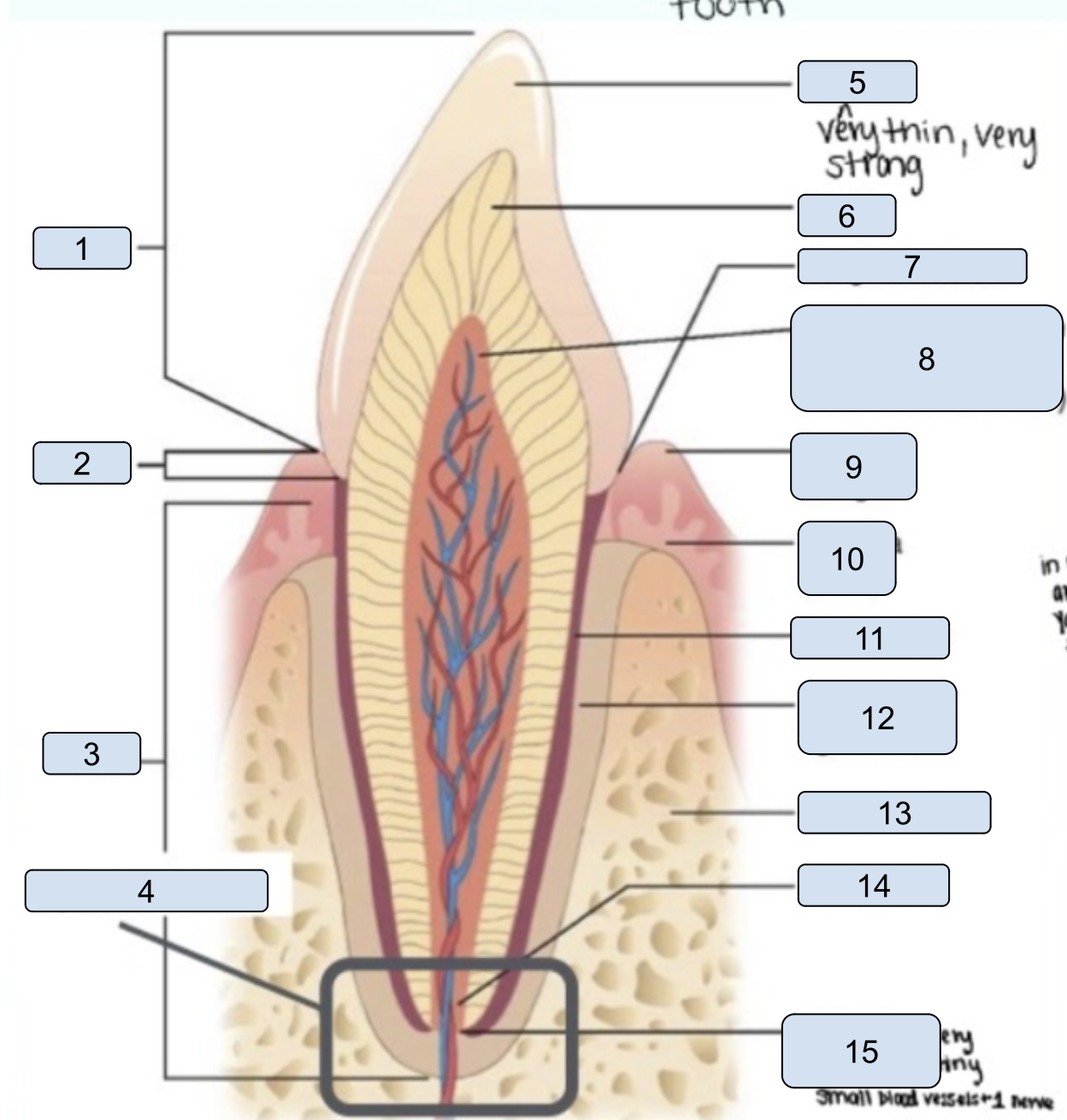
name the parts of the tooth
crown
neck
root
apex of root
enamel
dentin
gingival sulcus
pulp cavity
gingival margin
gingiva
cementum
periodontal ligament
alveolar bone
root canal
apical foramen
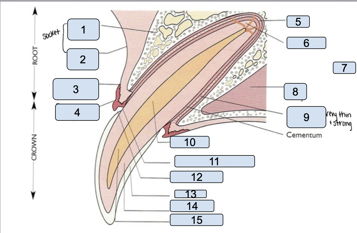
name the parts of the tooth
alveolar bone
alveolar mucosa
mucogingival junction
free gingival groove
apex
apical delta
nasal cavity
periodontal ligament
cementum
root canal
Cementoenamel junction
Gingival sulcus
Enamel
Dentin
Pulp chamber

name the directional terms
apical
coronal
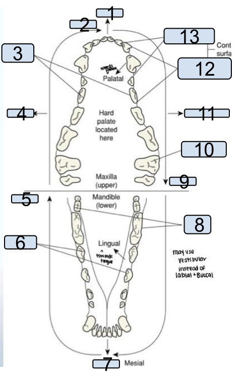
name the directional terms
labial
mesial
palatal surface
buccal
distal
lingual surface
labial
buccal surface
distal
occlusal surface
buccal
distal surface
medial surface
practice triadan and anatomic shorthand systems

name of this occlusion and description
-class I malocclusion (Neutroclusion)
-Jaw lengths are normal, 1+ teeth in abnormal position

name of this occlusion and description
-class II malocclusion (mandibular distortionclussion)
-“overbite”, “parrot mouth”
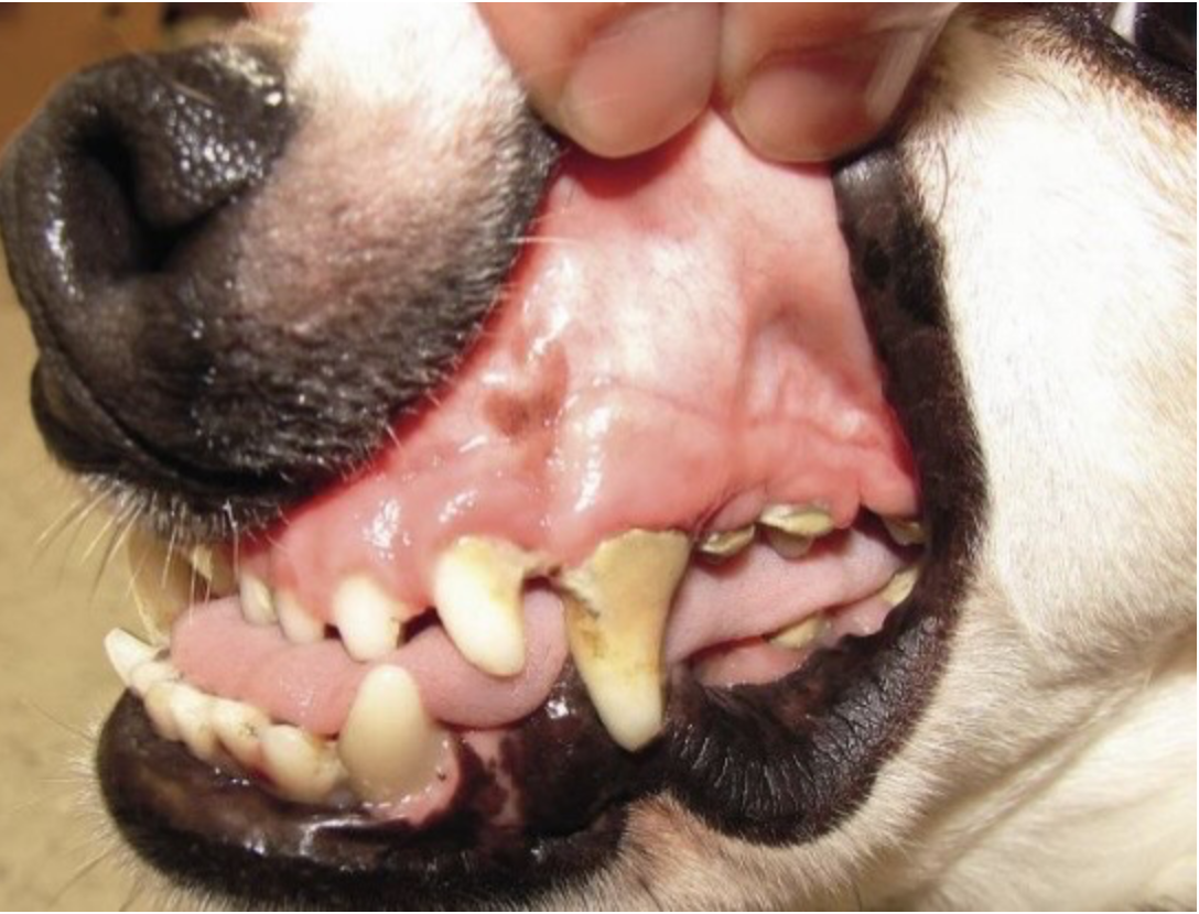
name of this occlusion and description
-malocclusion III (mandibular malocclusion)
-“underbite”, “monkey mouth”, considered “normal” in brachycephalics
What are retained deciduous teeth, and why are they a problem? What is the recommended treatment?
-malocclusion of surrounding teeth
-periodontal dx(due to debris caught between adult+primary)
-Usually extracted (interceptive orthodontics: extracting primary teeth to prevent malocclusions)
What are supernumerary teeth, and why might they be a problem?
-”Extra” adult teeth
-may cause issues similar to resistant primary teeth

Identify this pathology and explain its etiology
-Enamel hypoplasia
-Genetic & severe fever/infection during development
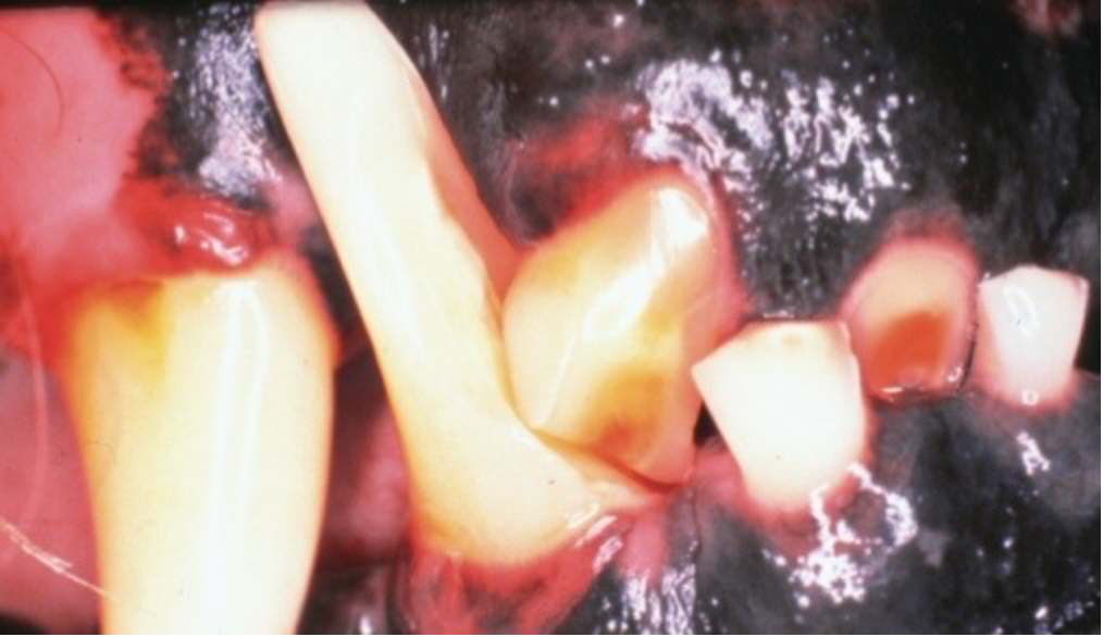
Identify this pathology and explain its etiology
-Attrition
-Wear of a tooth due to chronic abnormal contact with another tooth

Identify this pathology and explain its etiology
abrasion
-Wear of a tooth due to chronic contact with an object (chronically chewing on something; damage, common in dogs)
How is pulp exposure differentiated from tertiary dentin?
pulp exposure: Rapid wear results, explorer “falls” into pulp chamber, tx: extraction/root canal
tertiary(aka reparative) dentin: slow wear results, surface feels smooth as glass

What is this?
pulp exposure

What is this?
tertiary(aka reparative) dentin

Identify instrument and explain how it’s properly used
-explorer: sharp instruments used to detect plaque and calculus
-Used to explore cavities/check for exposed pulp chambers

Identify instrument and explain how it’s properly used
-periodontal probe: Used to measure the depth of the gingival sulcus/periodontal pocket
-Can also measure oral lesions of a diameter less than a centimeter

Identify this pathology and list how its typically treated
-caries:Aka cavities, breakdown of enamel +dentin by acidic bacterial waste, seen in dogs
-Tx:removal of decayed tooth, then then restoration (filling)

Identify this pathology and list how its typically treated
-Enamel Infraction(EI): incomplete fracture (crack)
-loss of tooth tissue

Identify this pathology and list how its typically treated
-Enamel Fracture(EF): Fracture with loss of crown substance, enamel only
-tx: filling

Identify this pathology and list how its typically treated
-Uncomplicated Crown Fracture(UCF): Fracture of crown only, pulp exposure
-tx: cap/filling

Identify this pathology and list how its typically treated
-Complicated Crown Fracture(CCF): fracture of crown only, pulp is exposed
-tx: extraction/root canal

Identify this pathology and list how its typically treated
-Uncomplicated Crown-Root Fracture(UCRF):Fracture of crown and root, pulp exposure
-tx: depends on how far tx extends subgingingivally-filling/root canal/extraction

Identify this pathology and list how its typically treated
-Complicated Crown-Root Fracture: Fracture of crown and root, pulp is exposed
-tx: extraction/root canal

Identify this pathology and list how its typically treated
-Root Fracture: Fracture of the root only
-tx: may heal on its own or extraction

Identify this pathology and list how its typically treated
-oronasal fistula: Communication between oral/nasal cavities, severe, chronic periodontal disease, sneezing when eating/drinking often observed
-tx: surgerical closure
How is the etiology of tooth discoloration determined?
Shine a light to the teeth to see if it is vital
What is Plaque and how is it removed?
-soft biofilm +removable w/brushing
-brushing, ultrasonic scalers and hand scalers

What is this and how is it removed?
-calculus: hardened, mineralized plaque that accumulates on the teeth
-tartar-removing forceps and ultrasonic scaling
What are the clinical signs of periodontal disease? How does it progress?
Renal, hepatic+cardiac dx, increased inflammatory markers, anemia of chronic dx, arthritis, diabetes melitius, untreated leads to periodontitis
gingivitis
-The 1st observable sign of periodontal dx
-stage 1 reversible w/brushing+home care
-Untreated leads to periodontitis
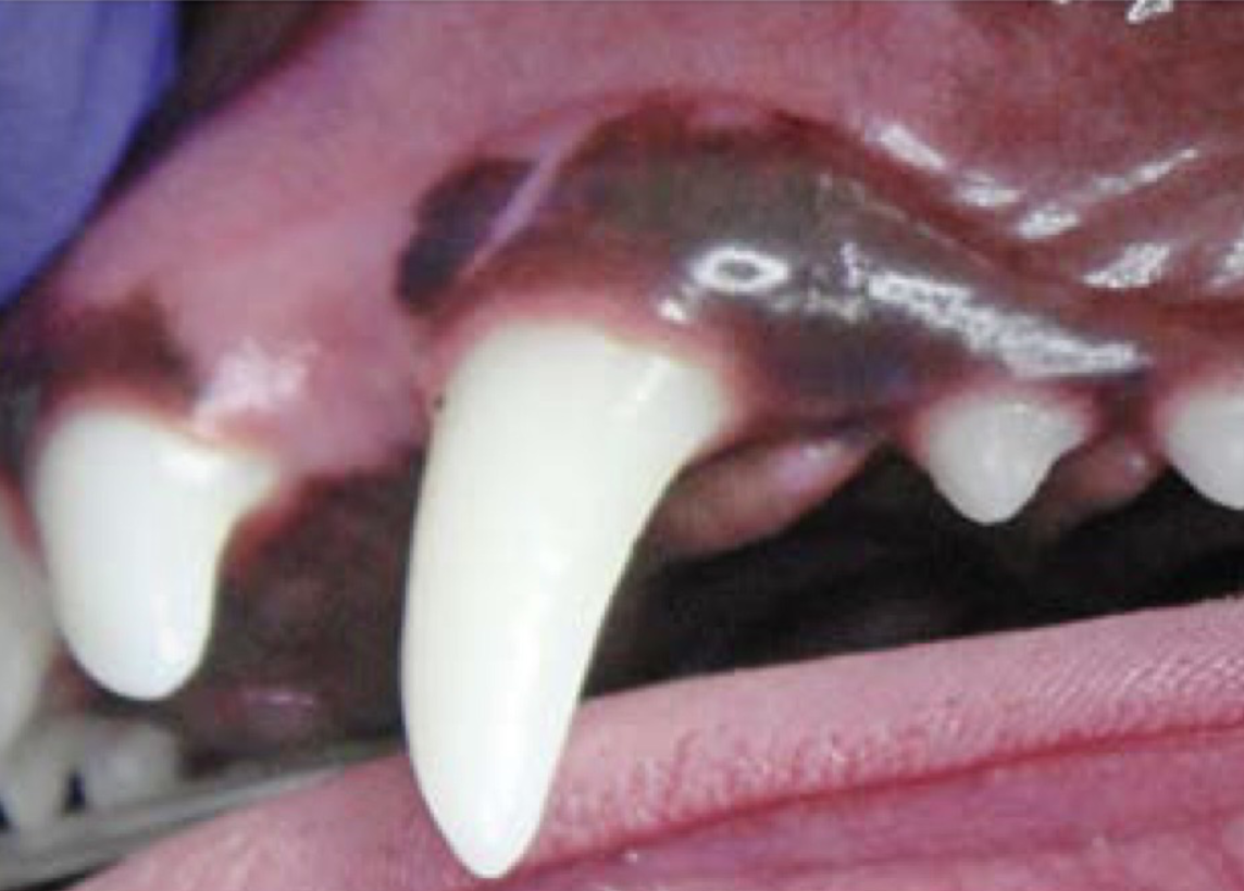
Gingival Index Grading: none
Grade 0

Gingival Index Grading: slight erythema/edema, bleeding
Grade 1

Gingival Index Grading: moderate erythema/edema, bleeding w/probing
Grade 2

Gingival Index Grading: severe erythema/edema, spontaneous bleeding, possible ulcers
Grade 3
Calculus Index grading: none
Grade 0
Calculus Index grading: thin band at free gingival margin only
grade 1
Calculus Index grading: 1/3 – 2/3 of crown is covered
grade 2
Calculus Index grading: >2/3 of crown is covered or bridging calculus is present
grade 3
mobility scoring: ≤ 0.2 mm
stage 0
mobility scoring: 0.2 – 0.5 mm
stage 1
mobility scoring: 0.5 – 1.0 mm
stage 2
mobility scoring: >1.0 mm, or any movement axially
stage 3
Furcation exposure index: < halfway under crown
stage 1
Furcation exposure index: > halfway but not through
stage 2
Furcation exposure index: all the way through
stage 3
Periodontal disease: Normal, no gingivitis, no signs of periodontitis observed
Stage 0
Periodontal disease: gingivitis only, no attachment loss, reversible with home care!
Stage 1 (PD1)
Periodontal disease: Early Periodontitis, <25% attachment loss, stage 1 furcation exposure, further destruction can be stopped only with professional periodontal cleaning under anesthesia)
Stage 2 (PD2)
Periodontal disease: Moderate Periodontitis, 25 – 50% attachment loss, stage 2 furcation exposure, treatment: extraction and advanced procedures by veterinary dentist (e.g., root canal)
Stage 3 (PD3)
Periodontal disease:: Advanced Periodontitis, >50% attachment loss, stage 3 furcation exposure, treatment – extraction
Stage 4 (PD4)

starts as a periodontal pocket
gingival recession/root exposure
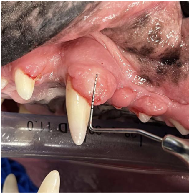
What is this? (also, what sequelae can occur? How is it treated?)
-gingival hyperplasia
-benign overgrowth of gingival tissue
-periodontitis
-surgically removed by DVM (gingivectomy)
How is periodontal pocket depth assessed?
“Walk” probe around tooth with short-up-and-down strokes every few mm
What are the normal pocket depth values for canines and felines?
-Dogs less than 3 mm
-Cats less than 1 mm

name and use
-calculus removal forceps
-Used for large chunks of calculus

name and use
-Ultrasonic Scalers
-convert electric/pneumatic energy to vibration to remove plaque and calculus

name and use
-beaver tail tip:
-for supragingival calculus

name and use
-periodontal tip
-for subgingival calculus

name and use
-furcation tip
-for calculus in furcations or between teeth

name and use
-hand scaler
-supragingival use only, may use in crevices

name and use
-curette
-Subgingival calculus removal, root planning, subgingival curettage
steps for instrument care
clean and disinfect
check sharpness and sharpen PRN
sterilize
How is instrument sharpness assessed? How are instruments properly sharpened? What tools/supplies are needed?
-Lightly drawing the instrument working-end along the plastic stick causes the blade to "bite" or grab the surface
-Drop of oil on stone, then wiped off, stone held between thumb + 1st finger(stoned moved back and forth), instrument placed at 90 degrees to stone, then rotated outward 10-15 degrees
-stone, oil
Why are teeth polished after scaling?
Decreases areas of plaque trapping + stagnation
What PPE must be used when performing a professional dental cleaning?
Gloves, mask, face shield/goggles,lab coat/old surgery gown/change of scrub top
What risks are associated with using mouth gags?
they can cause jaw trauma
How is ultrasonic cleaner w/ beaver tail tip used properly?
-Use the side of the beaver-tail tip, keeping it nearly parallel to the tooth surface.
-Use light pressure and short, overlapping strokes while keeping the tip moving
How is ultrasonic cleaner w/ periodontal tip used properly?
-Hold the tip nearly parallel to the tooth/root surface
-Use very light pressure and short, overlapping strokes, keeping the tip moving
How is polisher and paste used properly?
-Use a rubber prophy cup with veterinary dental polishing paste.
-Keep the cup moving over all tooth surfaces.
-Use light pressure and low speed to prevent heat buildup.
-Avoid holding the cup in one spot or polishing too long on one tooth.
-Rinse away the remaining paste when finished.
Why is water flow important when using ultrasonic scalers?
-Avoids overheating the tooth (thermal pulpitis)
-rinses away removed calculus/plaque
What is the purpose of fluoride treatment?
-slow reattachment of plaque
-steps: dry tooth surfaces w/gauze
-apply to all surfaces
-wipe/dry with compressed air(rinse)
the dental x-ray unit and discuss its unique qualities.
-Small focal spot: Produces detailed images of small tooth structures.
-Long, narrow cone (PID): Helps direct the X-ray beam toward the tooth.
-Low radiation output: Designed for imaging small areas, reducing unnecessary radiation exposure.
-Adjustable angulation: Allows images of different teeth and roots.
-Intraoral imaging: The sensor or film is placed inside the animal’s mouth for detailed views.
safe and proper use of the intraoral dental x-ray unit.
Position the patient: Place the animal under appropriate anesthesia and position the mouth for the desired view.
Position the sensor/film: Place it inside the mouth near the tooth being examined.
Align the X-ray beam: Direct the beam at the correct angle to capture the tooth, root, and surrounding bone.
Use proper exposure settings: Select the appropriate time and settings for the patient and sensor.
Follow radiation safety: Wear lead protection and a thyroid shield as required by protocol, use dosimetry badges, and stand behind a protective barrier or at least 6 feet away and out of the primary beam.
Never hold the sensor or animal by hand during exposure: Use positioning devices and appropriate restraint.
Check image quality: Ensure the entire tooth, including the root apex, is visible

what angle is this and how to use it
-Parallel technique
-Indicated to evaluate Mandibular molars of the dog+ mandibular premolars of the cat
sensor/plate is placed parallel to structure to be radiographed+between tongue/mandible
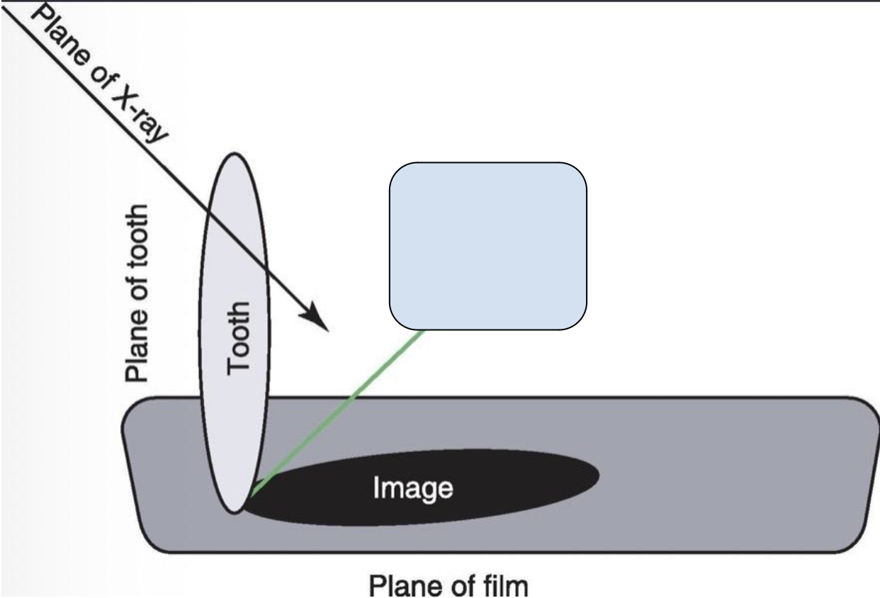
what angle is this and how to use it
-Bisecting angle:
-In areas where the parallel technique cannot be
What is the purpose of using Doxirobe? How is it applied?
-Used for deep Periodontal pockets
1)insert into cleaned/prepared pocket
2)application of water to harden gel
3)use of plastic instrument to pack gel into pocket
What is the purpose of using Clindoral? How is it applied?
-used for deep Periodontal pockets
1)completely dry all crowns with air syringe/gauze
2)Gently place cannula @ base of PP
3)Gently/slowly put small amounts into each pocket to fill gingival margin
4)allow to thicken, will not completely harden, but remain a gel matrix
What is the purpose of using Oravet? How is it applied?
-indivisible barrier to prevent bacteria from sticking to surface of teeth
1)Allow teeth to dry
2)Apply along gum line using applicator gun/prophy cup/finger/swab
3)spread sealant into sulcus/interdental spaces
What is the purpose of using Sanos? How is it applied?
-seals gingival sulcus against formation of plaque+promotes aerobic environment
1)applied after professional dental cleaning, dry teeth preferred (paint brush application)
FCGS
Etiology:
Whats happening to the patient:
Treatment of choice:
-feline chronic gingivostomatitis
-immune system becomes overly reactive to plaque
-oral inflammation beyond gingivitis and periodontitis
-extractions of premolars and molars, full mouth extractions if it involves all teeth, analgesia, soft food prior to SX
Tooth Resorption
What is the etiology?
What is the pathogenesis?
What are the 2 types and the treatment of choice for each?
-Loss of destruction of dental tissues
-the abnormal recruitment and activation of multinucleated clastic cells
-type 1: pulp and periodontal ligament intact, visible on rads (extraction), type 2: pulp and/or periodontal ligament not identifiable on rads (crown amputation)
oral neoplasia
Which are aggressive:
Which aren’t aggressive:
Which are metastatic:
Recommended treatment for aggressive neoplasia:
-oral malignant melanoma, squamous cell carcinoma, fibrosarcoma
-gingival hyperplasia, canine oral viral papilloma, granuloma
-oral malignant melanoma, squamous cell carcinoma, and fibrosarcoma
-surgical resection of the mass, temporary feeding tube