General Pathology
 Call Kai
 Learn
Learn Practice Test
 Spaced Repetition
 Match
Match Flashcards
 Knowt Play
1/114

There's no tags or description
Looks like no tags are added yet.
Name | Mastery | Learn | Test | Matching | Spaced | Call with Kai | Chat |
|---|
No analytics yet
Send a link to your students to track their progress
115 Terms
9 reasons to perform a PM exam
Determine cause of death
Compare with clinical diagnosis
Increase diagnostic accuracy
Assess concurrent disease and management problems for loss of production
Efficacy of medical/surgical therapy
Efficacy and toxicity of therapeutic agents
Educate
Obtain forensic or legal information
ID emerging diseases, zoonoses and disease surveillance
When should PM be conducted? What should be done if delay is necessary? Why?
ASAP after death
Chill the carcass to slow the rate of autolysis
Freezing and thawing causes tissue damage leading to difficult histological interpretation
Steps of performing a PM examination (11)
External examination
Reflect front and back limb and abdominal skin
ID last rib and cut into abdominal cavity
Cut open thoracic cavity with bone cutters caudal to cranial
Remove pluck and examine: Trachea, oesophagus, lungs, thyroid glands, heart
Oral cavity
Abdominal cavity examination: Liver, gall bladder (squeeze to confirm patency), intestines, spleen, stomach
Sample intestine (tie off intestine with two pieces of string -bonbon)
Urinary system: Adrenal glands, kidneys (cut surface), open bladder
Joints: Palpate and incise to assess joint fluid
Remove head (for brain examination)
3 types of sampling (+ handling requirements)
Histology = Fix in formalin (10%) to prevent autolysis
Samples ≤1cm thick
10 parts formalin : 1 part tissue
Whole brain → Large bucket of formalin (2 - 4d to fix)
Fix in-clinic, then transport in smaller container with just enough formalin to labeeeeeee
Use leak-proof screw top containers with wide necks
Can place multiple samples into SAME pottle
Formalin is hazardous (irritant gas affecting eyes and nose) → Make up in fume hood with mask
Microbiology, PCR, toxicology = Aseptic technique placed into sterile pottle/bag/swab
Parasitology
Whole parasites submitted in 70% alcohol
Faecal sample (≥5gm) for FEC
Whole abomasal contents + ≥10m small intestine for parasite counts → Mix with water to form aliquots for counting
7 details for sample submission form
Practice name, address and vet name
Owner's name
Animal ID and signalment
Date
Relevant clinical history
Nature and site of sample (diagram for histology and cytology)
Tests required
Describe 10 components of gross lesion description (+ examples)
Location = Tissue/organ, side of animal and which part of the organ
Arrangement = Grouping/distribution of lesion, scattered randomly? Symmetrical?
Multifocal vs. diffuse vs. coalesce
Number = Single vs. multiple
Miliary = Too many to count
Size
3D estimate in mm
Compare with common reference item
% of tissue involved in lesion
Shape
Raised/bulging/nodular vs. depressed/flat
Well- vs. poorly-demarcated
Colour
Consistency = Compare to normal
Lesion = Rubbery, firm, spongy, semi-solid, pasty, fluid-like, watery etc.
Pus = Gritty, dry, cheesy
Fluid = Smooth, greasy
Organ surface = Wrinkled, smooth, moist and glistening, dry and leathery, pitted, dull, opaque
Cut surface
Content = Describe normal for pregnant female, GIT and urinary bladder
Amount, nature, appearance, parasites, foreign bodies
Odour
Black leg = Rancid butter smell
Ketosis = Acetone breath
Not ALL may be applicable
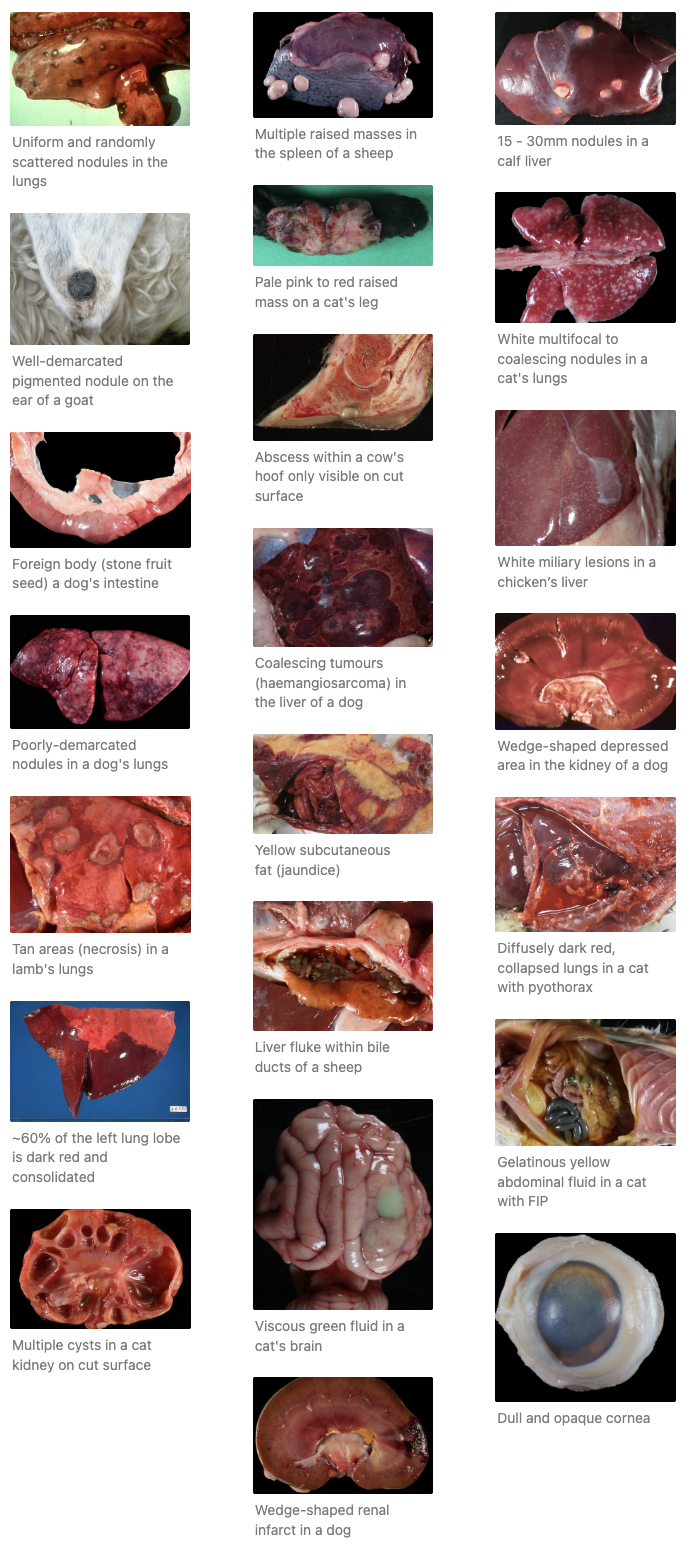
EXAMPLES: Provide a description and interpretation for the following images
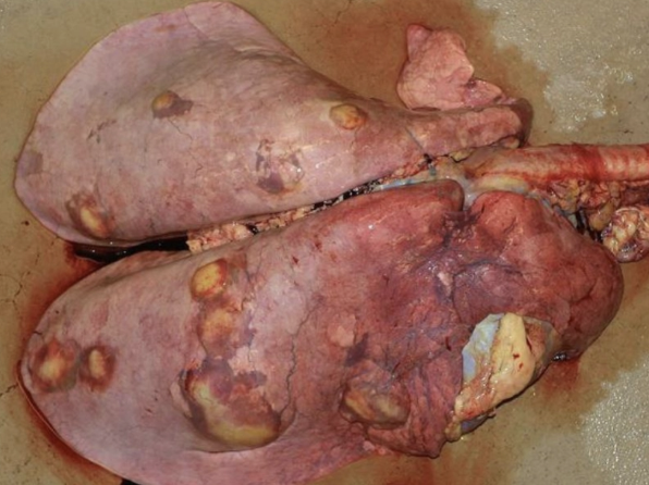

Description (LUNGS): Within the lung parenchyma and affected all lung lobes, there are multifocal to coalescing, variably sized (8mm - 5cm), pale yellow nodules, that on cut surface exude and thick, viscous, yellow fluid
Interpretation: Embolic pneumonia
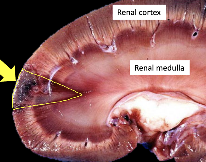
Description (KIDNEY): Within the renal parenchyma, there is a focal, triangular-shaped, well-demarcated, dark-red to brown area, that measures ~1 × 2cm and extends from the mid-medulla to the cortex
Interpretation: Renal infarct
Description (FEMUR): The marrow cavity is filled with a clear, pale yellow gelatinous material
Interpretation: Serous atrophy of fat
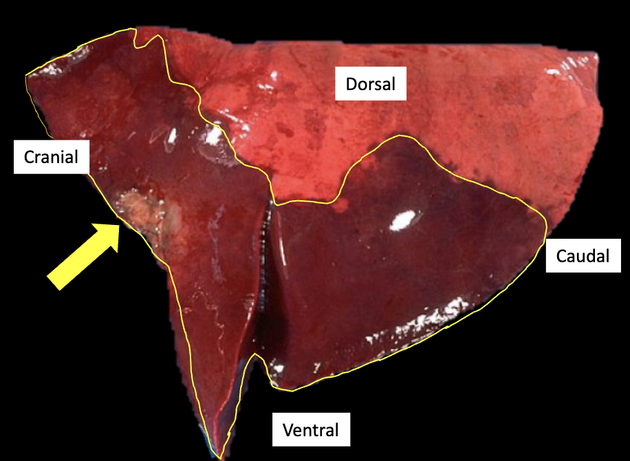
Description (LUNG): ~80% of the cranioventral portion of the left lung lobe and the ventral aspect of the caudal lung lobe are diffusely dark red to brown and firm. There is a single, focal 20 × 15mm dull grey, poorly-circumscribed area in the middle of the leading edge of the cranial lung lobe
Interpretation: Bronchopneumonia with focal abscessation
4 recommendations when photographing lesions
Good light to ensure image is clear and in focus
No glare over lesion in photo
Lesion in centre of photo
Include a scale (eg. ruler)
Death and Decay
Define somatic death
What is the order of cellular death (by organ)
Somatic Death: Disappearance of life from the entire individual (cardiac and respiratory function does NOT occur spontaneously)
Not all cells in the body die at the same time (eventually due to anoxia)
Cellular Death Order: First → Last
Brain and heart cells
Kidneys, lungs and liver cells
Mesenchymal cells (bone, connective tissue, cartilage)
List and describe 4 PM changes (+ examples)
ONE: ALGOR MORTIS
Definition: Cooling of body until it reaches ambient temperature
Depends on initial body temperature (eg. fever), insulation (fat/wool) and ambient temperature
Used to calculate range for time of death
TWO: RIGOR MORTIS
Definition: Stiffening of muscle that occurs after death
MoA: ATP is required to uncouple actin-myosin complexes. When ATP runs out after death, muscle fibres cannot relax and remain contracted
Onset of rigor mortis depends on glycogen stores (higher BCS → delayed rigor mortis) and ambient temperature (higher temperature → faster rigor mortis)
Onset: 1 - 9hr
Disappearance: 12 - 30hr later (autolysis of muscle fibres)
Order: Head → Heart → Diaphragm → Liver

THREE: BLOOD CHANGES
Hypostatic Congestion = Pooling of blood (dark) in dependent parts of body due to gravity
Blood clots and remains in place even when body is removed
Organs: Lungs and kidneys #1
DDx: Pneumonia

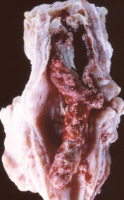
PM Clots
MoA: Heart stops beating → Blood stops flowing and clots → RBCs have time to sediment out to form pale clot of serum (“chicken fat clot”) over layer of red clot
Appearance:
Conforms to vessel = smooth and glistening appearance
Non-adherent (easy to remove from vessel)
Chicken-fat clot OR dark red
No lyaering

DDx: Thrombi = AM clot
Rough and granular (conforms to damaged blood vessel)
Does NOT separate into white and red layers
Adheres to vessel wall at site of injury
Colour depends on proportion of RBCs (usually dirty red-grey)
Fibrin, cells and platelets laid down in layers
FOUR: PM DEGENERATION
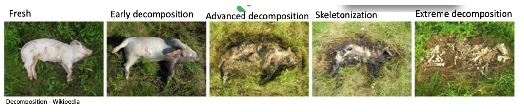
Autolysis = Breakdown of cells/tissues by body's OWN cellular enzymes (decay of organelle membranes)
Putrefaction = Breakdown of cells/tissues by BACTERIA from environment or within body
Depends on:
Temperature (warm = more active enzymes)
Bacterial species present
Neonates are slower to putrefy as they have no intestinal flora
GIT decomposes rapidly
Pseudo-Lesions
Definition
Describe 9 examples
Cause
DDx
Definition; Lesion on PM caused by autolysis and putrefaction
Examples:
Paleness and friability
DDx: Fatty liver (floats in formalin)
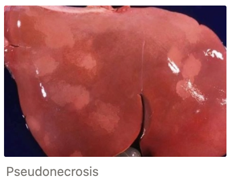
Pseudonecrosis = Discolouration and softening within organs
Cause: Pressure from other organs OR bacterial proliferation
DDx: Necrosis (should have line of demarcation)
Pseudomelanosis = Black staining of tissues
Cause: Production of iron sulphide (reaction between hydrogen sulphide from anaerobic bacteria, and iron from RBCs)

Gas production = Foul-smelling gas accumulates in body cavities OR tissue (emphysema)
Cause: Anaerobic putrefactive bacteria
DDx: Bloat

Hb imbibition = Red staining with Hb pigment
Cause: Hb escapes dead RBCs
DDx: Bruising or haemorrhage

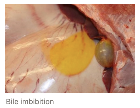
Bile imbibition = Yellow/green staining of tissues adjacent to gallbladder or bile ducts
Organ displacement
Cause: Diaphragmatic rupture
Mucosal sloughing in rumen
No sloughing = Metabolic issues eg. acidosis
Lens opacity
Cause: Low body temperature
DDx: Cataract (wait until fluid within eyes returns to ambient temperature → clear = PM change)
TWO Types of Cellular Damage
Definition
Cause
MoA
Morphological features on histology
ONE: CELLULAR DEGENERATION
Definition: Mild/brief cell damage which is REVERSIBLE once the stimulus has stopped
Reversible if membrane is NOT ruptured
Cause: Damaged sodium-potassium pump OR loss of ATP (eg. hypoxia) necessary for the pump to function
Required maintain cellular homeostasis by pumping out 3Na+ in exchange for 2K+ into the cell
MoA:
Damage to pump OR loss of ATP results in loss of function
Extracellular Na+ flows into the cell
Water is attracted to Na+ and follows it into the cell
Cell swells
Organelles swell = Ribosomes can detach from the ER
Impaired protein production and further compromised cell function results
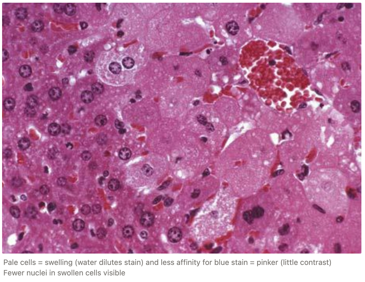
Morphological Appearance: Hydropic degeneration
Cell swelling = Pale cells
Eosinophilia = Less contrast and pinker than normal
MoA: Ribosomes detach from ER → Loss of affinity of blue-staining from acidic RNA
Cells are pinker than normal as ribosomes detach from the ER resulting in lost affinity for blue staining from the acidic RNA Results in relative increase in pinkness ALSO fewer nuclei in swollen cells visible
TWO: NECROSIS
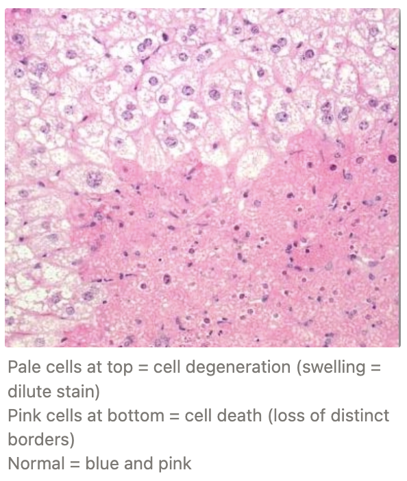
Definition: Irreversible cell injury resulting from severe or prolonged damage
Cause: Swelling becomes so severe → Points of no return
Mitochondrial damage preventing ATP function
Membrane rupture from lysed phospholipids (cellular AND organelle)
MoA: Membrane rupture is self-perpetuating through
Influx of intracellular Ca2+ activates endogenous phospholipidase which lyse the phospholipid membrane
Cytoskeletal damage (cell membrane detaches from cytoskeleton)
Free radical production
Lipid breakdown products have a detergent effect on the cell membrane
Morphological Appearance:
Eosinophilia (pinker than degeneration)
Shrunken cells
Loss of distinct cell membrane due to dissolution
Nuclear changes
Pyknosis = Shrunken, dark stain nucleus
Karyorrhexis = Fragmented nucleus
Karyolysis = Faded nucleus from lost affinity for haematoxylin
Hypoxia OR ischaemia?
Cardiac arrest
Anaemia
Thrombus
Suffocation
High altitude
Hypoxia = Lack of O2 supply → Decreased ATP production
Ischaemia = Lack of blood supply
Ischaemia
Hypoxia
Ischaemia
Hypoxia
Hypoxia
What is haematoxylin and eosin?
Common routine stain for histology (NOT cytology)
Haematoxylin is a basic dye which stains acidic structures blue (eg. nucleic acid)
Eosin is an acidic dye that stains proteins pink
Necrosis
Definition
Local response
Systemic response
Definition: Local death of tissue cells within a living individual caused by irreversible cell damage
Response: Mild local inflammatory reaction resulting from irritation (cellular debris within the ECF)
Leads to an accumulation of WBCs and a line of demarcation
Line of demarcation = Red rim hyperaemia (dilated blood vessels) that forms the border between living tissue and pale/friable necrotic tissue
Invaded by WBCs
Systemic Response: Release of inflammatory mediators (IL and PG) which cause fever, lethargy, depression and anorexia

FOUR types of necrosis
Definition
Causes
ONE: COAGULATIVE
Definition: Histological architecture is recognisable BUT cellular detail lost
Beginning of every other type of necrosis
Causes:
Ischaemia
Burns
Caustic chemicals

TWO: CASEOUS NECROSIS
Definition: Affected areas form a pale, granular mass (cheesy)
Loss of cellular AND architectural detail
Causes: Specific bacterial infection
Mycobacterium spp. and Corynebacterium pseudotuberculosis
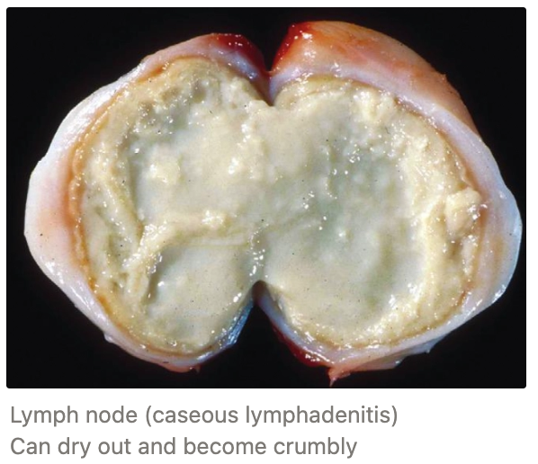
THREE: LIQUIFACTIVE
Definition: Disintegration of necrotic material into a liquid mass
Loss of cellular AND architectural detail
Causes: CNS malacia
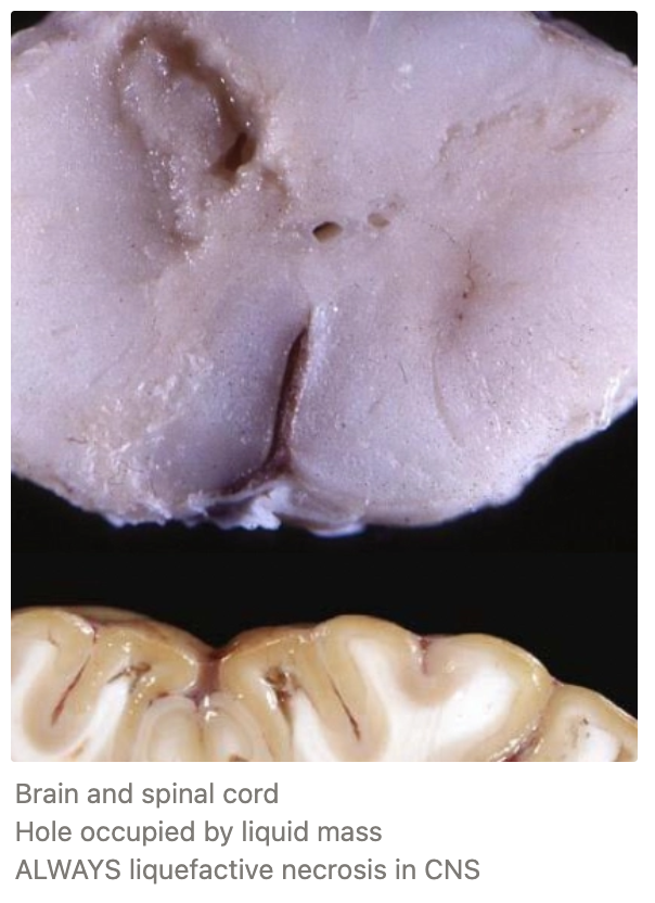
FOUR: FAT NECROSIS
Definition: Death of fat cells → Breakdown of fat into glycerol backbone and fatty acids
Cause: Fatty acids used in saponification = Combine with Ca2+/K+/Na+ salts to form soaps which precipitate
Common in mesentery and pancreas
Appearance: Opaque, dull, firm, white which are slightly basophilic
Some cases, peroxidation of lipids → deposition of ceroid (yellow pigment)
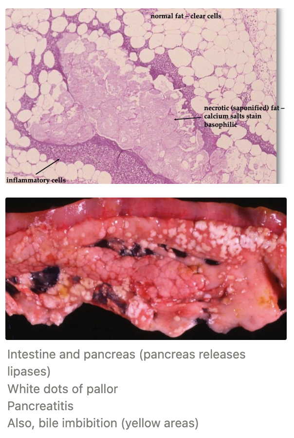
Sequelae of Necrosis
Importance
4 (5) Examples
Indication
Outcome
Importance: Remove dead material to prevent infection and reduce inflammation
Examples:
ONE: LIQUEFACTION
Indication: Small areas of necrotic tissue which are rich in fluid and enzymes for neutrophil and macrophage infiltration
Outcome: Resulting products are absorbed into the blood/lymphatics thus creating a small hole
TWO: SEQUESTRATION
Indication: Dry necrosis (eg. coagulative or caseous)
eg. cornea and bone
Outcome: Fibrous capsule surrounding a necrotic centre which can remain in the body (encapsulation without liquefaction)

THREE: ABCESSATION
Indication: Liquefaction of necrotic tissue → Suppuration and encapsulation
Outcomes:
Cold abscess (formation of thick, fibrous capsule around the pus)
Bursting to release necrotic, purulent material
FOUR: EROSION AND ULCERATION
Indication: Skin and luminal organs (bladder, ureter, bronchus, intestine)
Outcome: Sloughing (desquamate) of dead cells from underlying living tissue into surrounding lumen
Erosion = Loss of epithelial cells ONLY
Ulceration = Loss of epithelial cells AND basement membrane → Exposes underlying connective tissue (red due to vasculature)

FIVE: GANGRENE
Gangrene
Definition/indication
3 Types
Definition
Example
6 Morphological features
3 Reasons why gangrene is dark/green in colour
2 Sequelae
Treatment
Definition: Ischaemic necrosis of EXTREMITIES → Increased risk of bacterial infection due to lack of blood cells for normal defence
Types:
Dry Gangrene = Uncomplicated ischaemic necrosis eg. frostbite
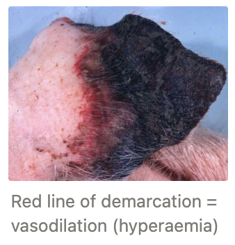
Wet Gangrene = Ischaemic necrosis in areas of fluid/blood → Complicated by bacterial infection resulting in liquefaction of necrotic tissue (eg. mammary gland)
Gas Gangrene = Special type of moist gangrene in which ANAEROBIC, gas-producing, saprophytic bacteria (eg. clostridia) establish in necrotic tissue due to low O2. The bacteria also release exotoxins which also induce necrosis in nearby, healthy tissue (eg. black leg caused by Cl. chauvoei)
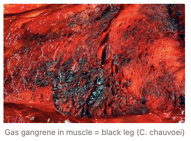
Morphological Features:
Cold to the touch
Unresponsive to stimuli
Does NOT bleed on incision
Dark/green colouration
Line of demarcation
Foul-smelling
Dark/Green Colour Due to:
Anoxic blood is dark red
Pigments (eg. haemosiderin) from RBC breakdown
Iron sulphide production (-SH containing AA breakdown and release H2S which reacts with iron from RBC breakdown to produce iron sulphide)
Sequelae:
Sloughing of dead tissue followed by scarring
Toxaemia = death
Bacterial toxins (septicaemia) and endogenous products from protein decomposition (endotoxaemia) are absorbed into the lymphatics and bloodstream of living areas to cause damage
Treatment: Debridement or amputation
Apoptosis
Programmed cell death that does NOT trigger an inflammatory process
Apoptotic bodies have markers to be targeted for phagocytosis
Physiological vs. pathological cellular adaptations
Physiological Cellular Adaptations = Changes to cells under NORMAL conditions in response to metabolic or physical demand
Pathological Cellular Adaptations = Changes to cells in response to sublethal, chronic cellular injury which enables the cell to survive
Types of Cellular Adaptation and Growth Changes
Definition
Examples
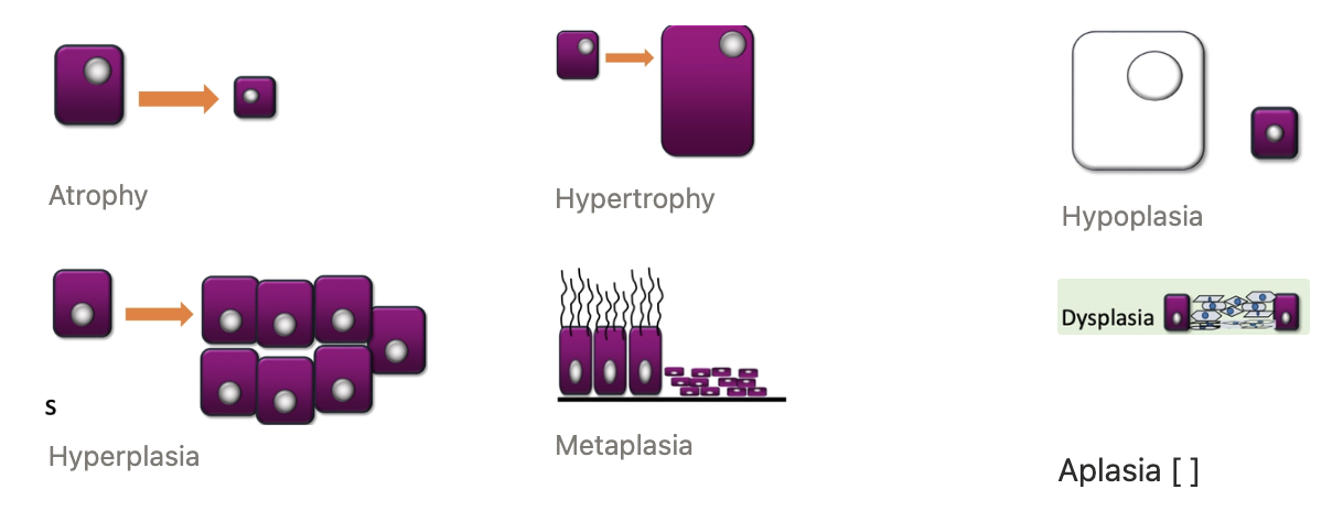
ONE: ATROPHY
Definition: Decreased cell SIZE (from normal size) which causes the whole organ/tissue to shrink
Less cellular activity results in fewer cell organelles that produce less
Examples:
Disuse atrophy (eg. muscles)
Denervation atrophy (eg. sweeny)
Loss of nutrition/hormonal stimulation (eg. small thyroid)
TWO: HYPERTROPHY
Definition: Increase in cell SIZE which causes the organ/tissue to get bigger (can be specific cells)
Cells are more active and require more organelles
ONLY way for post-mitotic cells (eg. cardiac muscle) to increase in size as cells cannot divide
Examples:
Physiological
Increased skeletal muscle bulk from prolonged exercise
Enlarged smooth muscle cells in the uterus during pregnancy
Pathological = Increased ventricle wall thickness due to a stenotic valve
THREE: HYPOPLASIA
Definition: Incomplete growth of an organ/tissue that NEVER reached full size
Fewer cells produced
Caused by congenital conditions (NOT true cellular adaptations)
Examples:
Aplasia = Complete absence of tissue/organ (extreme hypoplasia) eg. aplasia of epididymis
Atresia = Absence of the lumen eg. atresia ani (no opening of anus)
FOUR: HYPERPLASIA
Definition: Increased NUMBER of cells which may increase gross size
Stimulated in the same way as hypertrophy (can be both)
Usually normal function
Examples:
Goitre (thyroid hyperplasia)
Nodular hyperplasia of the spleen
BPH
FIVE: DYSPLASIA
Definition: Lack of normal histological architecture in the tissue/organ
Results from developmental/congenital condition OR acquired (scar tissue)
Examples:
Hip dysplasia
Kidney displasia
SIX: METAPLASIA
Definition: A fully differentiated cell type changes into another cell type from the same germline
Usually changes into a more durable cell type in response to chronic irritation
Example: Squamous metaplasia
Change from secretory ciliated columnar epithelium in the bronchi (loss of mucociliary blanket)
Change from secretory columnar cells in the mammary glands
What is a hamartoma? Give an example
Congenital condition resulting in improper mix of tissues within an organ
eg. Excess of vascular tissue in the skin causing strawberry birthmarks
Resemble neoplasm grossly and microscopically
Haemosiderin
Definition
Formation
4 Locations of accumulation
Definition: Yellow/brown insoluble pigment found in macrophages which can be broken down later to reuse iron
Also, ferritin = Iron complexed with apoferritin protein (soluble storage pigment)
Stains blue/black with Prussian blue-based stains (contains iron)
Formation: Made from iron (derived from Hb breakdown to heme) → Complexed loosely with protein
Locations:
Small amounts in splenic macrophages (NORMAL)
Local accumulation in old bruises and vascular congestion (eg. heart failure cells of lungs and liver)
Haemosiderosis = Systemic accumulation of excess haemosiderin in macrophages due to:
Severe haemolytic disease
Impaired iron utilisation
After blood transfusions
Haemochromatosis = Rare iron storage disease due to defects in iron regulatory genes (i.e. excess iron in bird diets)
Bilirubin
Formation
Process of elimination
Formation: Yellow/orange pigment formed from the breakdown of heme AFTER iron is released
Bilirubin Excretion:
Free (unconjugated) bilirubin is released into the bloodstream following haemolysis and loosely coupled with albumin (still free)
It travels to the liver to be taken up by hepatocytes and converted into the more soluble: conjugated bilirubin (ester with 2 glucuronic acids)
Conjugated bilirubin is excreted into bile via the bile duct
It enters the small intestine to be converted to urobilinogens
Some urobilinogens are reabsorbed to oxidised to urobilin and excreted in the urine (yellow colour)
Most urobilinogens are oxidised to stercobilin in the intestine to be excreted in faeces (brown colour)
Overview of Melanin
Definition
Normal synthesis
Cause of varied pigmentation
Function
Definition: Yellow/brown granules secreted by melanocytes
Responsible for the brown/black pigmentation of skin, hair, iris and choroid
Synthesis: Tyrosinase is used to oxidise and polymerise tyrosine which is complexed with sulphur-containing proteins to form melanin
Tyrosinase = Copper-dependent enzyme necessary to produce melanin
Varied Pigmentation: Variation in EFFECTIVENESS of melanocytes to produce melanin (NOT the number of melanocytes)
Function: Transferred to epithelial cells of the skin and hair, or taken up by macrophages → Melanin surrounds the nucleus to protect the DNA from UV damage
TWO Aetiologies of Decreased Melanin Production (Pallor)
Definition
Examples
Albinism = Congenital mutation where no melanin is produced in the WHOLE body (usually a result in tyrosinase deficiency)
Determine as eyes are red
Acquired hypopigmentation
Hormonal imbalance
Local loss of melanocytes due to trauma or chemical injury (eg. scarring = increased fibrous connective tissue and freeze branding)
Copper deficiency

Describe 4 causes of increased melanin production (darker)
UV light = UV light stimulates melanin production in exposed skin to protect nucleus from damage
Congenital = Uneven dispersion of melanocytes during embryonic development resulting in discrete focal areas of hyperpigmentation (eg. liver and lungs)
Moles = Dermal accumulations of melanoblasts which can transform into melanoma (neoplasm)
Plants = Accumulation of melanin precursors in various organs

Fat accumulation
3 Tissues fat normally accumulates in
4 Tissues fat should NOT accumulate in
Normal:
Adipose tissue
Adrenal glands
Sertoli cells
Abnormal:
Liver
Heart
Muscle
Kidney
Describe the process of fat metabolism
Lipids transported from the diet (as chylomicrons or FFAs) AND adipose tissue (as FFAs) to the liver
Most lipids converted to TAGs by hepatocytes (secreted from liver bound to apoproteins such as VLDLs)
Remainder is stored, or oxidised
Appearance of Fatty Liver
Gross appearance
Histological appearance
Acute
Chronic
Gross: Pale, greasy, friable and bulge out from the cut surface Liver may flat in water/formalin
Histology:
Acute = Numerous small fat globules in the cytoplasm around the central nucleus
Chronic = Globules coalesce which may displace the nucleus towards the edge of the cell

Terminal Vascular Bed
Structure
Function
2 Processes increasing blood to terminal vascular bed
Main process decreasing blood to terminal vascular bed
Structure: All capillaries are patent, but some not completely filled with blood (functional reserve maintained)
Function: Exchange of nutrients and waste products between the arterioles and venules
Increasing Blood:
Hyperaemia
Congestion
Decreasing Blood: Ischaemia
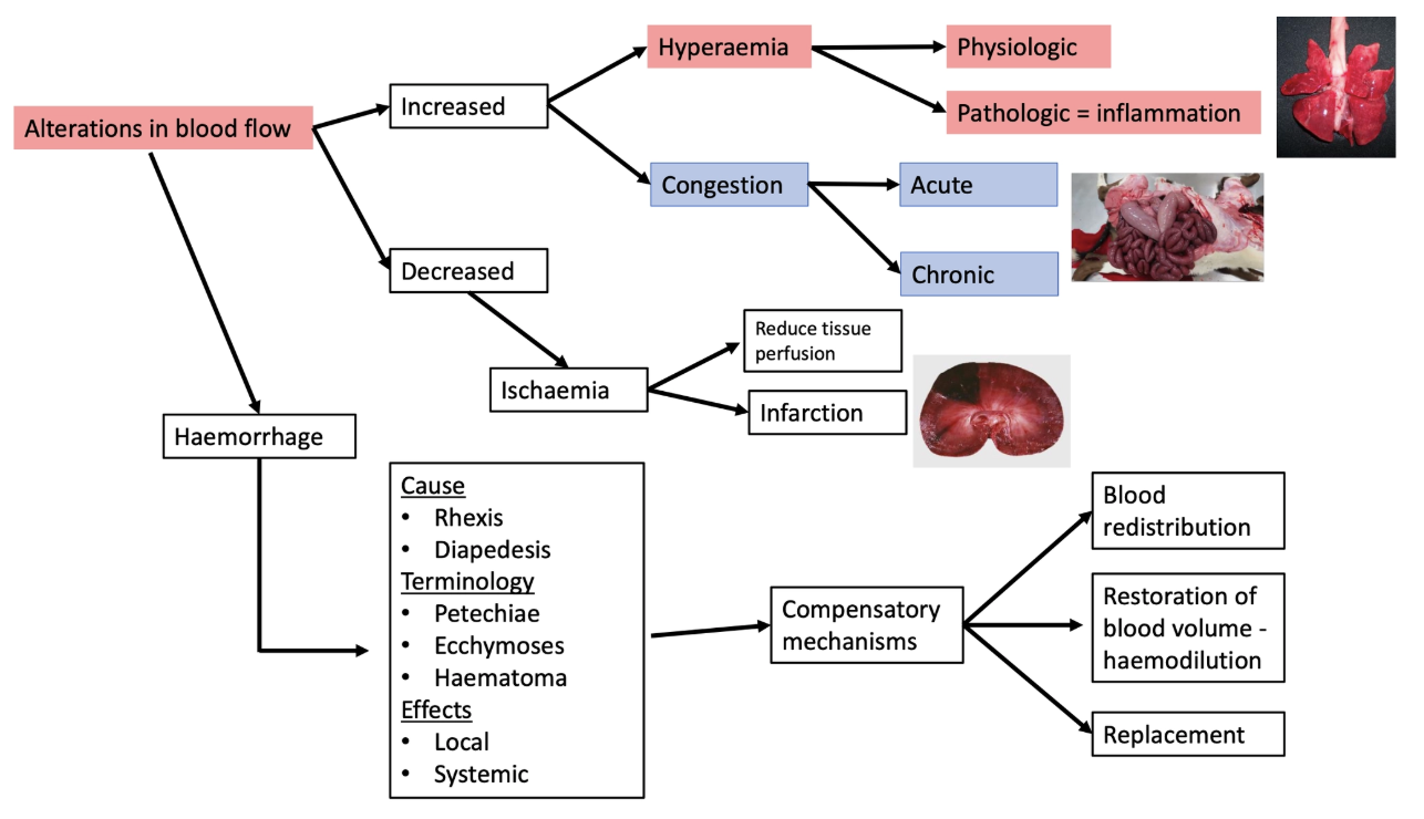

Hyperaemia
Definition
Appearance
2 Aetiologies (+ examples)
Definition: More blood to tissues due to increased arterial blood flow and more reserve capillaries flowing with blood
Usually an acute, active processes associated with inflammation
Appearance: Bright red tissues from accumulation of oxygenated Hb
Aetiologies:
Physiological hyperaemia
Increased arterial blood flow in the stomach and intestine during digestion
Exercise increases hyperaemia to muscles
Pathological hyperaemia = Hyperaemia as a result of inflammation
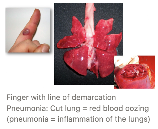
Congestion
Definition
Appearance
3 Types
Defintion
Examples
5 Effects of congestion
Definition: More blood to tissue caused by obstructed flow OUT of tissues at the venous side
Passive process
Appearance: Dark red/blue tissues from accumulation of deoxygenated Hb
Types:
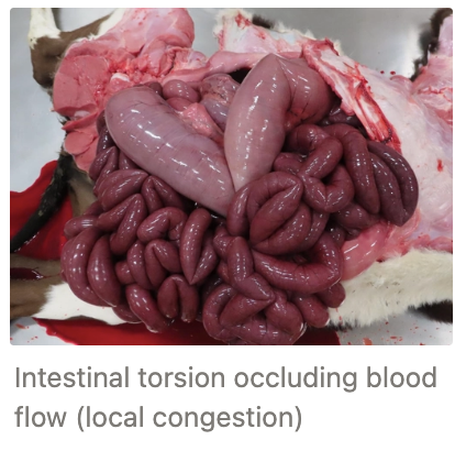
Localised = Local restriction of venous flow resulting in a BODY PART with bluish colour
Internal occlusion (eg. thrombus)
External compression (eg. tumour, tourniquet)
Systemic = Generalised congestion where the whole body is blue as a result of blood pooling in the great veins and affects a wide area of tissue eg. heart failure
Hypostatic = Blood pooling due to the force of gravity (PM examination)
Effects:
None (gradual onset of local congestion → time to develop adequate collateral circulation through other veins)
Oedema
Hypoxia and necrosis
Ischaemia and infarction
Diapedesis or rhexis
Diapedesis = Passage of blood cells through INTACT capillary walls
Rhexis = Haemorrhage caused by rupture of the vessel wall (usually due to trauma)
Ischaemia
3 Consequences
6 Causes
4 Factors modifying the effects of ischaemia
Consequences:
Hypoxia → necrosis
Local nutrient deprivation (not limiting)
Failure to remove waste products (eg. muscle ischaemia and pain)
Causes:
Cardiac arrest
Arterial obstruction
Thrombosis
Embolism
Arterial wall spasm (eg. ergot poisoning)
Arteritis leading to thrombosis (eg. bovine MCF or Strongylus vulgaris)
External occlusion (eg. bandage)
Venous obstruction
MoA:
Occlusion of the venules prevents blood flow out of the terminal vascular bed
Initially the arterial supply is not affected
Later, occlusion causes increased pressure in the venous side of the capillaries
Results in increased resistance to arterial blood flow and hence, ischaemia
Capillary damage/compression (eg. pressure sores)
Hypovolaemia
Severe vasodilation
Factors:
Specific tissue sensitivity to anoxia (brain vs. fibrous tissue)
Rate of development and duration (slow development = time to adapt by collateral circulation)
Well-oxygenated RBCs vs. anaemic (higher severity)
Temperature (low temp = decreased metabolic rate = reduced O2 demand)

Infarct
Definition
Most common type
2 Organs rarely affected (+ why)
7 Stages (artery occlusion → healing)
Definition: Localised are of coagulative necrotic tissue caused by ischaemia
Most Common: Arterial infarct (occlusion)
Organs Rarely Affected: Organs with dual blood supply
Lungs (pulmonary and bronchial arteries)
Liver (hepatic artery and portal vein)
Stages:
Artery occlusion causes decreased hydrostatic pressure within the capillary bed
Venous blood flows back into the tissue
Venous and capillary wall cells become hypoxic
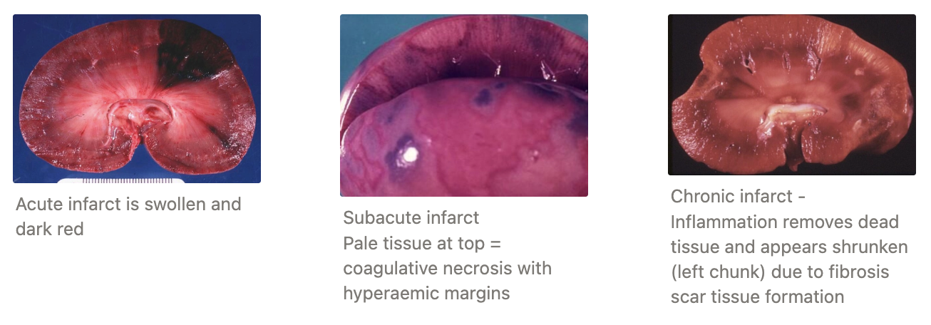
Acute infarct becomes swollen and dark red due to haemorrhage and fluid loss
Subacute infarcts become paler due to necrosis caused by hypoxia
Neutrophils and macrophages invade the infarct to remove necrotic tissue (line of demarcation)
Healing occurs by a fibrotic scar which appears as a depressed scar in a chronic infarct
Provide definitions for the following terms
Haematoma
Petechial haemorrhage
Ecchymotic haemorrhage
Purpura
Haematoma = Local accumulation of blood (usually clotted) that causes swelling in tissues
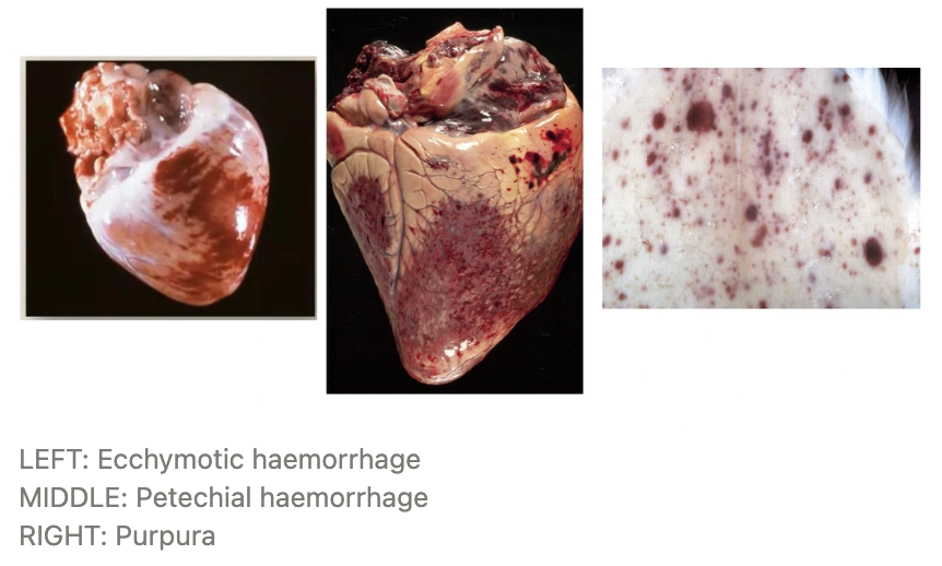
Petechial Haemorrhage = Pin-point haemorrhagic spots usually caused by haemorrhage per diapedesis
Ecchymotic Haemorrhage = Slightly larger foci of haemorrhage usually caused by haemorrhage per diapedesis
Purpura = Skin, MM, viscera haemorrhage
What are 5 causes of haemorrhage?
Trauma (rhexis)
Vascular diseases (eg. vasculitis)
Abnormally weak blood vessels (eg. vitamin C deficiency = lack of collagen to strength blood vessels)
Local high BP
Abnormal clotting
Effects of Haemorrhage
2 Local effects
4 Systemic effects
Local Effects:
Small haemorrhage = little effect (can be absorbed) unless vial area
Large haemorrhage produce space occupying lesions which can have severe mechanical effects
eg. Cardiac tamponade (pericardium), asphyxiation (lungs), pain (joints)
Systemic Effects: Depends on total volume lost AND rate
Chronic, small amounts → Iron deficiency anaemia (iron required for oxygen transport)
Bleeding GIT ulcers
Acute, at least 1/3 of total → Hypovolaemic shock and death
At least 24 hr period, at least 1/3 of total → Compensatory mechanisms and no death
Acute, less than 20% lost → Small systemic effect (fast compensation)
Describe the 3 phases of compensatory mechanisms for haemorrhage
PHASE ONE: REDISTRIBUTION OF BLOOD TO VITAL CENTRES
Rapid response (<4hr)
Splenic contraction releases large stores of RBC into circulation to maintain
Selective arteriole constriction (adrenergic nerves stimulated) so blood is redistributed to the brain, heart and lungs
PHASE TWO: RESTORATION OF PLASMA VOLUME
4 - 12hr
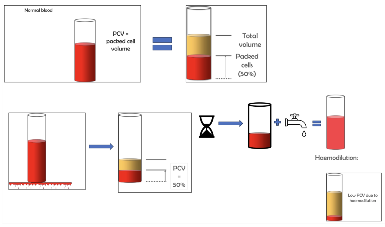
Arteriolar constriction causes intravascular pressure of capillaries to decrease which results in ECF moving into blood vessels
Haemodilution results as the ECF does NOT contain blood cells
Complete 2d after original haemorrhage → PCV indicates amount of original blood loss
PHASE THREE: REPLACEMENT OF LOST RBCs
Erythropoiesis is stimulated in bone marrow in response to RBC loss
Reticulocyte production begins 3 - 5 days post-haemorrhage, peaks 7d and diminishes by 12 - 14 days
What is shock? How is it caused?
Failure of circulatory system to adequately perfuse organs
Caused by disparity between the volume of circulating blood and the size of the vascular system
THREE Types of Shock
Definition
Examples
ONE: HYPOVOLAEMIA SHOCK
Definition: Dramatic decreased blood volume due to loss of fluid (dehydration) or whole blood (haemorrhage)
Examples:
Severe haemorrhage
Prolonged diuresis
Persistent emesis or diarrhoea
Burns
TWO: VASCULOGENIC SHOCK
Definition: Shock caused by widespread vasodilation or widespread endothelial damage
aka. Distributive shock
Examples:
Septic shock (G- bacteria produce endotoxin causing vasodilation)
Hypoxia
Anaphylactic shock
Neurogenic shock (loss of sympathetic innervation)
THREE: CARDIOGENIC SHOCK
Defintion: Shock resulting from decreased cardiac output
Examples:
Acute myocardial dysfunction (eg. large infarct)
Diastolic dysfunction (decreased filling eg. cardiac tamponade)
Systolic dysfunction (decreased emptying eg. increased vascular resistance and ruptured chordae tendinae)
Irreversible Shock
Definition
5 Effects on the body
Definition: When compensatory mechanisms are unable to sustain the patient leading to systemic hypoxia and death
Effects:
Widespread endothelial damage (leaking plasma from vessels causing sludging and stasis of blood flow)
Organs switch to anaerobic metabolism causing cellular and systemic acidosis
Hepatic and renal necrosis
Myocardial failure → Arrhythmia
Severe depression of the CNS → Loss of consciousness
Effect of Temperature on Shock
2 reasons why a shocked patient is cold
Why is overwarming. shock patient bad?
Shock Patients Cold Because:
Selective arteriole constriction (warm blood away from peripheral structures)
Decreased heat output due to interference of -CHO metabolism
Overwarming Shock Patient is Bad: Causes dilation of skin vessels and further reduces blood volume to vital organs
Overview of Thrombosis
Defintion
3 Causes (Virchow’s Triad)
Definition: Formation of a thrombus (blood clot) via coagulation
Normal occurrences during life
Causes (Virchow’s Triad):
Endothelial Injury = Abnormal blood vessel wall #1
Causes: Trauma, inflammation, chemical irritants etc.)
MoA: Activation of tissue factor which triggers the clotting cascade
Causes a blood clot to attach to the vessel wall at the site of damage
Abnormal Blood Flow = Influences progression of thrombosis
Blood stasis
Varicose veins
Aneurysm
Poor peripheral circulation
Turbulence = Disturbance in laminar flow (most commonly occurs at vessel branches or areas of irregularity in the vessel wall)
Hypercoagulability = Abnormal blood composition which influences progression of thrombosis
Haemoconcentration (eg. dehydration)
Higher conc. of procoagulants (genetic or contraceptive)

Occluding vs. Non-Occluding Thrombus
Definition
Sequelae
Occluding Thrombus
Definition: Blood clot fills the entire blood vessels which cannot lyse or recanalise
Sequelae: Leads to ischaemia and infarction
Non-Occluding Thrombus
Definition: Partially fills blood vessel
Sequelae:
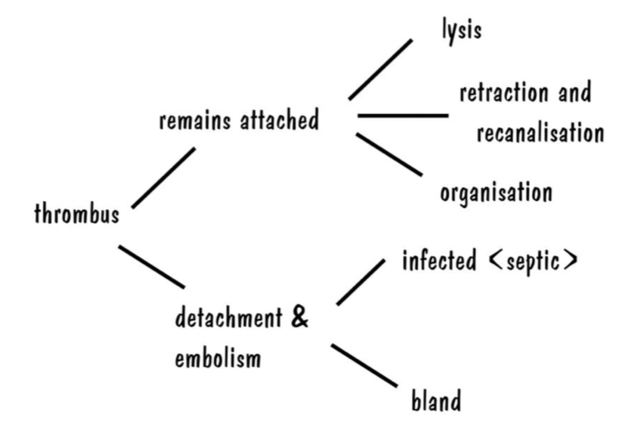
Lysis = Lysed in SMALL blood vessels by plasminogen-plasmid system
Causes little damage to surrounding tissue
Retraction and recanalisation = Multiple canals form within the thrombus which is lined with endothelial cells (edge of the thrombus AND canals) for blood to flow to distal tissues
Organisation = Occluding thrombus in larger vessel shrinks over time and is lined with endothelial cells to allow passage of blood through
Thromboembolism = Blood flows over the thrombus which dislodges part of it to become an embolus
Embolism
Define embolus vs. embolism
6 Types of emboli (+ examples)
3 Sequelae of emboli
Embolus: Abnormal mass circulating in the bloodstream
Embolism: Embolus arrested in a vessel which occludes it (vessel is same diameter as embolus)
Types:
Thromboembolic (bland or septic)
eg. Septic endocarditis
L = Myocardial or renal abscessation
R = Pulmonary abscessation
eg. Saddle thrombus in cats
Gas ("The Bends")
Fat (common after orthopaedic surgery or trauma)
Neoplasm (can cause metastasis)
Foreign body
eg. Migrating Strongylus vulgaris
eg. Bovine pulmonary embolic aneurysm
Cartilage (intervertebral disc fragments lodge in spinal cord blood vessels)
Sequelae:
Septic emboli spreads infection (eg. valvular endocarditis caused by infected IV catheter)
Tissue infarction if embolus fully occludes vessel
Nothing
Provide definitions for the following terms
Oedema
Anasarca
Oedema: Excessive accumulation of ECF
Anasarca: Generalised oedema
Cat = Hydrothorax, dog = ascites, ruminant = SC
Describe the normal formation and drainage of tissue fluid within the terminal vascular beds (3 key steps)
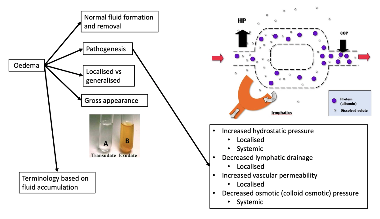
HP (and osmotic pressure) in the arterial end of the capillary bed causes fluid and solutes (not protein) to flow OUT of the capillaries into the ECF (via gaps between the capillary endothelial cells) to supply tissues with nutrients, O2 and minerals
Some fluid reabsorbed at the venous end due to COP (and hydrostatic pressure)
Most fluid taken up by lymphatic system which is eventually returned to the bloodstream
FOUR Mechanisms of Oedema
Definition/MoA
Outcome (localised or systemic oedema)
Examples
ONE: INCREASED HYDROSTATIC PRESSURE
Definition: Capillary pressure is determined by venous pressure, and not arterial pressure
Compression of venous end
Increased fluid in capillaries
Increase HP in capillaries
Overcomes COP of proteins
Net increased in fluid outflow in ECF
Outcome: Either localised OR systemic oedema
Examples:
Localised Oedema
Dystocia
Tourniquet
Tumour/abscess/thrombus occluding vein
Torsion of luminal organ
Generalised Oedema = Cardiac disease

TWO: DECREASED COLLOID OSMOTIC PRESSURE
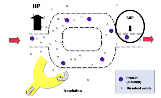
Definition: COP = Osmotic pressure exerted by proteins (albumin) in circulation which draws fluid back into the capillaries
Outcome: Systemic oedema
Examples: Hypoalbuminaemia
Increased albumin loss
Chronic haemorrhage (eg. Haemonchus contortus)
PLE
PLN (eg. nephrotic syndrome)
Decreased albumin production
Protein malnutrition (insufficient AA to synthesise albumin)
Chronic liver disease (hepatocytes cannot synthesise albumin)
THREE: LYMPHATIC BLOCKAGE
Definition: Decreased lymphatic drainage
Outcome: Localised oedema
Examples:
Extensive tumour invasion or regional lymph node
Removal of lymph nodes surgically
External mass compressing the lymphatic vessel

FOUR: INCREASED INTRAVASCULAR PERMEABILITY
Definition: Local inflammation → Widen gaps between endothelial cells to allow larger cells to enter tissues (eg. proteins and WBC)
Outcome: Localised oedema and exudate

Describe the gross appearance of oedema in solid tissues vs. body cavities
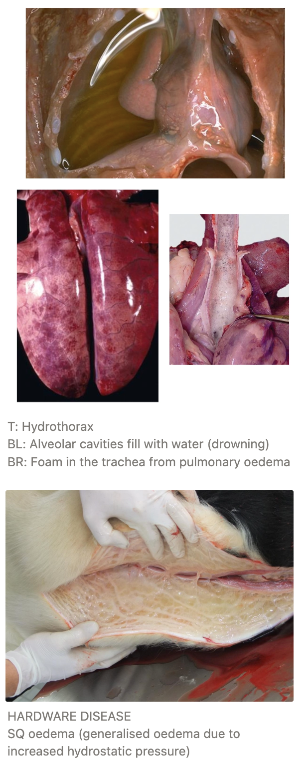
Solid tissues = Wet, heavy and enlarged
SC = Swellings that pit on pressure and gravity-dependent (bottle jaw and brisket oedema)
Incised tissues may seep fluid
Body cavities = Free fluid
Overview of Inflammation
Definition
4 Components required to induce an inflammatory response
5 Advantages
4 Disadvantages
5 Cardinal signs of acute inflammation
4 Outcomes
Definition: A complex cascade of LOCAL tissue reactions which consists of vascular and cellular responses
Active response to inciting event which enables survival and tissue homeostasis
Dynamic process with overlapping events
Components:
Inflammatory inducers (bacteria, activation of mast cells, trauma, necrosis)
Sensors to detect inflammatory inducers (dendritic cells, mast cells and macrophages eg. TLRs)
Inflammatory mediators induced by sensors (eg. TNF, ILs, histamine)
TLRs (Toll-like receptors) = MOST important family of pathogen recognition receptors (PRRs)
Specific TLRs interact with specific microbial products (PAMPs = pathogen associated molecular patterns) to activate a signalling pathway that stimulates production of inflammatory mediators
Target tissues that are affected by inflammatory mediators (cellular level OR tissue level)

Advantages:
Dilute and/or inactivate biologic and chemical toxins
Kill/sequestra microbes, foreign material, necrotic tissue, and neoplastic cells
Provide wound healing factors to damaged tissues
Restrict movement of appendages and joints to allow time for healing and repair
Increased temperature in body/locally to inhibit replication of some microbial agents
Disadvantages;
Tissue injury
Systemic effects of inflammation
Scar tissue formation
Loss of function (due to scar tissue formation)
Cardinal Signs of Inflammation:
Redness (rubor) via vasodilation
Swelling (tumor) via increased vascular permeability to allow larger healing proteins to enter site
Heat (calor) via vasodilation
Pain (dolor) via stimulation of neuronal pathways by inflammatory mediators
Loss of function (functio laesa)
Outcomes:
Return to normal
Abscessation
Fibrosis = Healing via connective tissue replacement
Progression to chronic inflammation
FOUR Classes of Inflammation (by Duration)
Duration
Cells present
Vascular response
Extracellular material
ONE: PERACUTE
Duration: Immediate
Caused by potent stimulus → No time to respond morphologically
Cells: Few WBCs (neutrophils) but likely normal
Vascular Response: ± Hyperaemia
Extracellular Material:
Oedema
Haemorrhage
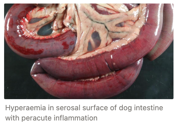
TWO: ACUTE
Duration: Minutes to 48hr
Cells: Variable WBC infiltration (neutrophils #1)
Vascular Response: Increased permeability, vasodilation and hyperaemia
Extracellular Material:
Oedema
Fibrin

THREE: SUBACUTE
Duration: Poorly defined between 3 - 4 days (transition between acute and chronic)
Cells: Proportion of WBC begins to change (neutrophils > macrophages > lymphocytes)
Vascular Response: Regressing hyperaemia and endothelial hypertrophy/hyperplasia
Extracellular Material:
Regressing oedema
Fibrin
No evidence of repair
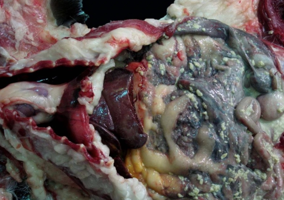
FOUR: CHRONIC
Duration: 7d - years
Caused by persistent inflammatory stimulus which the host failed to eliminate cause (eg. Mycobacterium hiding in macrophages)
Cells: Mononuclear #1 (macrophages, lymphocytes, plasma cells) and neutrophils
Vascular Response: Angiogenesis and neovascularisation
Extracellular Material: Collagen (fibrous granulation tissue)
Overview of Morphological Patterns of Inflammation
Importance
2 Determinants of morphological classification
Morphological classification based on (2)
Importance: Provides clues about aetiology, time course of disease, prognosis, treatment and DDx
Determinants:
Tissues involved
Type of aetiology inciting the inflammatory reaction
Morphological Classification:
Type of fluid leaking from vessels (exudate vs. transudate)
Cells and proteins migrating from vascular lumen to ECF
SEVEN Morphological Inflammatory Patterns
Definition
Location/Source
Appearance
Examples
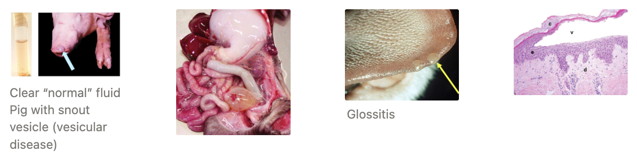
ONE: SEROUS INFLAMMATION
Definition: Pattern of acute inflammation characterised by accumulation of fluid with low concentration of plasma proteins and rare WBC
Source: From plasma leaking from damaged blood vessels or secretions from mesothelial cells lining and serous glands in the peritoneal, pleural and pericardial cavities
Appearance: Clear, thin fluid which cannot be easily differentiated from non-inflammatory oedema (eg. heart failure, hypoalbuminaemia)
Examples:
Thermal injuries (burns)
Vesicles (skin blisters eg. FMD)
Acute allergic response
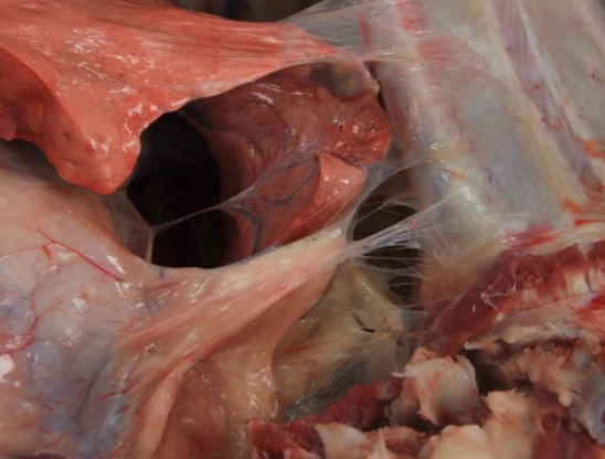
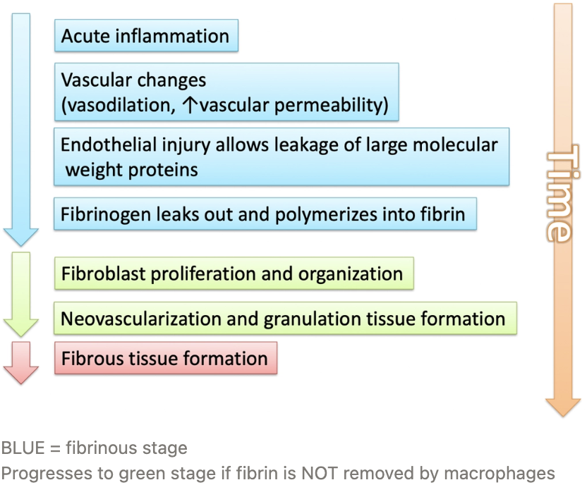
TWO: FIBRINOUS INFLAMMATION
Definition: Pattern of acute inflammation characterised by the accumulation of fluid with high concentration of plasma proteins and low to moderate numbers of WBCs
Source: Increased vascular permeability allows large proteins such as fibrinogen to travel between endothelial cells into the extravascular space
Fibrinogen polymerises to form delicate strands of fibrin which promotes wound healing through formation of a meshwork
Located around mucosa/serosa such as the mesenteric, pleural and pericardial surfaces
Appearance: Fluid is exudative and appears yellow due to the presence of fibrin
Outcomes:
Fibrous exudates removed by fibrinolysis and other debris by macrophages OR
Fibrin remains and stimulates proliferation and organisation of fibroblasts as well as neovascularisation and granulation tissue formation to form fibrous scar tissue
Examples:
Shipping fever
Peritonitis (eg. FIP)

THREE: DIPHTHERITIC INFLAMMATION
Definition: Fibrinous exudate (pseudomembrane) is so firmly attached to the underlying tissue in severe cases, that it cannot be removed unless torn off with superficial layer of bleeding tissue
Pseudomembrane = False membrane formed from sheets of dense fibrin containing blood, bacteria and cellular debris (WBC and necrotic mucosal epithelium) Forms after severe cases of fibrinous inflammation
Appearance: Solid cores of fibrin in tubular organs
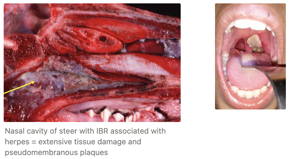
Examples: Infectious bovine rhinotracheitis (IBR) associated with herpesvirus
Forms pseudomembranous plaques in the nasal cavity
FOUR: SUPPURATIVE/PURULE
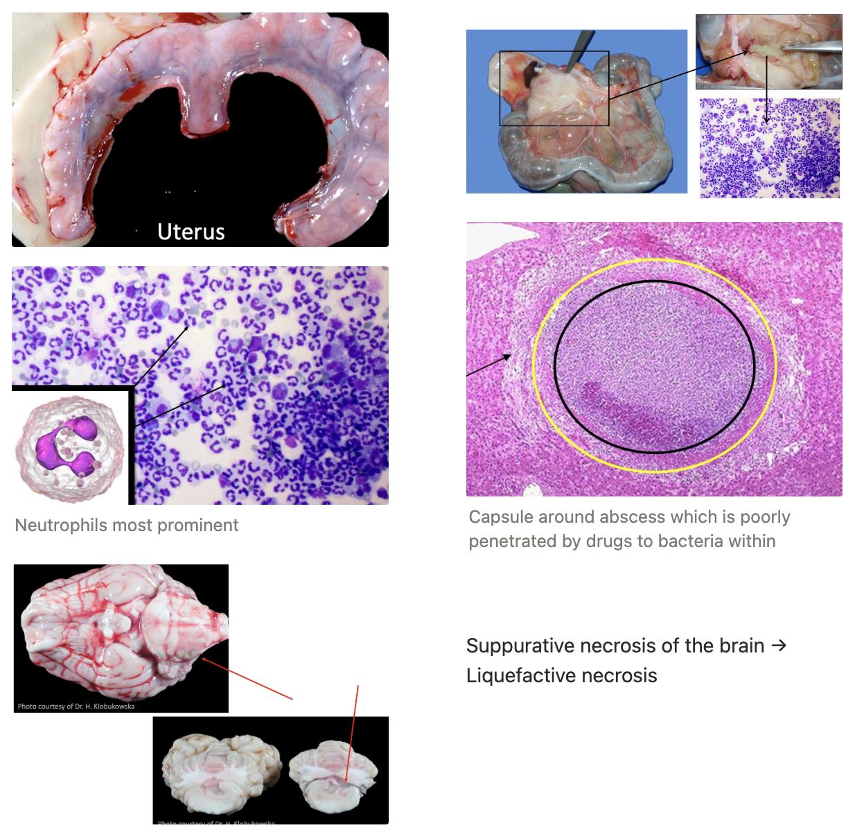
Definition: Pattern of acute inflammation where tissues respond by accumulation of fluid with high concentration of plasma proteins AND neutrophils
Source: Pyogenic bacteria
Appearance: Milky white and foul-smelling fluid which may form and abscess
Abscess
Central necrotic region (necrotic leukocytes and tissue cells) = BLACK line in diagram
Zone of preserved neutrophils (between yellow and black)
Outer region of vascular dilation and parenchymal + fibroblastic proliferation (arrow)
Examples:
Cat bite abscess
Any condition with pyo- (eg. pyometra)
FIVE: CATARRHAL/MUCOID INFLAMMATION
Definition: Exudate contains mucus from cells which function to
Entrap bacteria
Stimulate ciliary movement to expel mucus and debris
May contain lysozyme and IgA
Source: MM (GIT and RT) with abundant goblet cells and mucus glands
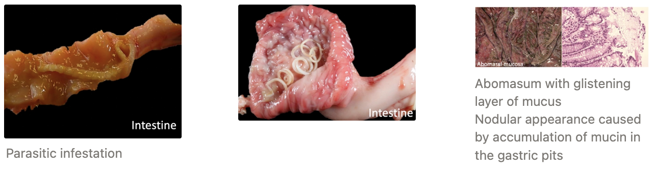
Appearance: Thick and gelatinous fluid with abundant mucus and mucin
Examples:
Low-virulence bacterial/viral infection
Mildly irritating chemicals
Parasitic infections
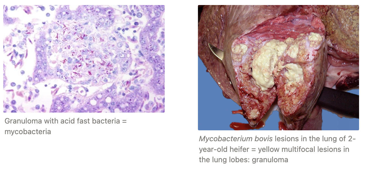
SIX: GRANULOMATOUS INFLAMMATION
Definition: Distinctive morphological pattern of chronic inflammation in a several infectious and some non-infectious conditions defined by macrophage presence
Source: Cellular attempt to contain offending agent that is difficult to eradicate
Strong activation of T lymphocytes leading to macrophage activation which may result in tissue injury
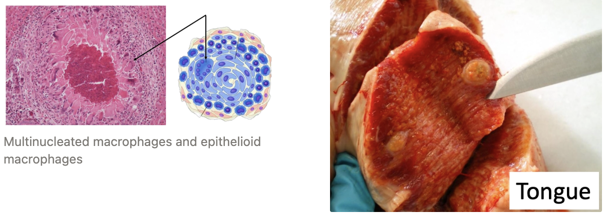
Appearance: Granuloma = Microscopic aggregation of macrophages which are transformed into epithelioid macrophages (epithelium-like cells)
Surrounded by mononuclear WBCs (lymphocytes and plasma cells)
The epithelioid macrophages may fuse to form giant cells present in the periphery or centre of the granuloma
Examples:
Fungal infection
Infection with Mycobacterium spp.
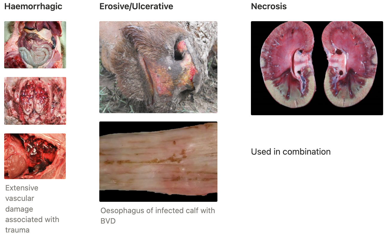
SEVEN: OTHER
Haemorrhagic
Erosive/ulcerative
Necrosis
Fibrinous vs. fibrosis
Formed from what?
Adherence to underlying tissue
Acute or chronic process?
Examples of each
Fibrinous | Fibrosis |
|---|---|
From fibrinogen which leaks from blood vessels | From fibroblasts (tissue repair) |
Separates and breaks off easily | Firmly adhered |
Acute process | Chronic process |
eg. acute peritonitis | eg. scar tissue |
Acute Inflammatory Response
Function
List 2 phases (+ definitions)
Function: To get the players (eg. leukocytes and proteins) from the vascular system to the site of injury
Phases:
Vascular Phase (histamine = #1 inflammatory mediator)
Vasoconstriction (very short duration)
Alterations in vascular caliber → Increased blood flow (vasodilation)
Structural changes in microvascular which permit plasma proteins and leukocytes to leave the circulation
Cellular Phase = Emigration of leukocytes from microcirculation → Accumulate in area of injury → Activation of leukocytes to eliminate offending agent
Marginalisation
Rolling
Adhesion to endothelium
Emigration across endothelium
Migration into tissues
THREE Processes of the Vascular Phase of Inflammation
Vasoconstriction = Very short duration to stop haemorrhage
Vasodilation = Arteriole dilation → Open new capillary beds in area → Hyperaemia
Induced by histamine (also nitric oxide, bradykinin, prostaglandins and leukotrienes)
Increased Vascular Permeability = Escape of protein-rich exudate into ECF → Oedema via
Retraction of Endothelial Cells = Gaps between endothelial cells increase, allowing larger proteins and leukocytes to migrate into the tissue
Immediate, transient response
Endothelial Injury = Direct damage to the endothelium (stimulated by severe injury, microbial toxins, endotheliotropic viruses etc.) which results in endothelial cell necrosis and detachment
Occurs rapidly after stimulus
Likely long-lived response (endothelium needs to regenerate and repair)

Describe how the vascular phase of inflammation causes blood stasis and the cellular phase
The vascular changes:
Increased blood vessel diameter
Loss of fluid
→ Slower blood flow, increased [RBC] in small vessels, increased blood viscosity (stasis) which
Causes leukocyte (neutrophil/macrophage) accumulation along vascular endothelium
Endothelial cells are activated by mediators produced at the site of inflammation → Increased adhesion molecules
Leukocytes adhere to endothelium and migrate through vascular wall into interstitial tissue
Describe the FIVE steps of leukocyte recruitment
ONE: MARGINALISATION
Leukocytes take a peripheral position along the endothelial surface
Occurs in venules
Due to blood stasis caused by acute vascular changes
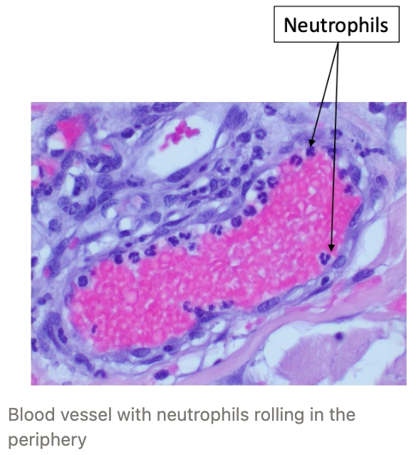
TWO: ROLLING
THREE: ADHESION TO ENDOTHELIUM
Adhesion Molecules = Proteins present on the TWO cell types (leukocyte/platelet and endothelial cell) which mediate adhesion of the two cells
Selectins = Adhesion molecules which mediate the initial/weak interaction between the endothelium and leukocytes to allow rolling
L-selectin (expressed on leukocytes)
E-selectin (expressed on endothelial cells)
P-selectin (expressed on platelets and endothelial cells)
Integrins = Proteins on leukocytes which mediate firm adhesion to the endothelial cells at the site of inflammation
Leukocytes normally express integrins in the low-affinity state
When the integrins are activated, they convert to the high affinity state to allow firm adhesion
Adhesion molecule expression enhanced by cytokines (TNK, IL-1 and chemokines)


FOUR: EMIGRATION ACROSS ENDOTHELIUM
Chemokines stimulate cell migration through the inter-endothelial spaces (via adhesion molecules between the endothelial spaces) towards the site of injury (towards the chemical concentration gradient)
Leukocytes penetrate the basement membrane and enter the extravascular tissue
In connective tissue, leukocytes can adhere to the ECM via integrins to be retained at the site where needed
FIVE: MIGRATION INTO TISSUES
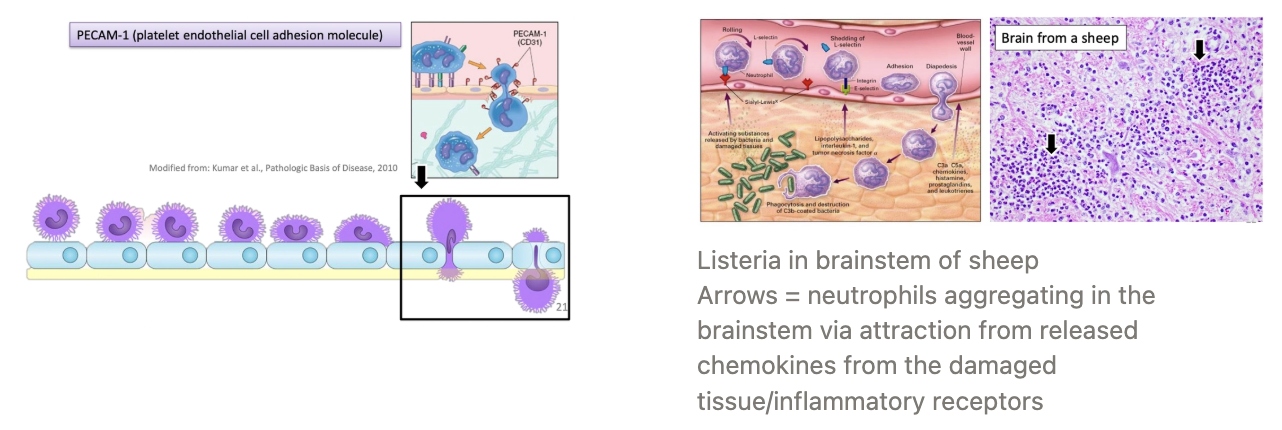
Chemotaxis of WBC
Definition
4 Chemoattractants
Definition: Mechanism responsible for the migration of leukocytes through tissue towards the inflammatory stimulus in the direct of the chemical gradient of locally produced chemoattractants
Chemoattractants:
Cytokines (esp. chemokine family = IL-8)
Components of complement system (eg. C5a)
Arachidonic acid metabolites (eg. leukotriene B4)
Exogenous = Bacterial products and some lipids
Describe the THREE steps of phagocytosis
ONE: RECOGNITION
Phagocytes have numerous receptors to recognise and bind specific microbes for phagocytosis
Enhanced by opsonisation of foreign material (triggers phagocytosis)
Opsonins = IgG, C3b, plasma lectins
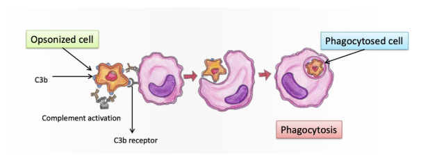
TWO: ENGULFMENT
The phagocyte's plasma membrane forms a vesicle around the bound microbe to create a phagosome
Phagosome fuses with a lysosome to form a phagolysosome which results in discharge of the lysosomal granules to breakdown and kill the microbe
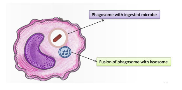
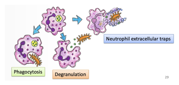
THREE: KILLING AND DEGRADATION
Respiratory burst: More reactive oxygen and nitrogen species derived from nitric oxide produced upon phagocytosis
Degranulation of neutrophils which release lysozymes into the ECF to kill bacteria extracellularly
Neutrophil extracellular traps
3 instances when leukocytes may cause injury to NORMAL cells
Prolonged inflammation leading to collateral damage
Autoimmune disease (inflammatory response directed against host tissues)
Allergic reaction (excessive host reaction against harmless environmental substance)
What 2 factors result in termination of the acute inflammatory response?
Inflammatory mediators are produced in rapid bursts and have short half-lives
Development of inflammation also triggers signals to terminate the inflammatory reaction:
Change of AA metabolites produced (from pro-inflammatory leukotrienes to anti-inflammatory lipoxins)
Liberation of anti-inflammatory cytokines from macrophages
Production of anti-inflammatory lipid mediators that inhibit production of TNF in macrophages
Overview of Mediators of Inflammation
Definition
2 Types
Definition
Examples
Definition: Any messenger that acts on blood vessels, inflammatory cells or other types of cells to contribute to an inflammatory response
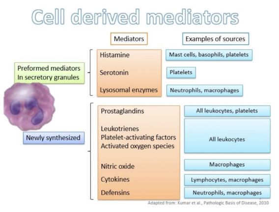
Types:
Cell-Derived = Mast cells, dendritic cells and platelets #1
Pre-Formed = Present normally in intracellular granules and 1st secreted during inflammation
Histamine
Serotonin (PLT)
Newly-Synthesised = Synthesised de novo (eg. arachidonic acid metabolites)
Eicosanoids
Cyclooxygenase (produce prostaglandins)
Lipoxygenase (produced leukotrienes and lipoxins)
Platelet-activating factor (PAF)
Reactive O2 species
Nitric oxide
Cytokines
Tumour necrosis factor (TNF0
Interleukin 1 (IL-1)
Plasma-Derived = Inactive precursors synthesised by liver and may be activated
Complement system
Fibrinolytic system
Coagulation system
Kinin system

Histamine
Sources
Type of mediator
Function
Degranulation stimulated by
Source: Mast cell degranulation (also basophils and platelets)
Type: Cell-derived, pre-formed inflammatory mediator
Function: Immediate transient phase of vascular response
Vasodilation of arterioles
Increased vascular permeability
Endothelial activation
Stimulation:
Physical injury
Binding of antibodies and complement
Neuropeptides
Cytokines
Arachidonic Acid Metabolites (eg. Eicosanoids)
Source
Type of mediator
2 Enzymes responsible for production
5 Functions (+ example eicosanoids)
Source: Fatty acid stored in the phospholipid membrane and released by phospholipases
Type: Cell-derived, pre-formed inflammatory mediator
Enzymes: Synthesise eicosanoids from AA
Cyclooxygenase → Prostaglandins
Lipoxygenase → Leukotrienes and lipoxins
Functions:
Action | Eicosanoid |
|---|---|
Vasodilation | PGI2 (prostacyclin), PGE1, PGE2, PGD2 |
Vasoconstriction | Thromboxane A2, leukotriene C4, D4, E4 |
Increased vascular permeability | Leukotriene C4, D4, E4 |
Chemotaxis, leukocyte adhesion | Leukotriene B4, HETE |
Inhibit inflammation | Lipoxin |

Platelet-Activating Factor (PAF)
Sources
Type of inflammatory mediator
3 Functions
Source: WBC and mast cells
Type: Cell-derived, pre-formed inflammatory mediator
Functions:
Platelet aggregation
Stimulates most vascular and cellular reactions of inflammation
Boosts synthesis of other mediators
Reactive O2 Species
Source
Type of inflammatory mediator
Function
Source: WBC
Type: Cell-derived, pre-formed inflammatory mediator
Function: Amplify inflammatory response
Nitric Oxide (NO)
Source
Type of inflammatory mediator
5 Functions
Source: Endothelium and macrophages
Type: Cell-mediated, pre-formed inflammatory mediator
Functions:
Vasodilation
Inhibit cellular response to inflammation
Reduce platelet aggregation and adhesion
Inhibit WBC recruitment
Killing microbes
Cytokines
Definition
Type of inflammatory mediator
2 Examples
Source
Function
Definition: Proteins produced by many cell types that modulate functions of other cells
Type: Cell-derived, pre-formed inflammatory mediator
Examples:
Tumour Necrosis Factor (TNF)
Source: Macrophages, mast cells, T lymphocytes
Function: Stimulates expression of endothelial adhesion molecules and secretion of other cytokines (systemic effects)
Interleukin-1 (IL-1)
Source: Macrophages, endothelial cells, some epithelial cells
Function: As for TNF (greater role in fever)
Importance of factor XII
Triggers ALL four systems
Intrinsic pathway of coagulation
Kinin system
Fibrinolytic system
Complement system
Complement System
Definition
4 Functions
Definition: Group of soluble proteins and membrane receptors which plays a key role in innate and adaptive immune defences
Activated to become proteolytic enzymes that degrade other complement proteins to form an enzymatic cascade
Functions:
Inflammation (C3a, C4a and C5a = anaphylatoxin)
Anaphylatoxin = Stimulates histamine release from mast cells (increases vascular phase of inflammation) Similar to allergy antigens = anaphylaxis
Chemotaxis (C5a for neutrophils, monocytes, eosinophils and basophils)
Phagocytosis (C3b = opsonin)
Cell lysis (MAC: C5b - 9)
MAC = Membrane attack complex which makes bacteria permeable to water and ions = cell death
Describe THREE outcomes of acute inflammation (+ circumstances of each)
Complete Resolution = Tissue site restored to normal (removal of cellular debris and microbes by macrophages and resorption of oedema by lymphatics) which requires:
Short-duration of stimulus
Minimal tissue damage
Damaged cells have high proliferative capacity and can regenerate
Healing by Fibrosis (Repair) = Tissue site repaired with fibrous connective tissue which requires:
Substantial destruction (esp. to ECM or stromal framework or crypts or basal membrane)
Destruction of cells incapable of regeneration (eg. heart and brain)
Abundant fibrin in exudate that cannot be clear
Chronic Inflammation = Inflammation of prolonged duration in which tissue injury, inflammation and attempts at repair, coexist in varying combinations due to:
Persistent infection of microorganisms that are difficult to eradicate (eg. Mycobacterium spp.)
Immune-mediated inflammatory diseases (eg. type III hypersensitivity)
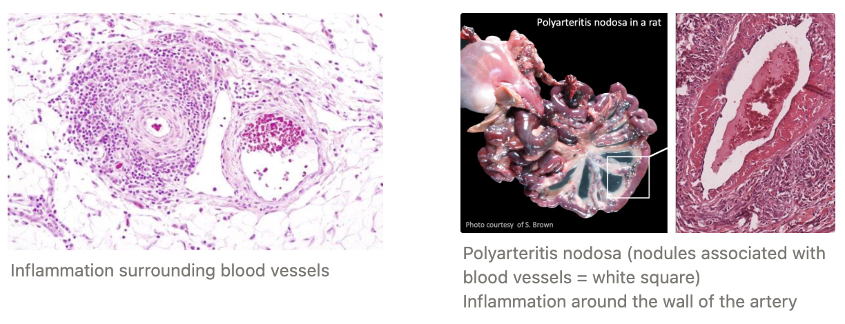
Prolonged exposure to toxic agents
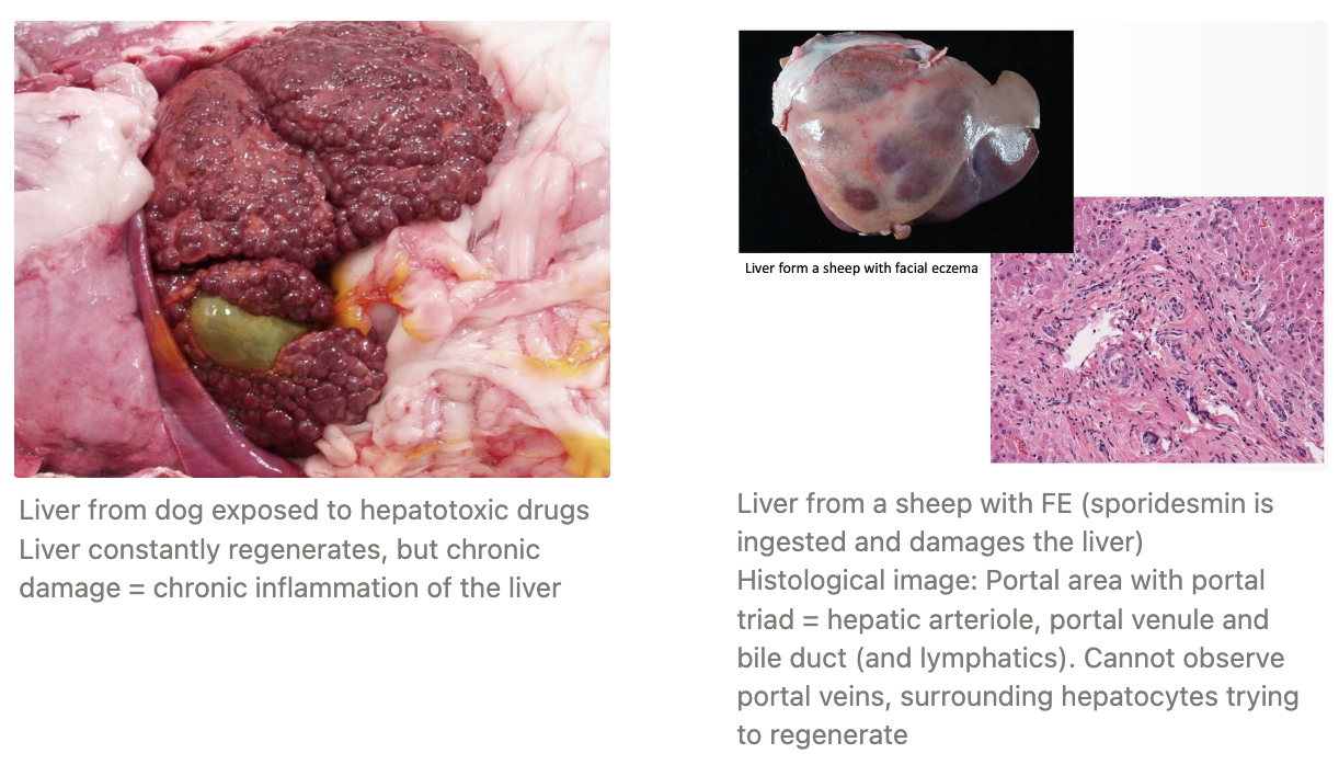
What are 3 morphological features of chronic inflammation?
Infiltration with mononuclear cells (macrophages, lymphocytes and plasma cells)

Healthy tissue destruction by inflammatory cells and persistent offending agent

Attempted healing by replacing damaged tissue with connective tissue (granulation tissue formation)
Proliferation of small blood vessels to vascularise new tissue formation

How does granulation tissue (immature) differ from fibrosis (mature granulation tissue) on histology?
Immature = Disorganised mesenchymal cells with tiny blood vessels and oedema in the tissue
Mature = Fibrosis with organised fibroblasts and blood vessels
Macrophage
Importance
6 Functions
Importance: #1 Cell player in chronic inflammation as they have pro-inflammatory properties to stimulate leukocyte recruitment, phagocytosis and microbial killing AND anti-inflammatory properties to stimulate healing
Functions:
Highly phagocytic
Antimicrobial activity (NO)
Expresses tissue factor which may stimulate extrinsic coagulation pathway
Important Ag-presenting cell to drive the immune response
Stimulates chemotaxis and activation of inflammatory cells and fibroblasts
Secretes cytokines and growth factors to stimulate fibroblast infiltration and angiogenesis = REPAIR

What are 7 systemic effects of inflammation?
Fever
Leukocytosis = High WBC count
Acute-phase proteins
Increased pulse and BP
Shivering
Chills
Anorexia
3 Factors influencing balance of regeneration vs. repair
Proliferative capacity of the cells
Cells with high proliferative capacity continuously renew their cells and are able to regenerate after injury which means the organ is more likely to have restored normal function
Integrity of the ECM
Resolution OR chronicity of injury and inflammation

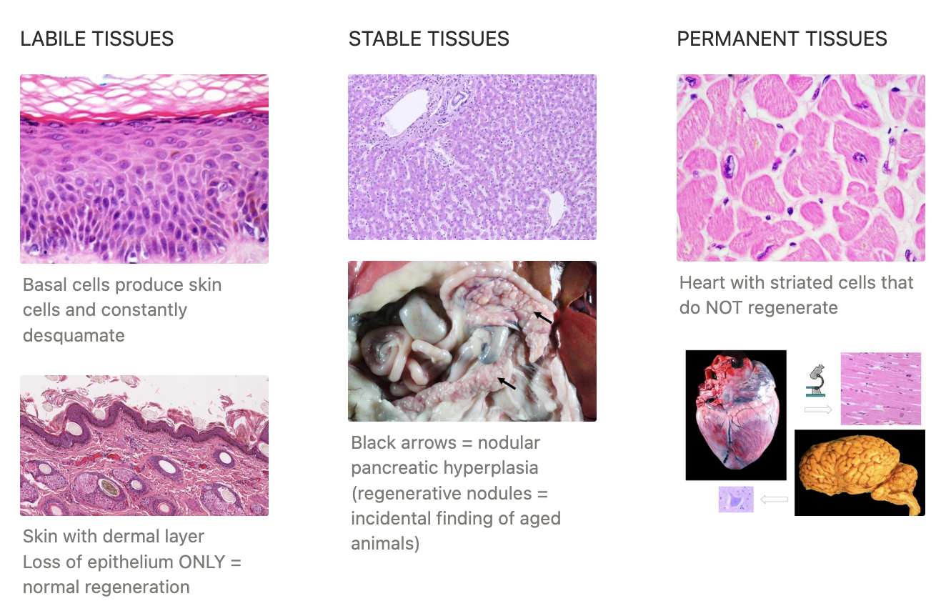
THREE classes of tissues based on proliferative capacity
Definition
Examples
ONE: LABILE TISSUES
Definition: Contain undifferentiated stem cells that are continuously diving and proliferate throughout their life, replacing those that are lost
Examples:
Mucosal epithelium
Bone marrow cells and haematopoietic tissues
TWO: STABLE TISSUES
Definition: Contain quiescent cells with low levels of replication
The cells are usually in the rest phase of the cell cycle, but undergo rapid division in response to injury (stimulated by growth factors)
Examples:
Mesenchymal cells
Vascular endothelial cells
Parenchymal cells of the liver, kidneys and pancreas
THREE: PERMANENT TISSUES
Definition: Terminally differentiated non-proliferative cells
Damage to these cells will result in loss of function as they repair by scar tissue formation
Examples:
Cardiac cells
Nervous tissue
Describe FIVE stages of healing by repair/scar tissue formation/fibrosis
Inflammation
Angiogenesis via vascular endothelial growth factor (VEGF)
Branching and extension of adjacent PRE-EXISTING vessels
Recruitment of endothelial progenitor cells from bone marrow
Migration and proliferation of fibroblasts
Scar formation = Deposition of connective tissue containing collagen and other ECM components
Connective tissue remodelling
What are the 3 phases of cutaneous wound healing?
Inflammation
Proliferation
Maturation
Granulation Tissue
Appearance
Structure
Proud flesh
Appearance: Pink, soft, granular appearance due to the presence of new small vessels and proliferation of fibroblasts
Oedema as new vessels are leaky to allow passage of plasma proteins into ECF
Structure:
Capillaries
Fibroblast
ECM (with collagen)
Proud Flesh: Exuberant granulation tissue formed on the distal limbs of horses which inhibits epithelialisation and potentially results in loss of function

Factors that Influence Wound Healing
4 Systemic factors
4 Local factors
Systemic Factors:
Poor nutrition (eg. protein or vitC deficiency)
Metabolic disorder (eg. diabetes mellitus = microangiopathy)
Poor tissue perfusion
Hormones (GC are anti-inflammatory and can inhibit collagen synthesis)
Local Factors:
Infection #1
Mechanical (compressing blood vessels and wound dehiscence)
Foreign bodies
Size, location and type of wound
What are five disadvantages of repair?
Deficient scar formation resulting in dehiscence
Ulceration due to inadequate vascularisation
Excessive formation of repair components (exuberant granulation or hypertrophic scars and keloids)
Deformed tissue due to excessive contracture
Fibrosis impairs function (depends on tissue type)
Overview of Plasma Proteins
2 Types
4 Functions
Age-related changes
Types:
Albumin
Globulin
Functions:
Exert COP
Participate in immune/inflammatory responses and clotting processes
Nutritive functions
Aid maintenance of acid-base balance
Age-Related Changes: Younger = Lower TP as colostrum is metabolised
Consider use of adult RR
Albumin
% of protein serum
Source
Half-life
6 Functions
Serum: 30 - 35% albumin (65 - 70% other protein)
Source: Synthesised in liver
Half-Life: Differs between species
Dogs: ~8 days
Horses: ~19 days
Cattle: ~2 - 3 weeks
Functions:
Maintains COP (oncotic pressure)
Assists transport of sparingly soluble substances to prevent their loss through the kidney
Transports 40% total serum Ca
Source of amino acids in peripheral tissues
Antioxidant and buffer
Negative acute phase protein
THREE Groups of Globulins
Source
Function
Alpha-Globulins
Source: Liver
Function: Transport proteins and inactivate proteases (anti-inflammatory proteins)
Beta-Globulins
Source: Liver
Function: Transport, immunoglobulins (IgM and IgA) and complement (C3)
Examples: Fibrinogen, prothrombin and plasminogen (plasma NOT serum)
Gamma-Globulins
Source: B lymphocytes/plasma cells
Function: Increase IgG during immune response
Refractometry
Function
4 Disadvantages
Function: Measures the total solid concentrations in different fluids (plasma, serum, urine, exudates)
For plasma, it measures a refractive INDEX as an estimate for the total protein
-ve:
High concentration of glucose, urea, Na or Cl → falsely high TP
Lipaemia (milky serum) = turbidity alters light transmission → falsely high levels
Haemolysis (true or fractious animal or poor preservation of sample) → blurred line
Bilirubin (icteric) → altered colour NOT reading
Serum Protein Electrophoresis (SPE)
Function
MoA
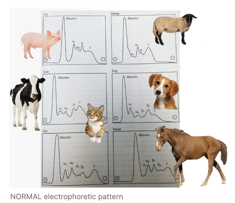
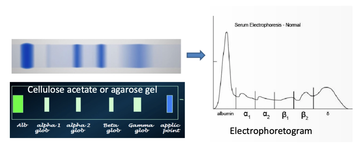
Function: Used to separate proteins into multiple bands (fractions) based on their size and charge → Determine cause of hyperglobulinaemia
Innate/acquired IMMUNE RESPONSE
Neoplastic lymphoid proliferation
MoA: When exposed to an electrical current across a gel, the proteins migrate across it at varying amounts depending on size and charge (eg. albumin is the smallest with the largest charge = further migration)
Protein bands are then translated by a densitometer into tracing to produce an electrophoretogram
Most commonly caused by dehydration (assess PCV to determine)
Main cause of hyperalbuminaemia
Relative hyperproteinaemia
Increase in albumin AND globulin concentration (NORMAL A:G ratio)
Dehydration #1 (assess PCV to determine)
4 Causes of hypoalbuminaemia (+ examples)
Decreased Production
Maldigestion (exocrine pancreatic insufficiency) and malabsorption
Insufficient supply of amino acids = decreased albumin production
Malnutrition/cachexia (dietary deficiency or chronic parasitism)
Insufficient supply of amino acids
Advanced hepatic disease
Catabolic states (pregnancy, lactation, malignant neoplasia)
Increased Loss/Catabolism
Renal disease with chronic proteinuria (protein-losing nephropathies)
Severe exudative skin diseases
Sequestration within body cavities in protein-rich effusions
PLE
Fluid Therapy = Dilutes plasma
Acute Inflammation
Albumin = Negative APP (mild decrease to divert resources away from albumin production to the production of globulins like fibrinogen and immunoglobulins)
3 causes of hyperglobulinaemia
Haemoconcentration (dehydration)
Increased alpha and beta globulins during an acute phase reaction (increased APPs) OR gamma globulins during a chronic phase reaction
Neoplasia (polyclonal gammopathy)
Acute Phase Proteins (APPs)
Definition
Importance
Functions
When does synthesis begin?
Stimulated by
Classification
3 Examples
Definition: Plasma proteins that are increased (+) or decreased (-) during any type of inflammatory response due to ANY type of injury, infectious disease OR non-infectious disease
Alpha-globulins #1
Importance: Monitoring APPs overtime can help monitor acute inflammation/chronic disease over time (if there is resolution or not)
Functions: Immune response, protect against oxidative stress and have anti-infective properties
Synthesis: Hepatic synthesis begins 24hr after acute injury
Stimulated By: Cytokines (IL-1, IL-6 and TNF)
Classification: Major/moderate/minor depending on the speed and magnitude of increase
Major = Increase in concentration by 100 - 1,000x after stimulation with peak in 24 - 48hr (fall rapidly on recovery)
Moderate = Increase in concentration by 5 - 10x with peak in 2 - 3d (decreases more slowly on recovery
Examples:
Fibrinogen (Fb)
Serum amyloid A (SAA)
C-reactive protein (CRP)

Fibrinogen (Fb) as an APP
When does it increase?
Class of APP
Importance
Species
Time: Increases during acute AND chronic inflammation
Class: Moderate APP and late responder
Importance: Best know APP as it is the greatest proportion of plasma protein produced during acute phase response
Species: Superior to TWBCC (hyperfibrinogenaemia precedes neutrophilia in large animals)
Cattle
Horses
Serum amyloid A (SAA) as an APP
When does it increase?
Class of APP
Importance
3 Species
Time: Rises in 12 - 24hr and peak 2d after injury (returns to normal after 5 - 7 days)
Class: Major APP for many species (rapid responder)
Species:
Horses for inflammation, monitoring efficacy of therapy and post-op recovery
Cattle and dogs as magnitude of SAA = severity of inflammation (not tests offered in NZ)