Lecture 10
 Call Kai
 Learn
 Practice Test
 Spaced Repetition
 Match
 Flashcards
 Knowt Play
1/62

Earn XP
Description and Tags
vision part 2
63 Terms
intrinsically photosensitive ganglion cells express ____
melanopsin
melanopsin
enables direct depolarization
rods express ___
rhodopsin
cones express ___
opsins
rhodopsin convert ___ —> ___ when exposed to light
11-cis-retinal, all-trans-retinal
phototransduction in the light
__
__
__
light activates transducin, cGMP is hydrolyzed, closure of Na and Ca channels
transducin is a _______
2nd messenger
phototransduction in the darkness
__
__
__
increased cGMP is present, opening of Ca and Na channels, NT release
ON-center ganglion cells have ___ APs with light in receptive field
more
ON-center ganglion cells have ___ APs with decreased light in receptive field
no
OFF-center ganglion cells have ___ APs with light in receptive field
no
OFF-center ganglion cells have ___ APs with decreased light in receptive field
more
ON/OFF ganglion cells
__
__
respond transiently to increases and decreases in light, located at receptive field center
ON bipolar cell in increased light in center
__
__
__
__
__
center cone cell hyperpolarized, ON bipolar cell depolarized, glutamate released, Glutamate binds to NMDA and AMPA receptors, ON center ganglion cell fires
ON bipolar cell in decreased light in center
__
__
__
__
center cone cell depolarized, glutamate released, glutamate binds to mGluR6 receptor, ON bipolar cell hyperpolarized
OFF bipolar cell in increased light in center
__
__
center cone cell is hyperpolarized, OFF bipolar cell is hyperpolarized
OFF bipolar cell in decreased light in center
__
__
__
__
__
__
__
center cone cell depolarized, glutamate released, glutamate binds AMPA, bipolar cell depolarized, glutamate released, glutamate binds AMPA and NMDA, ganglion cell fires
horizontal cells use ___
GABA
amacrine cells use ___
GABA, glycine, Ach, dopamine, glutamate
lateral interactions in the retina allow _____
ganglion cells to be sensitive
retino-recipient brain regions
process image forming info
retino-no-recipient brain regions
process non image forming info
list retino-recipient brain regions
LGN, V1, superior colliculus
list retino-no-recipient brain regions
superchiasmatic nucleus, olivary pretectal nucleus
the superchiasmatic nucleus is located in the _____
hypothalamus
retina
decodes info about color, contrast, luminence, and motion
retinal ganglion cells
transmit feature specific info in parallel channels
parallel channels
__
__
__
__
__
__
__
__
__
retina, retinal ganglion cells, optic nerve, optic chiasm, optic tract, ganglion cell axons terminate on retino brain regions, optic radiation, V1
retino-geniculo-cortical pathway
__
__
__
retina, LGN, layer 4 of V1
thalamocortical pathway: ____ to ____
LGN, V1
visual field deficits
results from damage to retino-geniculo-cortical pathway
anopsias
large visual field deficits
hemianopsia
half of both visual fields are affected

where would this deficit be?
right optic nerve

where would this deficit be?
optic chiasm
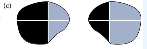
where would this deficit be?
right optic tract

where would this deficit be?
right V1
the LGN has __, __, and __, ganglion cells
P, M, K
K ganglion cells are _____
in between layers
P ganglion cells are projected from_____
cones
M ganglion cells are projected from ____
rods
cerebral cortex
integrates info arriving at V1
the V1 has ____ arrangement
columnar
V1 Layer 1:
outermost
V1 layer 6:
innermost
V1 layers 2, 3, 5, 6:
__
__
hold pyramidal neurons, release glutamate to subcortical and cortical targets
V1 layer 4:
__
__
__
has spiny stellate neurons, target of LGN axons, appears as stripes (striate cortex)
preferred orientation
__
__
peak of tuning curve, orientation of a stimulus to which a cortical neuron maximally responds
edges
neuron’s preferred orientation
extrastriate visual areas
other cortical areas devoted to visual processing of info from V1
ventral stream
__
__ to __
__
recognize object or scene, V1 to inferior temporal cortex (temporal lobe), what pathway
dorsal stream
__
__ to __
__
analysis of motion and spatial relationships, V1 to medial temporal area (parietal lobe), where pathway
Pupillary light reflex purpose
let in more light in dark conditions
Pupillary light reflex steps
__
__
__
__
__
retina, pretectum, EW nucleus, ciliary ganglion, pupillary constrictor muscle
accomodation is controlled by ____
EW neurons
accomodation when viewing something near vs far:
ciliary muscle thickens and round up lens
how lens shape is determined
zonule fibers exert tension vs lens elasticity
near response
when viewing a close object, accommodation, convergence, and constriction are required for optimal activity
extraocular eye muscles
redirect focus
conjugate eye movements
coorperating extraocular eye muscles so that binocular fixation is maintained
smooth pursuit
eyes shift position in a slow, smooth, continuous motion to track a moving object, ie watching a plane
saccadic eye movements
rapid eye movements to redirect focus, ie watching a baseball in a game
saccadic suppression
prevents blurring of object as it sweeps across retina