Neuropsych Exam 2
 Call Kai
 Learn
 Practice Test
 Spaced Repetition
 Match
 Flashcards
 Knowt Play
1/52

There's no tags or description
Looks like no tags are added yet.
53 Terms
What is histology?
What do the Golgi, Nissl stains, myelin staining procedures show of the microscopic structure of the brain?
Histology - the study of the structure and morphology of tissue
Gogli Stains - silver chromate and brain tissue stains some neurons black, just neurons can’t see in brain
Nissl Stains - doesn’t stain axons, you can only see cell bodies
Myelin Stains - distribution/intensity of myelin sheath on neurons
How do cranial X-rays work in general? What structures or damage show up how?What are the advantages and disadvantages of cranial X-rays?
Cranial X Rays - discrimination of low and high density regions in the body
High density → white Low density → dark
Advantages - picks up tumors, skull fractures, hemorrhages, cheap and easily available
Disadvantages - poor discrimination between brain tissue and CSF, only 2D images, poor structural resolution, cumulative radiation
Explain how angiography works and what it is used for
Angiography - visualization of cerebral vasculature
catheter inserted via an external artery and guided close to entry of arterial supply of brain
contrasting dye is injected → of diagnostic use only
Aneurysms and tumors (weak walls, mesh tube inflate balloon
What is the general process in computerized axial tomography, how is it different from an X-ray? What disadvantage of X-rays goes for CT scans also? Which has the higher radiation dose: a skull X-ray or a head CT? why is that
Computerized Axial Tomography (CT) - X-ray scanner rotated slowly until a measurement has been taken at each angle and computer constructs an image
Thin X-ray beam → slices of brain constructed along each planes of axis
CT has more radiation as it is multiple X-rays combined (Head CT 100 Chest X-rays)
How in general does an MRI work? What are the advantages of MRIs? What types of things can be measured with MRIs, and how can it be used in research?
MRI - application of powerful magnetic field to image the brain
magnetic field aligns the axes of the natural spin/rotation of atoms in water molecules → radiofrequency causes axes to spin, termination → stop spinning
Released energy is measured and used to visualize the structure of the brain
Advantages - no radiation, non-invasive, soft tissue contrast
Disadvantages - high cost, long scan times, claustrophobic
Explain what DTIs can image, and in what types of disorders this is interesting information. What descriptive research can be done with DTIs?
Diffusion Tensor Imaging (DTI) - MRI in which water diffusion at a location is calculated
DTI shows preferred direction of diffusion → shows visualization of directional fibers
look for reduced/changes in diffusion in specific brain regions → demyelinating disorders
detect areas that might indicate partial/complete disconnection between brain regions
Diffusion Tractography - uses DTI to build an image of probable white matter tracts
What does an fMRI measure and how does it work in general? What process is used to visualize brain processing via BOLD imaging? What is difficult about an fMRI for patients, and what effect does this have for ability to use fMRI?
Functional Magnetic Resonance Imaging - uses oxygen consumption in the brain to provide a moving and dtailed image of the functioning brain
BOLD Signal - using subtraction methods to see which task/stimulus is responsible for activity in particular area of the brain
Can show level of activity and size/appearance of various areas of the brain
Disadvantages - claustrophobic, loud, scary for young infants/parents → only used for certain types of people
What are the various fMRI research approaches? What different things are revealed by task-based and resting-state research approaches? Why do resting state approaches allow for testing of different groups of people?Why would brain connectivity be of interest to researchers?
Task Based Approaches - activation levels and Multivoxel pattern analysis
Resting State Approaches - several netowrks in brain are active when you are just lying there
shorter period of time no tasks need to be done
Brain Connectivity - which areas connect
complex thoughts/behaviors come from the connection between brain areas
How does a PET work? What is PET used for? What are the advantages and disadvantages of PET? What is it that PET does that fMRIs do better? Which two types of PET imaging are used in research for Alzheimer’s disease?
Positron-Emission Tomography (PET) - functional, records emission of radioactivity from injected radioactive chemicals to produce a high resolution image
uses radioactively labelled glucose
Advantages: early detection of tumors (high metabolism)
Disadvantages: low spatial and temporal resolution (FMRI Better)
Amyloid PET imaging and rCBF → alzheimers research
What is the general process in single cell recordings? Who is this process used with?
Single Cell Recording - measures electrical activity in the brain (within neuron data)
electrode is inserted into brain adjacent to a neuron to record its activity
Most common with animals
Can record a single action potential or many action potentials
What is the general process in an EEG? What can be studied with EEGs? ERP, what is it, how does it work? What is it used for? What is function of averaging in ERPs? How is that like the subtraction process in fMRIs?
Electroencephalographic Recording (EEG) - records electrical potentials/brain waves in the brain
Reflects the collective and synchronous activity of neurons in the cortex using a generator
Used for - sleep studies, epilepsy diagnosis, depth of anesthesia, studying normal brain function (consciousness differences)
Event-Related Potentials (ERP) - brief change in a slow-wave EEG signal in response to a discrete sensory stimulus
Averaging signals takes out the background noise (just like subtraction in fMRI)
What is magnetoencephalography? What is it used for? What is optical imaging? In which types of cases can this be used when fMRI cannot?
Magnetoencephalography - records magnetic potentials produced by brain activity
requires special facilities - magnetic brain writing
used to help diagnose epilepsy
Optical Imaging - Information about source and time course of neural activity
thought to reflect blood flow to active areas
less scary/intense than fMRI (babies)
What types of recordings are part of a polysomnogram? What are they measuring, and why is this important?
EEG - main sleep measure
EOG. electricalocculogram (eye writing)
EMG - electromyogram (muscle writing)
What kind of waves during normal awake time on EEG? (amplitude, frequency, name) What kind of waves during relaxation on EEG? (amplitude, frequency, name) What 2 phenomena during stage 2 sleep on EEG? What kind of waves during deeper sleep (3 & 4) on EEG? (amplitude, frequency, name) What is NR3 or N3?
Alert/Awake State (1) - Beta Rhythm → high frequency, low amplitutde
Calm/Resting (2) - Alpha Waves → lower frequency, higher amplitude
Deep Sleep (3+4) - Delta Waves → slower waves, lower frequency, high amplitude
NR3/N3 is deep restorative sleep

How are the waves on an EEG during coma? How are they different from delta waves?
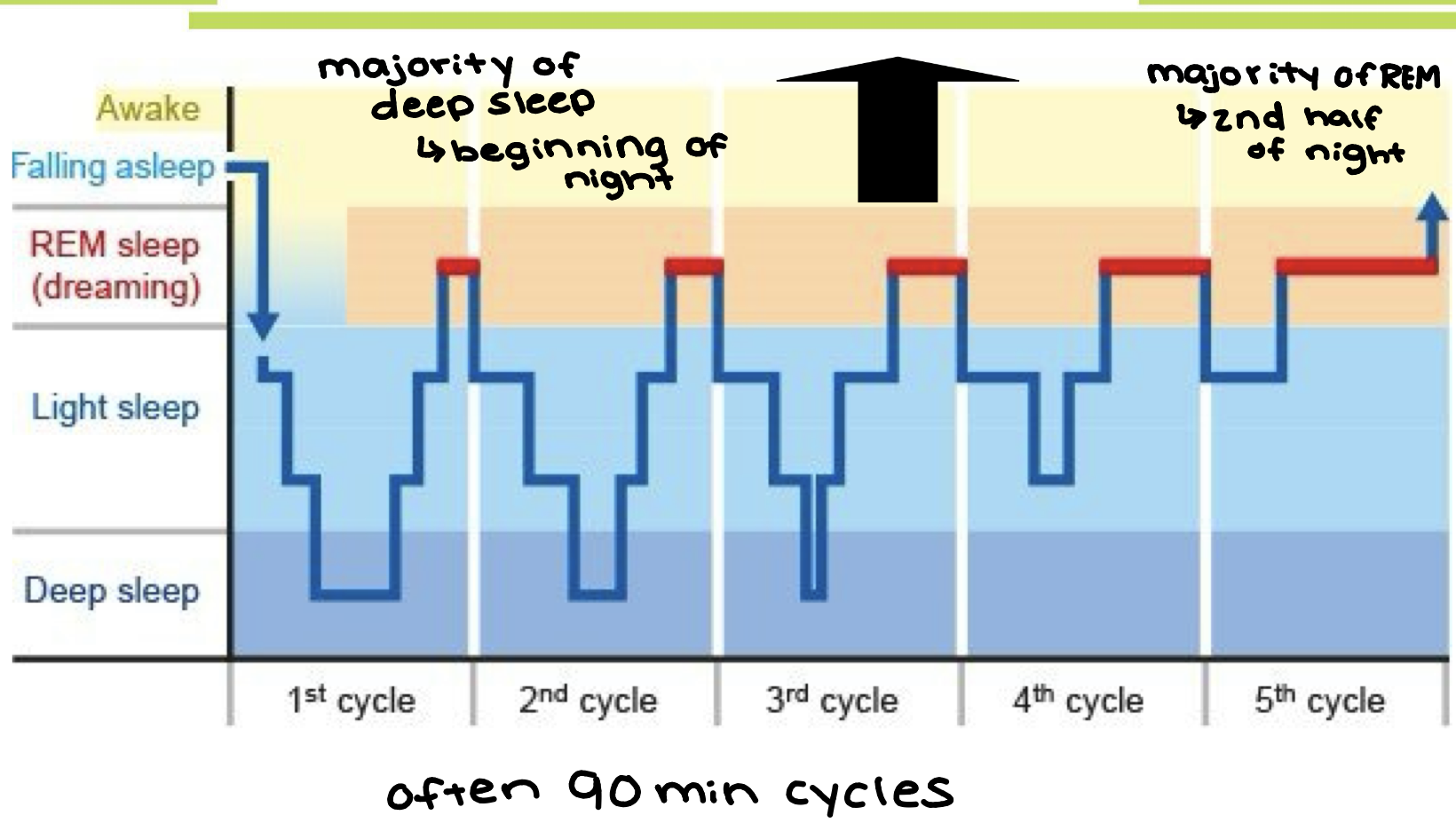
Explain the sleep cycles that one goes through in a normal night (both stages and REM/NREM alternation).
What is the trend for Slow wave sleep and REM sleep during the night? How many REM cycles during a ‘normal’ night?
Coma - NOT deep sleep → very slow low amplitude waves
Sleep Cycles - 90 min (NREM - 75%, REM - 25%)
majority of deep sleep at the start of night
majority of REM sleep 2nd half of the night (close to consciousness, neck down movement inhibited, rapid eye movement)
About 5 cycles of REM during the night
How does the % of REM sleep change during your lifespan? (infants, grandparents), How does the % figure obscure REM duration differences?
Babies - lots of REM (50% of their sleep is REM)
Adults - 25% of their sleep is REM
Older People - shorter sleep with less REM (18-20% REM but less sleep)
What is the circadian rhythm? Which sensory system acts on which part of the brain? What does the pineal gland have to do with the circadian rhythm? How does entrainment of the circadian system work (generally)?What is sleep pressure? What is it thought to be due to?
Circadian Rhythm - natural internal 24 hour clock that tells you when its time to sleep
Superchiasmatic Nucleus (SCN) in hypothalamus sends signals
Light-entraining → synching bodily clock to light sources outside
Melatonin from the pineal gland
Sleep pressure and adenosine
Sleep pressure → longer you stay up the more your body needs sleep
What are the four main views on the function of sleep? Are they exclusive of each other?
What are the two processes related to consolidation of memories happens during sleep?
What were the findings recently with respect to CSF circulation and sleep? What does this have to do with Alzheimer’s disease? What is the glymphatic system?
Memory Consolidation - extreme fast forward/fragments of events in hippocampus + V1, replay of events and file transfer to remember what you need
Neil deGrasse Tyson movie → memory consolidation during REM (sleep before test)
Cleaning the Glymphatic System - more active CSF circulation during sleep, glymphatic system cleans → helps break down beta-amyloid, which leads to alzheimers
Recuperation + Repair - most growth as a kid happens during sleep, when sick you should sleep to let the body recover
Evolutionary Reasons - humans have bad night vision so it is safer to sleep at night, more you sleep higher on the food chain you are
What did your instructor mention was the ‘prelearning’ value of sleep? What are the two processes in which consolidation of memories happens during sleep? What was the fourth way in which you improve ‘knowledge’ during sleep? What was the fifth way in which sleep has possibly long term effects on your cognitive functioning?
Prelearning - need to be well rested in order to best learn new info
Consolidation of Memories
NR Replay
Spindles memory
Emotion Consolidation processing
Sleep “Clears” Hippocampus (facilitates learning the next day)
Alzheimers (beta-amyloid)
What are some of the cognitive effects of sleep deprivation? What are some physical effects of sleep deprivation? What are the emotional effects of sleep deprivation
Sleep Deprivation → increased sleepiness, faster sleep onset
Poor Mood, Poor Vigilance, Poor Executive Functioning/Cognitive Control
Physiological - temp decreases, BP increases, immune function decreases, hormonal changes, metabolic changes (hungrier)
How do alcohol and caffeine influence sleep? What is the significance of 90 minutes when you manipulate sleep?
Alcohol + Caffeine - delayed sleep onset, reduced sleep duration, loss of deep sleep, long half-life
Sleep Manipulation - 90 minutes so you can get a full sleep cycle in!
What are three types of insomnia based on how long they last? What are three sleep disturbance patterns that count as insomnia? What is the prevalence of insomnia? What sleep hygiene measures were discussed in class? How does blue light have a poor effect on sleep?
Trouble falling asleep at the start
Waking up in the night and trouble going back to sleep
Waking up too early
Acute - less than 1 month, Transient - less than 1 week, Chronic - more than 1 month
Prevalence - 1 in 4 adults
Sleep Hygiene - regular bedtime, cool/quiet/dark, relax prior to bedtime, no screens, no food 2-3 hours, no exercise 2-3 hours, watching caffeine/alcohol
can help treat insomnia
What happens in sleep apnea (in general)? What is the prevalence of sleep apnea? How does obstructive sleep apnea ‘work’? What is central sleep apnea? How much of sleep apnea is obstructive as opposed to central (9 out of 10)? What is the standard therapy for sleep apnea? What is the main problem with this therapy?
Sleep Apnea - blockage of airway during sleep
Obstructive - tongue blocks airway (most common sleep disorder 25% of people)
Central - CO2 so low, body has no incentive to breathe
9/10 cases obstructive
CPAP - continuous positive airway pressure → pressed air lifts tongue to cure blockage
problem is its uncomfy and nobody wants to wear it
How common is narcolepsy? What are the symptoms of narcolepsy that were discussed in class? Explain the difference between cataplexy and sleep paralysis. Which parasomnias are REM and NREM parasomnias
Narcolepsy - excessive daytime sleepiness
sleep attacks (falling asleep randomly), cataplexy (lose muscle tone), hypnagogic hallucinations (hallucinations right before sleep), sleep paralysis
Cataplexy - fully awake; Sleep Paralysis - transitioning from sleep to awake and generally shorter
Parasomnias
NREM - sleepwalking, sleep terrors, confusional arousals, sleep-related eating disorder
REM - REM sleep behavior disorder (common symptom of Parkinsons → use of limbs during REM), Nightmare disorder, Recurrent sleep paralysis (most people have it 1 or 2 times in life)
Explain the distinction between epilepsy and seizures. What is the prevalence of epilepsy?
Describe the grand mal and petit mal seizure (old) classification. What types of seizures are these on the new classification?
What is the difference between a focal and generalized seizure? Where do they originate?
Epilepsy - not all seizures are a result of epilepsy, epilepsy = seizures generated by own brain dysfunction Prevelance - 1-4% of the population will have multiple seizure episodes (difficult to diagnose)
Grand Mal - loss of consciousness/equilibrium, tonic-clonic convulsions, rigidity (high tone) and tremors (big movements), resulting hypoxia may cause brain damage
Petit Mal - absence seizure, not associated with convulsions, disruption of consciousness, cessation of ongoing behavior
NEW CLASSIFICATIONS
Focal Seizure - does not involve the whole brain (starts in 1 part → focus)
Generalized Seizure - involves the entire brain (comes from thalamus → relay to rest of brain)
In which order are the brain lobes "epileptogenic"? How does a focal to bilateral tonic-clonic seizure progress?
Brain Lobes - temporal - 30-50%, frontal - 20-25%, parietal - 6%, occipital - 5%
Focal Preserved Consciousness - no alterations in consciousness, person is responsive and aware
Focal Impaired Consciousness - altered awareness AND sensory or motor symptoms
Focal to Bilateral tonic-clonic - begins in one place and spreads to both hemispheres
What are the stages of an epileptic attack? What modalities can the aura be associated with? What does tonic, clonic, atonic, and myotonic mean?
Stages - aura (lucky → tells you to lie down and predicts seizure), seizure, post-ictal
aura can be sensory, physical, emotional
Tonic - rigid, clonic - convulsions, myotonic - losing muscle tone, atonic - losing all tone
Why is the diagnosis ‘epilepsy’ or ‘seizure disorder’ a lengthy process to establish? What are the differential diagnoses? What types of therapy were discussed for epilepsy in class?
Diagnosis - careful history, what type of seizure was it, differential diagnosis means you have to rule out any other conditions (syncope, narcolepsy, migraines, panic disorder)
Therapy - surgery → take out focus, ketogenic diet could help symptoms
How many layers can most parts of the cortex be divided into? What are thought to be the functions of these layers?
How are the columns in the cortex organized, how is this different from the layers?
What is Brodmann’s division of the cortex into separate areas based on (i.e. why did he say these areas were different?)
Cortex is arranged into 6 layers → I, II, III do inputs and V, VI, do outputs
Neurons with similar properties for a column → each column consists of several laminae
Layers = function; Columns = content
Broadmann’s Map - cytoarchitectonic map → based on organization, structure, and distribution of cortical cells
What are the three parts of all sensory systems? Explain what it means that receptors only respond to a range of stimuli.
What is transduction when you are talking about receptors? What is the "common language" that stimuli get transduced to?
1. Sensory Receptors, 2. Neural Relays, 3. Central Representations in the Neurocortex
Sensory Receptors only respond to narrow band of stimuli/energy
some respond only to certain shape
Transduction - receptors transduce energy into action potentials our brains can use
Transduction in visual, auditory, somatosensory, taste, olfaction, and pain systems
Explain how receptors locate events, detect change and constancy and help to distinguish internal from external stimuli.
How does receptor density determine the sensitivity? Explain how stimulation of other types of receptors around a pain receptor can make pain appear less, and how this is an example of an interaction among different senses at the relay level?
Receptive Fields - locate sensory events, allow identification of change/constancy, allow distinction between self and other, determines sensitivity
most receptors tell you about outside world but interoceptive receptors tell you about self (vestibular and proprioceptor system)
Through Neural relays
hierarchy of motor responses
message modification takes place at relays (change perceptiveness of sensation → bruise there?)
sensory interactions (hit head + rub it helps because competing stimuli)
What does it mean that sensory systems have multiple representations in the brain?
1 sensory system is distributed through multiple parts of the brain
Ex. Visual System → SCN, eyes, pretectum, pineal gland, superior colliculus, optic nerve, and visual cortex all receive representations
Explain the functions of cornea, iris, lens, and retina. Be able to point out the above structures and the sclera and conjunctiva. Which structures refract light, in what proportions to they take care of the refraction?
Cornea - bends and refracts 80% of light entering the eye
Iris - contracts to change the size of pupil and adjusts amount of light coming in
Lens - bends and refracts 20% of light, highly variable in shape (accommodation → far away = flat) makes image sharp on retina
Retina - back of the eye that converts light signals into action potentials for brain
Sclera - white stuff outside cornea with blood vessels → pink eye
Conjunctiva - thin clear membrane that covers the front surface of the eye and lines the inside of the eyelids

Explain what the fovea and the blind spot on the retina are. How are they related to sensitivity of these areas? What is the macula?
What are the three cell layers of retina, and how does the light travel to get to the receptor layer? Why does this appear counterintuitive?
Fovea - high concentration of cones in the center of the retina, point fovea at what you are looking at to get clearest image
Blind Spot - where the optic nerve attaches to eye and nerves exit → creates blind spot
3 Layers of the Retina - Photoreceptor Layer, Bipolar Cell Layer, Ganglion Cell Layer
Light travels backward: ganglion → bipolar → photoreceptor and back out

What is the main pathway for vision called, what is its course? What % of fibers go this way?
What is the tectopulvinar pathway and what is its course?
Geniculostriate Pathway - most important! (90% of signaling)
axons convey information to lateral geniculate nucleus (thalamus)
then relayed to striate cortex (V1, primary visual cortex, BA 17)
Tectopulvinar Pathway - phylogenetically older (10% of signaling)
eye → superior colliculus → pulvinar (thalamus) → temporal and parietal lobe visual areas
pathway for pupilary constriction
What are the optic nerve, optic chiasm, optic tract, LGN, optic radiation, striate cortex, V1?
Which parts of the thalamus take part in the geniculostriate pathway and which in the tectopulvinar pathway?
Optic Nerve - bundle of nerves that connects the back of each eye to the brain
Optic Chiasm - crossover point for optic nerve fibers
results in contralateral projections from nasal ganglion cells
Optic Tract - relays information from optic chiasm to LGN
LGN - first key relay point in optic transduction
Optic Radiation - bundle of nerve fibers in the brain that carry visual information from the thalamus to the primary visual cortex
What is the visual field? Explain the overlap between the visual fields of the R and L eyes.
Explain which fibers from the two “hemifields” or "hemiretinas" or halves of the retina go where before/after the optic chiasm. Which hemifield is nasal and which is temporal?
What do “ipsilateral” and “contralateral” mean?
Visual Field - what you can see with one eye at any given point
overlap - what is seen by both eyes when looking at something
Temporal Hemiretina - inside visual field - info does not cross
Nasal Hemiretina - information from outside of visual field
Ipsilateral - stays within same side; Contralateral - Information crosses over
What does it mean that the representation in V1 (BA 17) is retinotopic? What is cortical magnification?
Retinotopic Representation - information as it is represented = exact correspondence to how it appears on the retina
Cortical Magnification - representation correlated to amount of receptors receiving stimuli
more cortical real estate for stuff on fovea
What do the simple, complex and hypercomplex cells respond to (see book)?
What is processed in MT (V5), and V4?
What is processed in the fusiform face area, the fusiform body area and the parahippocampal place area?
Simple - bars/lines/edges with a specific orientation at a fixed position
Complex - orientated bars or edges with movement
Hypercomplex - lines or bars with specific optimal length, corners, edges, or angles
MT (V5) - motion, important for action
V4 - color, important for deciding what something is
Fusiform Face Area - face analysis
Fusiform Body Area - body parts
Parahippocampal Place Area - landscape/landmarks
What is a scotoma? What is hemianopia?
Know what the effect for the visual field is of lesions along the optic tract
Scotoma - loss of vision in one local area, a blindspot (parts of visual field missing)
Hemianopia - loss of vision for one side of the visual field
Lesions on the optic tract → information does not get to striate cortex → blindness in areas
What are the symptoms of akinetopsia, what are they thought to be the result of? How common is this disorder?
Akinetopsia - inability to identify objects in motion
dysfunction of V5 cortex
Result of brain damage/TBI/Stroke
Extremely rare
What are the symptoms of achromatopsia, what are they thought to be the result of in the cerebral form? How common is this disorder?
What is the apparent function of V4 in terms of color perception?
Achromatopsia - loss of ability to detect color
Damage to V4 cortex → lack of integration of color and shape, lack of color constancy
1 in 30-50,000 people
Know the anatomy of the outer, middle and inner ear, be able to identify ear drum, oval window, round window, cochlea. Know the names of the middle ear bones, hammer, anvil, stirrup.
What are the receptors for hearing, and how are they situated with respect to the basilar membrane and the cochlea?
What is the relationship between the basilar membrane and tonotopic organization?
Outer Ear - Pinna
Middle Ear - Hammer, anvil, stirrup, (malleus, incus, stapes) and eardrum
Inner Ear - Cochlea and auditory nerve
Hair Cells → wider areas of ear canal hairs are wider and stiffer and respond to higher frequencies, floppier and thinner further in respond to lower frequencies
Tonotopic Organization → where cells are stimulated = frequency type

What is the name(s) of the nerve that transmits information from the cochlea?
What is the progression and transformation of information from the pinna onwards to the primary auditory cortex (i.e. outer, middle, inner ear (w/ details)
VIII - vestibulococlear, auditory, statoacoustic nerve transmits info from the cochlea
Pinna → eardrum → hammer, anvil, stirrup → cochlea
Cochlea → auditory nerve → cochlear nucleus → Ipsi/controlateral superior olivary nucleus → inferior colliculus → medial geniculate nucleus → primary auditory cortex → secondary auditory cortex
The auditory brainstem response test of newborn hearing (ABR) is a version of which test that we learned about?
What does it mean that the primary auditory cortex has a tonotopic organization?
What are other names for A1?
Auditory brainstem response test - EEG
neurons are arranged in an orderly map according to the sound frequencies they respond to best
A1 - herschl’s gyrus, Broadmann’s 41 and 42, transverse temporal gyri
Where is the secondary auditory cortex? What are the core, belt and parabelt areas?
Secondary Auditory Cortex - (belt) superior temporal gyrus in temporal lobe
Tertiary Auditory Cortex - (parabelt) adjacent to belt in temporal lobe
How does the ‘theory of object perceptions” in the slides match up (very generally) with the functions in the various parts of the primary, secondary visual cortex, lateral occipital complex, what may be happening in the (ventral) visual stream? (V1 to hippocampus)?
Visual Association
Primary Cortex → striate occipital cortex (BA 17) V1
Secondary Cortex → extrastrate occipital cortex (BA 18 + 19)
Tertiary Cortex → inferior temporal and posterior parietal cortex (NOT OCCIPITAL)
2 streams:
DORSAL - goest to posterior parietal cortex (the what pathway)
VENTRAL - goes to inferior temporal lobe (the where pathway)
What (generally) is the difference between the sparse coding and population/ensemble coding views of object perception?
What types of evidence are there for object-specific (grandmother) cells? What are some of the problems with hypothesizing object-specific neurons in shape perception?
Sparse Coding - one/few neurons represent one idea/item
Population/Ensemble Coding - many neurons and different levels of each represent items/ideas
Grandmother Cells
Evidence - halle berry neuron
Problems - what if neuron dies? what if grandma changed?
What is the difference between view-dependent and view-invariant shape perception?What is the evidence for these types of shape perception?
View-Dependent - you must see an object from a specific view/vantage point to recognize it
Evidence: reaction time studies and error rates
View-Invariant - no matter where/how you see the object you are able to recognize it
Evidence: structural component analysis, fast basic-level recognition
What does ‘agnosia’ mean?
What are the differences between apperceptive and associative visual agnosia both in how they present in patients?
Agnosia - failure to know
Apperceptive Visual Agnosia - no recognition of objects
Associative Visual Agnosia - inability to name what is seen
What is biological motion perception and what parts of the temporal lobe seems to be involved in this? What is the role of biological motion perception in social cognition?
Biological Motion Perception - motion based on biological entities
superior temporal sulcus (STS) analyzes biological motion (mouth/eyebrows) and respond to approach motions of body
Damage STS → impaired recognition and recall of faces and impaired perceptions of social signals
What parts of face processing are carried out in the Fusiform face area, the superior
temporal sulcus, the striate cortex and the occipital face area?
FFA - inferior temporal lobe → invariant aspects of faces (perception of unique identity)
STS - changeable aspects of faces (perception of eye gaze, expression, lip movement)
Striate Cortex - occipital lobe → initial processing of basic visual features
OFA - occipital lobe → early facial feature extraction (structural coding, identifying specific parts of the face)
What is prosopagnosia? Are there differences in degree? Is it congenital or
acquired?
Prosopagnosia - inability to recognize faces
Different levels of impairment in different people
Congenital and Acquired forms both exist
Some have severe face blindness since birth; others get from TBI