Anatomy Exam 2
 Call Kai
 Learn
Learn Practice Test
 Spaced Repetition
 Match
Match Flashcards
 Knowt Play
1/82

There's no tags or description
Looks like no tags are added yet.
Name | Mastery | Learn | Test | Matching | Spaced | Call with Kai | Chat |
|---|
No analytics yet
Send a link to your students to track their progress
83 Terms
What are the functions of the nervous system?
collect information, process and evaluate information, and respond to information
What is the difference between grey matter and white matter?
grey matter is darker in color, regions of the CNS made mostly of cell bodies and axons.
white matter is lighter in color due to its myelin content, regions of the CNS made mostly of axons.
Where is grey and white matter found?
grey matter - mostly outside the brain, inside spinal cord, ganglia (cell bodies) in PNS
white matter - mostly inside the brain, outside spinal cord, nerves (axons) in PNS
What are the parts of the CNS?
the brain and spinal cord
What are the structures of the PNS?
cranial nerves, spinal nerves, ganglia, peripheral nerve endings/receptors, nerve plexuses (cervical, brachial, lumbar, sacral)
What are the four divisions of the nervous system?
somatic sensory, visceral sensory, somatic motor, autonomic motor (visceral motor)
know the parts of a neuron
What are the glia and their functions of the CNS?
astrocytes, oligodendrocytes, microglia, ependymal cells
What are the glia and their function in the PNS?
Schwann cells, satellite cells
Know the parts and connective tissue layers of a nerve.
What are the four divisions of the brain?
Cerebrum, Diencephalon, Cerebellum, and Brain Stem
Identify structures on the external and internal views of the brain
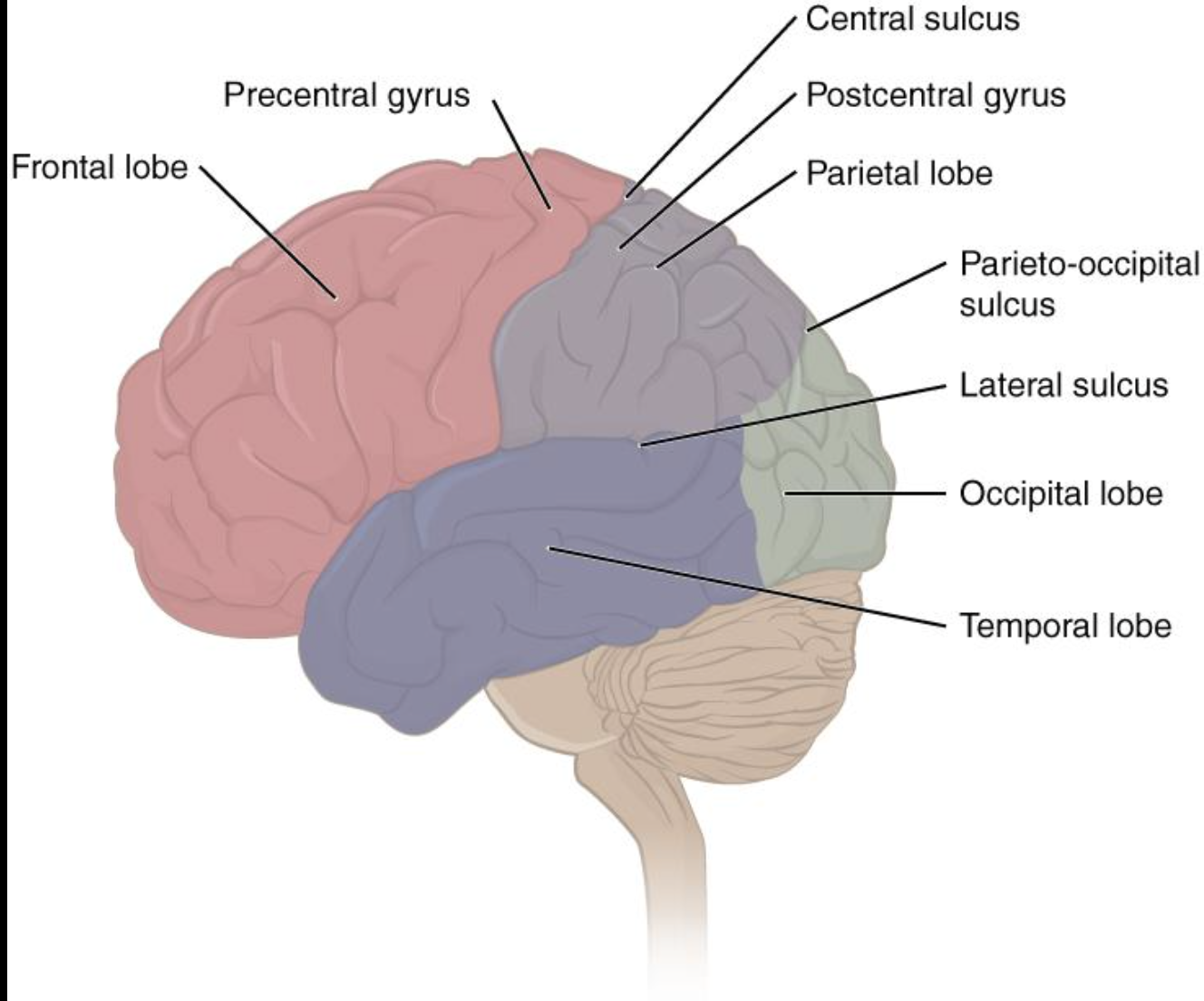
What are the lobes of the cerebrum, etc? Know the major sulci and hyri
frontal, temporal, parietal, occipital, insula lobes.
Hypothalamus
helps maintain homeostasis
Epithalamus
houses the pineal gland (endocrine gland which secrets melatonin)
Thalamus
Relays all somatosensory, visual, and auditory information (except for smell)
Brain stem (in general)
Connects cerebrum to spinal cord, start of cranial nerves, autonomic and reflex centers.
Cerebellum
fine tunes and tweaks body movements, coordinates body movements, maintain posture and equillibrium
Cerebral nuclei
Stops and regulates the intensity of voluntary movements.
Corpus Callosum
Provides the major pathway for communications between the left and right hemispheres of the brain
Broca’s area
Motor speech control
Wernicke’s area
Recognition and understanding of speech
Amygdala
Memory, decision making, and emotional response
Cingulate gyrus
Emotional response (limbic system)
Hippocampus
Memory storage (limbic system)
Primary somatosensory cortex
Receives information from somatic senses, enables conscious understanding of these
Primary motor cortex
Controls voluntary skeletal muscle activity
Primary visual cortex
Receives visual information from the retina of the eye.
Cerebrum (in general)
thinking, voluntary movement, sensory processing, memory, language, and emotions
Know the meningeal layers
dura mater - tough, outermost protective layer
arachnoid mater - middle, web-like layer
pia mater - thin, innermost layer that tightly follows the surface of the brain and spinal cord
What are the 3 layers of meninges?
Dura mater, Arachnoid mater, Pia Mater
Dura mater
outer meningeal later, thick and fibrous, strong protective sheath around the brain and spinal cord.
Arachnoid Mater
middle meningeal layer, thin, gauzy and fibrous, subarachnoid space is filled with mesh (arachnoid trabeculae)
Pia mater
deepest meningeal layer, touches the brain and spinal cord directly, conforms to the shape of underlying structures.
Know the vessels that supply the CNS with blood. Identify elements of the cerebral arterial circle.
supply the CNS with blood - internal carotid arteries, vertebral arteries
CAC: anterior cerebral arteries, anterior communicating artery, internal carotid arteries, posterior communicating arteries, posterior cerebral arteries
What are the arteries in the cerebral arterial circle?
R & L posterior cerebral arteries, R & L posterior communicating arteries, R & L internal carotid arteries, R & L anterior cerebral arteries, anterior communicating artery.
Identify the other arteries supplying blood to the brain. Where do they branch off from? Where do they travel to?
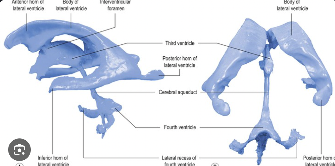
Know the parts of the ventricular system
What is CSF? What is its function?
cerebrospinal fluid
brain and spinal cord “float” in CSF
resists compressive forces and cushions against trauma
feeds brain, removes waste, and carries chemical signatures

Identify the dural venous sinuses
What is the pathway of CSF from production to the drainage via the internal jugular vein
Clinical Application: Multiple Sclerosis
autoimmune disease in which the immune system attacks myelin in the CNS
impaired vision, muscle weakness and spasms, urinary and fecal incontinence, mood swings.
Clinical Application: Alzheimer’s Disease
a progressive neurodegenerative disease that damages and destroys neurons in the brain, most common cause of dementia
memory loss, confusion and disorientation, difficulty thinking and making decisions, personality and behavior changes, difficulty communicating.
Clinical App: Meningitis
inflammation of the meninges, the protective membranes surrounding the brain and spinal cord
fever, stiff neck, sensitivity to light, extreme fatigue or lethargy
]
What are the functions of the spinal cord?
sensory and motor innervation of the body
two-way conduction (communication) pathway for signals between the body and the brain
center for reflexes

Identify the structures of the spinal cord (including cauda equina, conus medullaris, and filum terminale)
What are the 3 layers of meninges protecting the spinal cord?
Dura mater, Arachnoid mater, Pia mater
What are the spaces associated with the 3 layers of meninges?
epidural space - between dura mater and surrounding bone
subdural space - between dura mater and arachnoid mater
subarachnoid space - between arachnoid mater and pia mater
What are the anchors for the spinal cord?
denticulate ligament - side-to-side stabilization
filum terminale - anchors the bottom of the spinal cord
Identify structures of the spinal cord

What are the 5 steps of the reflex arc?
1.) stimulus activates a sensory receptor (external or internal stimuli)
2.) nerve impulse travels through the sensory neuron to the CNS (usually to spinal cord, rarely to brain)
3.) information processed by interneurons
4.) motor nuerons send action impulse to effector
5.) effector responds to nerve impulse
What are dermatomes? Why are they important for health care professionals to know?
areas of skin that receive sensory information from one specific spinal nerve level. each dermatome corresponds to a spinal nerve level.
we identify nerve damage, diagnose spinal injuries, perform neurological exams, etc.
How many cervical spinal nerves?
8
How many thoracic spinal nerves?
12
How many lumbar spinal nerves?
5
How many sacral spinal nerves?
5
How many coccygeal spinal nerves?
1
How are the spinal nerves numbered relative to the vertebrae?
named for the vertebrae that they exit near
Does C1 spinal nerve emerge superior or inferior to C1 vertebrae?
superior
What about the C8 spinal nerve? Since there are only 7 cervical vertebrae
emerges between C7 and T1 vertebrae
Cervical Plexus
C1-C5
Phrenic nerve - diaphragm (C3,C4,C5 keeps the diaphragm alive!)
Ansa cerivcalis - C1-C3: infrahyoid muscles
Brachial Plexus
C5-T1
Musculocutaneous - elbow flexion (biceps brachii)
Axillary - arm abduction (deltoid)
Radial - elbow, wrist and finger extension
Median - wrist/finger flexion and thumb opposition
Ulnar - finger abduction and adduction
Roots → Trunks → Divisions → Cords → Branches (Remember To Drink Cold Beverages)
Lumbar Plexus
L1-L4
Femoral nerve - motor function: hip flexion and knee extension (front of thigh), sensory function: anterior/medial thigh and medial leg (knee extension)
Obturator nerve - motor function: thigh adduction (bringing thighs together), sensory function: medial thigh
Sacral Plexus
L4-S4
Sciatic nerve - largest nerve in the body
Tibial nerve - plantarflexion (points toes down)
Common fibular nerve - dorsiflexion and foot eversion
Pudendal nerve - supplies perineum and external urethra and anal sphincters
Identify the proximal and distal ends of the brachial plexus. (Identify the roots, trunks, branches, cords and divisions)

List the Brachial and Cervical Plexus nerves (5)
List the Lumbar and Sacral Plexus nerves (4)
What is homeostasis? How does the body monitor its own function?
steady state of body systems that living organisms maintain, it monitors its own function through negative feedback system.
What is the ANS?
general visceral motor system of the PNS
What is the function of the ANS?
involuntary control of visceral functions, regulates systems’ homeostasis, controls all of the routine functions of our physiological systems.
How is the ANS different from the somatic motor nervous system?
What are the general effects of the sympathetic nervous system
maintain homeostasis when under pressure, increase heart rate, respiration, and adrenaline, dilates pupils and slows digestion, liver releases sugars (energy into blood)
What is the sympathetic chain/trunk?
interwoven structure of preganglionic neuronal axons and ganglia
What are the general effects of the parasympathetic nervous system?
maintains homeostasis when at rest, slows respiration and heart rate after exertion, increases blood flow to digestion, constricts pupils
Effects of the sympathetic NS
Effects of the parasympathetic NS
What are some functions of the endocrine system?
Hormone secretion - directly into blood or interstitial fluid for transport throughout the body
Homeostasis Maintenance - ensure the body environments are operating within optimal functional ranges.
Where are the hormones released in the endocrine system?
Through the bloodstream
Different endocrine glands, hormone produced, and target
What is the master gland?
The hypothalamus
What is another function of the pancreas?
What are the secondary endocrine glands?