Neurons, the Spinal Cord, and Spinal Nerves
 Call Kai
 Learn
 Practice Test
 Spaced Repetition
 Match
 Flashcards
 Knowt Play
1/56

Earn XP
Description and Tags
Chapter 19
57 Terms
What are the three major parts of a motor neuron?
The soma
Dendrites
Axon
Soma
Highlighted in blue

Dendrites
Highlighted in purple
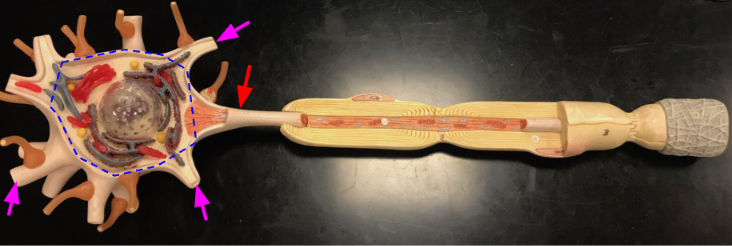
Axon
Highlighted in red

What are the major structures of the soma?
The nucleus
Nissl bodies
Nucleus
Highlighted in blue

Nissl body
Highlighted in purple

Dendrites
Emanate from the soma
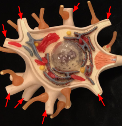
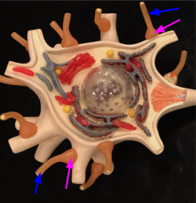
Telodendria
Highlighted in blue. They attach (synapse) their axon terminals onto dendrites and soma

Axon terminals
Highlighted in purple
Axon hilock
Emanates from the soma, just before the axon

Axolemma
Cell membrane of the axon

Neurolemmocytes (Schwann cells)
Makes myelin sheaths for axons in the PNS
Nodes of Ranvier
Gap between schwann cells that allow for messages to travel faster down the axon
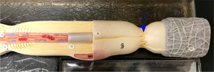
Neurilemma
Plasma membrane of a neurolemmocyte

Endoneurium
Loose connective tissue that covers the neurolemmocytes

Anterior median fissure
Highlighted in blue

What are the regions of the spinal cord?
Cervical
Thoracic
Lumber
Sacral
Coccygeal
Cervical spinal cord

Cervical enlargement of the spinal cord
A thickened portion of the spinal cord; due to many motor and sensory neurons that serve the upper limbs

What is a plexus?
A network of intersecting nerves, blood vessels, or lymphatic vessels in the body
Cervical plexus
Network of nerves formed by the anterior rami of spinal nerves C1-C4. Supplies nerves for the back of your head and the upper cervical muscles
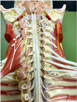
Phrenic nerve
Emanates from the cervical plexus and takes motor signal to the diaphragm

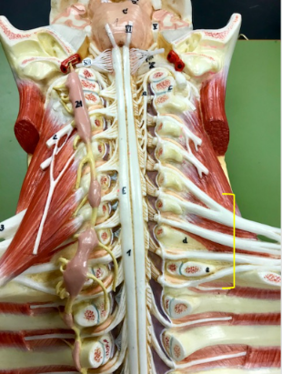
Brachial plexus
Network of nerves formed by the anterior rami of spinal nerves C5-T1. Serves your chest, shoulder, and arms
Thoracic spinal cord
Highlighted in blue. Sits alongside the ribcage

Lumbar region of the spinal cord
Highlighted in blue

Sacral region of the spinal cord
Highlighted in red

Coccygeal region of the spinal cord
Highlighted in purple

Lumbosacral enlargement of the spinal cord
Thickened area that contains numerous motor and sensory neurons serving the lower limbs
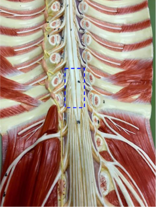
Conus medullaris
Tapered inferior portion of the spinal cord. Found generally at the L1 vertebra

Filum terminale
A strand of pia mater that anchors the conus medullaris to the posterior coccyx

Cauda equina
Collection of lumbar and sacral spinal nerves running through the vertebral canal

Lumbosacral plexus
Network of nerves formed by the anterior rami of spinal nerves

Obturator nerve
Takes motor and sensory signal to/from the medial thigh

Sciatic nerve
Take motor signals to the hamstring muscles and carries sensory information from most of the posterior thigh, leg, and foot

Spinal dura mater

Arachnoid mater

Subarachnoid space
Continuous with the 4th ventricle and contains CSF

Pia mater
Actually surrounds the spinal cord

Posterior median sulcus

Posterior funiculus
White matter region of the spinal cord located on the dorsal side. Conveys information on touch and proprioception

Lateral funiculus
White matter region. Provides sensory signals to the cerebellum and thalamus and motor commands from the cortex and brainstem

Anterior funiculus
Provide sensory signals to the thalamus as well as motor commands involved in balance and coordination
Anterior median fissure

Ventral horn of the spinal cord
Contains cell bodies of somatic motor neurons. Their axons will travel to skeletal muscles

Dorsal horn of the spinal cord
Contains axons from sensory neurons. Provides information from the body regarding touch and proprioception
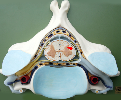
Grey commissure
Connects the 2 halves of the spinal grey matter

Central canal of the spinal cord
Contains CSF and is continuous with the 4th ventricle
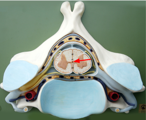
Dorsal root
Contains sensory axons bringing sensory information into the dorsal horn

Dorsal root ganglion
Contains cell bodies of the sensory neurons bringing sensory information into the spinal cord
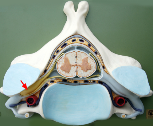
Ventral root
Contains the axons of the ventral horn motor neurons that will travel to skeletal muscles
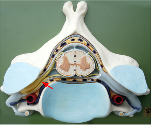
Spinal nerve
Contains sensory axons of the dorsal root and the motor axons of the ventral root and is therefore considered a mixed nerve
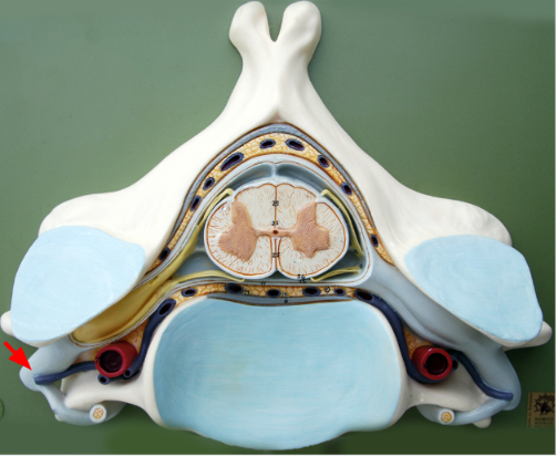
Lateral horn
Only found in the thoracic and upper lumbar parts of the spinal cord
Contain cell bodies of autonomic motor neurons that send axons from the ventral root to synapse with smooth muscle, cardiac muscle, and glands

Spinal nerve

What is the blue arrow
posterior ramus

What is the red arrow
anterior ramus

What kin of neurons are in this image?
multipolor motor meurons
