Introduction to Diagnostic and Medical Ultrasound
 Call Kai
 Learn
 Practice Test
 Spaced Repetition
 Match
 Flashcards
 Knowt Play
1/53

Earn XP
Description and Tags
Comprehensive practice flashcards covering the historical milestones, physical principles, transducer technologies, ultrasound imaging modes, and clinical scanning practices discussed in the lecture.
54 Terms
What fundamental concept of sound did Leonardo da Vinci discover around 1500 AD?
sound travels in waves and identified the angle relationship between a sound source and its reflection point.
What relationship concerning sound waves did Galileo discover in 1638?
He discovered the relationship between the frequency and pitch of sound waves.
How did Lazzaro Spallanzani demonstrate that bats navigate in the dark?
He showed that bats navigate using echo reflection from high-frequency inaudible sound (echolocation) rather than sight.
What discovery was made by Christian Doppler in 1842?
He discovered the connection between sound and motion.
Who discovered the piezoelectric effect in 1880?
The Curie brothers, Jacques and Pierre Curie.
What major contribution to medical ultrasonics did Dr. Karl Theodore Dussik make in 1942?
He used ultrasound to investigate the brain, producing the first published work on medical ultrasonics.
What experiment did physicist Jean Daniel Colladon perform using an underwater church bell?
He used it as an early ultrasound transducer to calculate the speed of sound through water, proving that sound travels faster in water than in air.

What is the direct piezoelectric effect? And how does it contribute to ultrasound?
pressure on a crystal creates an electrical charge. It helps convert returning sound waves into electrical signals to create images.
What is the reverse piezoelectric effect?
When alternating voltage is applied to a piezoelectric crystal, the crystal vibrates and produces high-frequency pressure waves known as ultrasound.
What type of energy does an ultrasound transducer send into the human body?
Mechanical energy.
How is reflected mechanical sound energy converted when it returns to the ultrasound transducer?
The piezoelectric crystals convert the returning mechanical pressure waves back into an electrical pulse to generate an image.
How is ultrasound defined in terms of human hearing?
Ultrasound is mechanically produced sound waves with frequencies higher than the upper limit of human hearing, exceeding 20,000Hz.
Why did diagnostic ultrasound develop much more slowly than X-rays?
Ultrasound imaging required advanced electronics and computers to process signals and produce images, which were not available early on.
What frequency range defines infrasound?
Frequencies below 20Hz.
What frequency range defines audible sound for human hearing?
20Hz to 20,000Hz.
What frequency threshold defines ultrasound?
Any frequency above 20,000Hz.
What frequency range is utilized in diagnostic ultrasound?
1,000,000to 20,000,000Hz (1MHz to 20MHz).
What is the clinical trade-off between high and low ultrasound frequencies regarding penetration?
Lower frequencies penetrate deeper into tissues, whereas higher frequencies have poorer penetration.
What is the clinical trade-off between high and low ultrasound frequencies regarding resolution?
Higher frequencies provide superior image resolution, whereas lower frequencies produce lower resolution.
What are the typical frequency range and penetration depth of a linear vascular transducer?
It operates between 3MHz and 9MHz and penetrates approximately 5to 6cm.
Which transducer is vascular?

What superficial anatomical structures are commonly imaged using a linear vascular transducer?
Carotid arteries, thyroid, testicles, breast tissue, and superficial lumps or bumps.
Why is the contact face of a cardiac transducer designed to be small?
It is designed small to allow acoustic access between the patient's ribs (intercostal scanning).
Why does a cardiac transducer use lower frequencies than a vascular transducer?
The heart is located deeper inside the body, requiring lower frequencies to achieve sufficient penetration depth.
Which one is a cardiac transducer?

What is the frequency range of the EC9-5 endocavity transducer?
5MHz to 9MHz.
What transducer shape and frequency range are typically used for general abdominal ultrasound?
A curvilinear transducer with a rounded face operating between approximately 5MHz and 8MHz.

How does acoustic attenuation in body fat compare to attenuation in muscle tissue?
Fat attenuates sound waves more than muscle tissue does.
What are the three echo sciences that contributed to the development of modern medical ultrasound?
Sonar, radar, and metal flaw detection.
What does the acronym SONAR stand for?
Sound Navigation and Ranging.
SONAR definition?
SONAR is a technique that uses sound propagation to navigate, communicate, or detect objects underwater, utilizing sound waves to measure distances and locate underwater features.
What maritime event accelerated the development of sonar technology?
The sinking of the Titanic.
What device did French physicist Paul Langevin develop in 1916 for military submarine navigation?
A high-energy ultrasound echo-sounding device (hydrophone / sonar transducer).
How does active sonar determine the distance to an underwater target?
It emits sound pulses through water, times how long it takes for the echo to return after reflecting off the target, and calculates distance using the known speed of sound in water.
What does the acronym RADAR stand for?
Radio Detection and Ranging.
What experiment did British physicist Edward Appleton conduct in 1924 using radio echoes?
He determined the height of the ionosphere.
Why can radar not be utilized underwater?
Radar uses radio waves in the microwave frequency range (wavelength approximately 1cm), which are strongly absorbed by seawater within feet of transmission.
In which medium does radar operate, and in which medium does sonar operate?
Radar operates in air; sonar operates in water (which is acoustically similar to soft tissue and blood in the human body).
Who was the first developer of metal flaw detection using ultrasound?
Sergei Sokolov.
What is pulsed- echo ultrasound?
A technique that uses short bursts of ultrasound waves that are emitted and then reflected back from tissues to create images of internal structures. It is commonly used in medical imaging to visualize organs and diagnose conditions.
What structural elements are evaluated using ultrasound metal flaw detection?
Ship hulls, armor plates of tanks, bridges, and pipeline welds.

What does A-mode ultrasound stand for, and what does it display?
Amplitude mode; it is a non-imaging display showing the amplitude (power/intensity) of reflected sound waves as spikes relative to depth/time.
What frequency range was delivered by transducers in early A-mode ultrasound?
Between 0.8MHz and 3MHz.
What causes the high initial spikes seen on an A-mode trace of the head?
Sound waves reflecting off dense bone (the skull).
How does therapeutic ultrasound generate heat in muscles and joints?
The transmitted sound waves cause the molecules within the cells of tissues to vibrate, which generates thermal energy (heat).
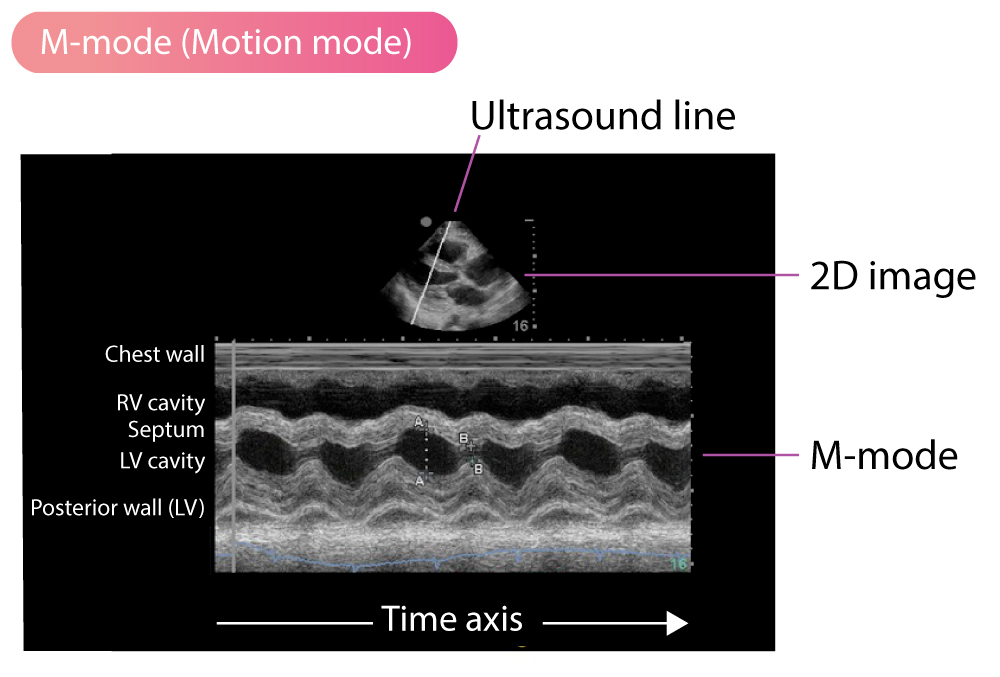
What does M-mode ultrasound stand for, and what does it demonstrate?
Motion mode; it displays the movement of anatomical structures over time along a single scan line.
Which anatomical heart structure characteristically forms an 'M'-shaped wave pattern on M-mode?
The mitral valve.
What physiological parameter can be calculated by measuring the distance between recurring peaks on an M-mode trace?
Heart rate.
How were early static B-mode images obtained in the 1960s?
Using an articulated arm with a stylus transducer moved across the patient's body (or submerged in a water bath) to trace an image onto an oscilloscope screen.
Which three biometric measurements are standardly combined to assess fetal gestational age?
Biparietal diameter (BPD), abdominal circumference (AC), and femur length.
Doppler motion
detection by the change in ultrasonic frequency due to motion of the source, observer or reflecting surface Christopher Doppler “1842”
Doppler Ultrasound Modes
Continuous Wave Doppler, Pulsed Doppler, Color Doppler, Power Doppler
Continuous Doppler
provides continuous measurements of blood flow and velocity by employing a transducer that emits and receives ultrasound signals simultaneously.
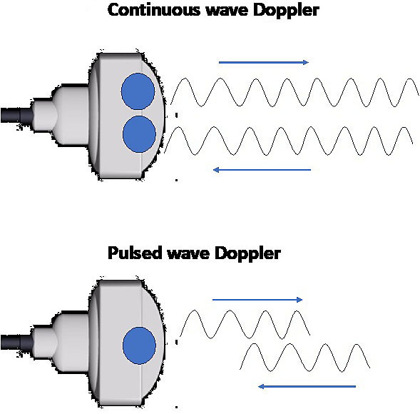

Pulsed Wave Doppler
measures blood flow and velocity at specific points in the body by sending out pulses of ultrasound waves and analyzing the reflected signals.