Cell biology
 Call Kai
 Learn
Learn Practice Test
 Spaced Repetition
 Match
Match Flashcards
 Knowt Play
1/80

There's no tags or description
Looks like no tags are added yet.
Name | Mastery | Learn | Test | Matching | Spaced | Call with Kai | Chat |
|---|
No analytics yet
Send a link to your students to track their progress
81 Terms
name the two types of light microscopes
compound microscopes and stereo microscopes
describe the difference between compound and stereo microscopes
Compound microscope relies on light passing through the specimen
Stereo microscopy is great for dissecting small subjects, however stereo is not great for imaging cells in a tissue
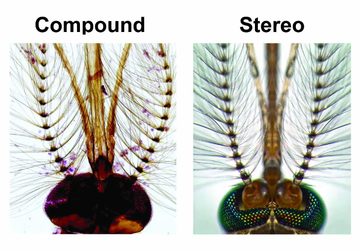
What type of microscope is able to see very small molecules and atoms?
Electron microscope
describe resolution
Resolution means how far apart two objects need to be to be able to see them separately. A shorter wavelength allows us to see things with a greater resolution.
Describe what about electron microscopes allow them to view smaller things with a greater resolution
Electron microscopes uses high energy electrons (Gamma rays) which have a much shorter wavelength (<0.01 nm) and higher frequency compared to photons which are used in a light microscope, allowing a greater resolution

What are requirements/ setbacks of using electron microscope?
Requires sample to be in vacuum
Samples must be dehydrated
Act of imaging damages subject
Only gives monochrome images
What are the two forms of electron microscopes?
Scanning (SEM) and Transmission (TEM)
what are the differences between SEM and TEM
SEM:
• Detector at an angle to stage
• Detects scattered electrons
• Gives ‘3D’ images (xref stereo)
• No need for sectioning
TEM
• Electrons pass through sample to
detector
• Gives ‘flat images’

Describe fixing
Fixing preserves the specimen
• E.g. soak in formaldehyde/glutaraldehyde
• Embed and dehydrate in resin/paraffin
Describe sectioning
Sectioning (microtome) takes a thin slice
of the specimen
Describe staining
Staining sections enhances contrast
• Dyes stain different cellular components
• Heavy metals (eg uranyl acetate) for EM
Describe the membrane of a eukaryotic cell
Made up of a bilayer of phospholipids (Amphipathic lipids) , Hydrophobic core, separating aqueous compartments
Describe the polarity of the phospholipid
Hydrophilic head, hydrophobic fatty acid tails
What can your membrane contain?
phospholipid bilayer, Cholesterol, glycoproteins, glycolipids
How are membranes dynamic?
They can undergo fission and fusion (dividing into two/ combining to form one)
What does fluorescence microscopy allow us to do?
Allow us to see things in high resolution with high detail.
What is the relationship between the energy of a wavelength and its colour
Blue light has more energy than red light
Blue (High energy) Red (Low energy)
define flouraphore
a chemical compound that absorbs light energy at a specific wavelength and re-emits light at a longer wavelength.
If a cell appears red, what colours would it absorb and reflect?
Absorb blue light, reflect red light
What happens when a molecule absorbs light
When a molecule absorbs light electrons gain energy and when this happens its excited and moves further from the nucleus
The excited electron will release this energy and will emit a photon and return back to its relaxed/ground state
What does the difference in energy between the ground state and the excited state reprisent?
The difference in energy between the ground state and the excited state is equivalent to the colour that molecule will look at, however the electron will lose thermal energy therefore some energy is lost and the photon emitted will be closer to a red colour.
What does fluorescence do?
Fluorescent methods use
different excitation and
emission wavelengths
Makes them very sensitive
Can be quantified accurately
Wavelengths are controlled using filters
Illuminates sample with a specific
colour of light (excitation)
Detects a higher wavelength of
light (emission)
What colour does DAPI stain?
Blue
What colour does Propidium Iodide (PI) stain?
Stains DNA red but can only enter dead cells
What colour does Acridine orange stain?
DNA green
RNA orange
What is the purpose of immuno fluorescence
Is able to identify specific areas of proteins
How does immuno fluorescence work?
Fluorophore stains conjugated to an antibody The antibody’s specificity localises the stain to particular cell components
What is a downside of immuno fluorescence
takes a long time
What is the purpose of green fluorescent protein?
Is encoded by a gene so we can use genetics to add this gene into organisms to produce a green fluorescent organism.
It was originally found in a jellyfish
The GFP gene can be fused with a target gene to study gene expression.
What is the purpose of Confocal Microscopy?
Can take visual sections of extremely thin focal plane through a specimen allows 3D reconstruction.

What does FRAP stand for ?
Flourescence Recovery After Photobleaching

How does FRAP work
It allows us to determine the speed that a lipid molecule can move through the membrane, as it burns through the organelle leaving only the membrane and then over time lipid which is also stained will return into the organelle and we can measure the time it takes for the fluorescence to return.

What does FRET stand for?
Forster Resonance Energy Transfer
What does FACS stand for
Fluorescence Activated Cell Sorting
How does FACS work?
Fluid flow creates a stream of single cells
Size and fluorescence measured of each cell Cells separated into droplets and charge applied
Droplets separated by charge

What is FACS useful for?
• Classifying immune cells
• Isolating stem cells
• Assessing cell viability
• Finding rare cancer cells
What are bacteria
simplest organisms
What are the shapes of bacteria?
Rod- shaped (Bacillus), Spherical (Coccus), spiral (Spirillium)
What type of cell are bacteria?
Prokaryotic
What are charecteristics of Prokaryotic cells
No nucleus - DNA in a nucleoid
• Inner membrane surrounded by
peptidoglycan cell wall in the
periplasm.
• Gram-negative bacteria have an
additional outer membrane.
• Capsule of polysaccharide (sugars)
• Flagella for motility
• Protein fibres – pili, fibrils, fimbriae
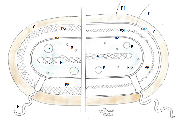

Label this prokaryotic cell
IM = inner membrane
PP = periplasm
PG = peptidoglycan cell wall
OM = outermembrane
C = capsule (sugar coat)
N = nucleoid
P = plasmid
R = Ribosome
F = flagellum
Pi = pilus
Describe eukarytoic microbes
Have a nucleus (with nuclear
membrane, pores and endoplasmic
reticulum)
• Mitochondria (endosymbiotic
bacteria)
• Fungi (including yeasts)
• Protists (everything eukaryotic which
isn’t plant, animal or fungus:
• Algae (plant-like protists)
• Protozoa (animal-like protists)
• Fungus-like protists
Describe characteristcs yeast (Fungi cells)
• Small (5 um) and simple eukaryote
• Contain the subcellular structures
common to all eukaryotic cells
• Chitin cell wall (like cellulose with N)
• Can divide like bacteria by binary
fission (fission yeast –
Schizosaccharomyces pombe)
• Some bud (budding yeast,
brewer’s/baker’s yeast –
Saccharomyces cerevisiae)
• Other fungi have complex life cycles

Describe charavteristics of protists (Eg. green algae)
• Photosynthetic (chloroplasts –
endosymbiotic bacteria)
• Can be flagellated (Euglena,
Chlamydomonas)
• Diatoms grow a silica shell (frustule)
What are the foour main types of Protists - Protozoa
Four main types (polyphyletic):
• Mastigophorans/Flagellates – move with flagella
• Sarcodines/Amoebae
• Apicomplexans – immobile parasites
• Ciliates – move with cilia
Describe cillia
• Cilia are essentially the
same across the
eukaryotes, including
those lining our lungs.
• Unlike flagella, they are
enclosed within the cell’s
plasma membrane
• Dynein is a ‘motor’ protein
which powers the flexing
of fibers within the cilium
Describe the cell boundry of ciliates
• An inner membrane under the plasma
membrane
• Alveolar spaces for strength
• An underlying cell cytoskeleton or ‘pellicle’
What is DNA composed of
DNA is composed of individual nucleotides
What are the three main compnents of a nucleotide
Phosphate group deoxyribose sugar, nucleobase
Describe the order of organisation within a chromasome
Phosphate group deoxyribose sugar, nucleobase make up a nucleotide. many nucleotides make up a strand of DNA and two strands hydrogen bond together to form a double helix of DNA. DNA then wraps around histones tightly forming chromatin fibres which are then coiled further to form a chromasome

Where is the 5’ end and the 3’ end on a nucleotide?
5’ is on the phosphate group and the 3’ end is on the empty hydroxyl group.

How are individual nucleotides bonded together?
Condensation reaction to form a Phosphodiester bond
(A phosphodiester linkage forms between the 3’ C of the sugar in thymine and the P of the phosphate group in guanine. Water is lost so it’s a condensation reaction)
How many hydrogen bonds do A and T/U form
2
How many hydrogen bonds do C and G form
3
What are the differences between DNA and RNA
RNA has uracil instead of thymine
On C2, Ribose has a hydroxyl (–OH) group
Uracil has a hydrogen (–H)
DNA has thymine instead of uracil
Deoxyribose has a hydrogen (–H).
thymine has a methyl (–CH3) group.
How does DNA stores the genetic code
Transcription
How does DNA transfer the code into RNA
Translation
What are the products of transcription and translation?
proteins that carry out the functions of the cell
What phase of the cell cyle does DNA replication occur?
S phase
What enzymes does DNA replication require
Topoisomerase and helicase
DNA polymerase and ligase
Topoisomerase and helicase
Unwind the DNA helix
What does DNA polymerase and ligase do?
join individual nucleotides and synthesise new DNA strands.
What name is given to the way DNA replicates itself
Semi-conservative
( One strand of the parental DNA is conserved in each of the new molecules formed.)
What are the two strands in DNA named during semiconservative replication?
Leading and lagging
What direction does DNA polymerase add new nucleotides
in the 5’ to 3’ direction only.
Describe replication in the leading strand
On the leading strand DNA polymerase is moving towards the replication fork so add nucleotides to the 3’ end of the new strand and can copy continuously

What is the name of the enzyme that unzips DNA to form a replication fork?
DNA helicase
What is the name of the enzyme that joins individual nucleotides to synthesise a new DNA strand?
DNA polymerase
At which end of DNA molecules are new nucleotides added?
5’ end toward the 3’ end
What is the difference between the way leading and lagging stands of DNA are synthesised?
Lagging strand must be replicated in short fragments called okazaki fragments, multiple primers and DNA polymerase are uses
What is the name of the enzyme that makes the lagging strand complete?
DNA ligase