HAPIB Module 2 - Digestive and Urinary Systems
 Call Kai
 Learn
Learn Practice Test
 Spaced Repetition
 Match
Match Flashcards
 Knowt Play
1/201

There's no tags or description
Looks like no tags are added yet.
Name | Mastery | Learn | Test | Matching | Spaced | Call with Kai | Chat |
|---|
No analytics yet
Send a link to your students to track their progress
202 Terms
What are the major organs of the GIT?
A series of hollow organs
Mouth/oral cavity
Oesophagus
Stomach
Small intestine (duodenum, jejunum, ileum)
Large intestine (cecum, ascending colon, transverse colon, sigmoid colon, and descending colon)
Rectum
A*nus
What are the accessory organs of teh GIT?
Produce secretions to assist digestion
Salivary glands
Liver
Gallbladder
Pancreas
what are teh functions of the digestive system?
The digestive system converts food into its simplest components
Absorbed into the bloodstream or excreted as waste
Ingestion
Mechanical digestion
Chemical digestion
absorption
elimination of waste
What is ingestion?
The entry of food/liquids into the digestive tract via the mouth
what is mechanical digestion?
Crushing/shearing of ingested food
the physical/mechanical breakdown of food into smaller components
what is chemical digestion?
(extraction of nutrients)
Enzymatic breakdown of food into substances that can be absorbed
what is absorption?
Movement of nutrients into the bloodstream -> sent to the liver for processing
what is elimination of waste?
Indigestible food is compacted into faeces
what are the organs of the upper GIT?
mouth/oral cavity
oesophagus
stomach
small intestine (duodenum only)
what is the function of the oral cavity?
first organ in the upper GIT
Mechanical digestion (mastication = chewing) and mixing of food with saliva
Enzymes in saliva begin the process of chemical digestion
what is the function of the oesophagus?
second organ of the upper GIT
A fibromuscular tube that transfers food (bolus) into the stomach
what is the function of the stomach?
fourth organ of the upper GIT
Chemical digestion - bolus is mixed with gastric juices (HCI and digestive enzymes)
Mechanical churning - facilitate digestion
Bolus is converted into chyme (liquified form of the food ingested mixed with acid and digestive enzymes)
what is the function of the small intestine (duodenum)?
last organ of the upper GIT
Duodenum = first 20-25cm
Chemical digestion -> aided by digestive juices from the pancreas and gallbladder
what are the organs of the lower GIT?
small intestine (jejunum and ileum)
large intestine
rectum/anus
what is the function of the small intestine (jejunum and ileum)?
first organ in the lower GIT
Jejunum and ileum = remaining ~3-6m of the small intestine
Where nutrient absorption occurs
what is teh function of the large intestine?
second organ in hte lower GIT
~1.5m long
Cecum, ascending colon, transverse colon, descending colon, sigmoid colon
Reabsorption of water
Compacts waste (faeces) for elimination
what is the function of the rectum/anus?
final organ of the lower GIT
Store and eliminate waste (faeces)
what are the functions of teh accessory organs of the GIT?
Secretion (to facilitate chemical digestion)
Salivary glands - salivary amylase
Pancreas - pancreatic enzymes, buffers (breaks down proteins, fats, carbohydrates)
Liver - bile (emulsify fats)
Detoxification
Liver - inactivates toxins
Storage
Gallbladder - bile
Liver - iron, glucose, fat soluble vitamins
what are teh four layers of the GIT wall?
The structure of the digestive tract varies slightly from region to region, but generally consists four major layers:
Mucosa (innermost layer)
Epithelial mucous membrane and connective tissue
In contact with the bolus/chyme moving through (in the lumen)
Submucosa
Dense irregular connective tissue
Contains blood vessels, nerves, glands
Muscularis externa
2-3 layers of smooth muscle
Facilitates movement of materials and mechanical digestion
Serosa/adventitia (outermost layer)

what is the mucosa of the GIT wall?
(innermost layer)
Epithelial mucous membrane and connective tissue
In contact with the bolus/chyme moving through (in the lumen)
three layers
epithelium
loose ct
muscularis
what is the epithelium layer of the GIT mucosa?
in direct contact with the contents of the lumen of the GIT
Epithelial composition depends on its function
Mouth + oesophagus -> stratified squamous (protection)
Stomach, small intestine + large intestine -> simple columnar (absorption)
Rectum + anus -> stratified squamous (protection)
Contains specialised goblet cells that secrete mucus to keep the epithelium moist
In the small intestine, contains microvilli -> increase surface area for nutrient absorption
what is the loose ct layer of the GIT mucosa?
(lamina propria)
Nutrients can travel through easily
Contains:
Blood vessels
Lymphatic vessels
Sensory nerve endings
Gut associated lymphoid tissue (GALT)
Some mucous glands
Fibroblasts
Macrophages
what is the muscularis layer of teh GIT mucosa?
(muscularis mucosa)
Thin layer of smooth muscle cells
Its function is still under debate
Believed to alter the shape of the lumen and move mucosal folds/villi
what is the submucosa of the GIT wall?
A layer of dense irregular CT (but still relatively loose)
Provides physical support to the mucosa
Connects the mucosa to the underlying muscularis
Contains:
Large blood vessels (receive absorbed nutrients)
Large lymphatic vessels
Submucosal glands (exocrine)
Secrete enzymes and buffers into the lumen via ducts
Submucosal plexus (Meissner's plexus) - sensory neurons and autonomic nerve fibres that innervate the glands
Controls glandular secretions, alters electrolyte and water transport, regulates local blood flow (i.e. controls fluid secretion and absorption)
what is the muscularis layer of teh GIT wall?
Multiple muscle layers
Organised in circular and longitudinal layers
Composition depends on location within the GIT
Oesophagus - contains 2 layers of muscle -> mixture of skeletal muscle (voluntary swallowing) and smooth muscle
Stomach - contains 3 layers of smooth muscle -> inner oblique, middle circular, outer longitudinal
Small intestine, large intestine, rectum - contains 2 layers of smooth muscle -> inner circular and outer longitudinal
Contraction of these muscle layers is essential for mechanical processing and moving materials along the GIT
The muscularis externa also contains the myenteric plexus (Auerbach plexus)
A network of sensory neurons + autonomic nerve fibres that runs between the two layers of muscle
Located between the circular and longitudinal muscle layers
Coordinates digestive muscle activity (i.e. controls motility)
Parasympathetic stimulation - increased muscle tone
Sympathetic stimulation - decreased muscle tone
what is teh serosa/adventitia layer of teh GIT wall?
Depending on the region of the GIT, the outermost layer of the GIT is either called adventitia or serosa
Serosa
Found in the intraperitoneal regions of the GIT (i.e. most of the stomach, parts of the small + large intestine and rectum)
A thin layer of loose CT covered with mesothelium -> lubricates the outer surface of the GIT -> decreased friction
Serosa = lubricates structures
Adventitia
Found in the retroperitoneal regions of the GIT (i.e. oral cavity, pharynx, oesophagus, parts of the stomach, small and large intestine, and the anus)
Contains a dense network of collagen fibres
Adventitia = binds/attaches structures
how does the GIT wall change?
Mucosa
Epithelial type
Stratified squamous/simple columnar
Physical appearance
Protrusions (villi)
Invaginations (glands)
Muscularis
Number of layers
2 layers/3 layers
Complete/incomplete layers
Muscle type
Smooth muscle/skeletal muscle


what are the structures of the oral cavity?
Lined by oral mucosa (stratified squamous epithelium) -> needs to be very protective
Hard palate - bony support structure at the top of the oral cavity
Soft palate - soft muscular structure, posterior to the hard palate
Tongue - skeletal muscle
Uvula - dangly piece of tissue that hands at the back of the oral cavity -> prevents food from entering pharynx prematurely
Teeth
Gingiva (gums)
Tonsils - lymphoid tissue

what are teh functions of teh oral cavity?
Mechanical digestion
Mastication (chewing)
Saliva + food = bolus
Chemical digestion
Digestion of carbohydrates and lipids begins here
Salivary amylase - carbohydrate digestion
Lingual lipase - lipid (triglyceride) digestion
what are teh salivary glands?
three major pairs of salivary glands
sublingual
submandibular
parotid

what are teh sublingual salivary glands?
Lie on either side of the tongue
Produce a mucous secretion that acts as a bufffer/lubricant
Produces ~5% of saliva entering the oral cavity
what are the submandibular salivary glands?
Lie on the inner surface of the mandible (jaw)
Produces a mixed serous/mucous secretion containing buffers, mucin, and salivary amylase
Secretions released under the tongue
Produces ~75% of the saliva entering the oral cavity
what are teh parotid salivary glands?
Large glands located just in front of the ears
Produce a serous secretion containing lots of salivary amylase
Produces ~20% of saliva entering the oral cavity
Mumps = viral infection of the parotid gland
what is saliva?
The salivary glands produce 1-1.5 litres of saliva every day
Saliva contains ~99.4% water
The remaining ~0.6% contains:
Electrolytes (mainly Na+, CI- and HCO3-)
Buffers (keeps mouth at pH 7.0 preventing the build up of acids produced by bacteria)
Antibodies (IgA)
Enzymes - salivary amylase
Lysozyme (antibacterial enzyme)
Lingual lipase
Salivary mucin (a slimy visco-elastic material)
what are teh functions of saliva?
Lubrication
Chemical digestion - enzymes
Protection - antibacterial lysozymes, buffers, IgA
Dissolves food - taste
what is teh structure of the oesophagus?
The oesophagus is a collapsed tube that runs posteriorly to the trachea
Structure of the oesophageal wall
~25cm long, ~2cm in diameter
Mucosa
Stratified squamous epithelium (protection; abrasion/bacteria)
Contains large folds - keep the lumen closed unless swallowing is occurring
Allows for expansion when swallowing and food is moving through
Submucosa
Contains large mucus glands (lubrication)
Muscularis
Upper 1/3 = skeletal muscle (swallowing is voluntary)
Middle 1/3 = mixture of skeletal and smooth muscle
Lower 1/3 = smooth muscle
Adventitia
Not serosa
Connective tissue layer lies outside the muscularis layer, anchoring the oesophagus to neighbouring structures
The oesophagus contains two sphincters
Upper oesophageal sphincter
Lower oesophageal sphincter
what is the upper oesophageal sphincter?
Upper oesophageal sphincter - prevents air entering digestive tract and the reflux of food into airways
what is teh lower oesophageal sphincter?
Lower oesophageal sphincter - prevents reflux of stomach contents
what is teh function of the oesophagus?
Convey food and liquids from the mouth into the stomach
No absorption!
what are the three phases of swallowing?
buccal phase (voluntary)
pharyngeal phase (mainly involuntary)
oesophageal phase (mainly involuntary)
what is the buccal phase of swallowing?
(voluntary) - first phase of swallowing
Contraction of the tongue to push bolus up against the soft palate and into the oropharynx
what is the pharyngeal phase of swallowing?
(mainly involuntary) - second phase of swallowing
Tongue blocks off the oral cavity
Epiglottis blocks the larynx/trachea
Upper oesophageal sphincter (UES) relaxes and opens
Food enters the oesophagus
what is the oesophageal phase of swallowing?
(mainly involuntary) - last phase of swallowing
UES contracts and closes
Bolus is forced through the oesophagus by peristalsis
Food enters the stomach
Once food has reached the stomach, LES closes (to prevent regurgitation
what is the stomach?
An expandable muscular organ that churns food into chyme
four main regions;
fundus
cardia
body
pylorus
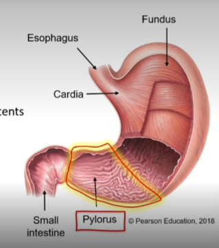
what is the fundus of the stomach?
superior to the esophageal junction
top part of teh stomach
what is the cardia of the stomach?
within ~3cm of gastroesophageal junction
Entry point for food/liquids into the stomach
Oesophageal sphincter keeps stomach contents from reentering the oesophagus
Abundant mucus glands - protection from acid
what is the body of the stomach?
the largest region
Lies between the fundus and the curve of the "J"
The "mixing bowl" for ingested food
what is the pylorus of the stomach?
the curve of the "J" leading to the small intestine
Can be further subdivided into the pyloric antrum and the pyloric canal
Pyloric sphincter regulates chyme -> small intestine
what is the structure of the stomach wall?
Mucosa - lined with simple columnar epithelium
Produces a carpet of alkaline mucus (protection)
Cells are replenished every 3-7 days
Contains deep folds that form gastric glands
Submucosa - fibrous connective tissue containing blood vessels, lymphatic vessels and nerve cells
Muscularis - made up of three layers
Inner oblique - responsible for creating the churning motion to aid mechanical breakdown of food
Not present in any other part of the digestive system
Middle circular
Outer longitudinal - responsible for moving the bolus towards the pylorus (through muscular shortening)
Serosa - a thin ct layer that secretes a lubricating fluid to reduce friction
Continuous with the visceral peritoneum
what are teh rugae of the stomach?
When empty, the mucosa and submucosa layers of the stomach wall form prominent folds/ridges called rugae
As the stomach fills with food these rugae flatten out to allow the stomach to expand
The mucosal layer of the stomach forms a series of gastric pits, each housing a number of gastric glands
what are the gastric glands of the stomach?
Mostly exocrine
Opens into the stomach through gastric pits in the mucosa
Secrete most of the acid and enzymes required for chemical digestion in the stomach (~1.5L per day)
Gastric glands contain specialised cells - the composition of these cells depends on the gland's location within the stomach

what are goblet cells?
Cells of the gastric glands:
secrete mucus - protects the mucosal wall from acid
what are parietal cells?
cells of teh gastric glands
secrete intrinsic factor - necessary for vitamin B12 absorption (small intestine)
Produce HCI - kill microbes, denatures proteins
what are G cells?
cells of the gastric glands
enteroendocrine cells - produces hormones
Produce gastrin - increases stomach motility
Stimulates HCI/enzyme production
Relaxes pyloric sphincter
what are chief cells?
cells of the gastric glands
secrete pepsinogen (inactive) which is converted into pepsin (active) by HCI -> degrades proteins
Secrete gastric lipase -> breaks down lipids
what are the functions of the stomach?
Temporary storage of ingested food
Stomach contents (up to 2-4L of food) are held for approximately 2 hours
Mechanical digestion of ingested food
Achieved by contraction/relaxation of the three muscle layers
Chemical digestion of ingested food
Stomach mucosa produces HCI and digestive enzymes (~1.5L of gastric juice produced each day)
Food is broken down into a thick, acidic, soupy chyme
Production of intrinsic factor
Help absorb Vitamin B12
The stomach doesn’t play a big role in the absorption of food, but it can absorb water (especially if the body is dehydrated), alcohol (~10-20% of ingested ethanol), some drugs (e.g. aspirin)
what is the small intestine?
Averages 3-5m in length and is divided into three segments
duodeunum
jejunum
ileum
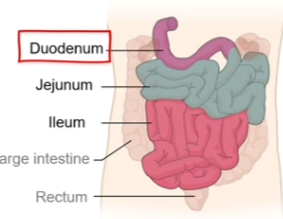
what is the duodenum?
First 20-25cm of the small intestine, proximal to the stomach
Receives chyme from the stomach and digestive secretions from the pancreas and liver
Considered the "mixing bowl" of the small intestine
Characteristic features:
Contains few circular folds
The villi are small
The submucosa contains lots of duodenal glands -> mucus
Why?
Structure = function!
The duodenum receives chyme from the stomach and must neutralise the acid before it damages the absorptive surfaces of the small intestin
what is the jejunum?
Approx. 1-2m long
Marked by a sharp bend at the beginning
Where the majority of chemical digestion and nutrient absorption occurs
Characteristic features:
Contains lots of circular folds
The villi are abundant and very long
Why?
To maximise nutrient absorption
what is the ileum?
The longest segment - approx 2-3m long
Where absorption of vitamin B12, fats (eps. Fatty acids and glycerol) and bile salts occurs
Characteristic features:
Contains fewer circular folds than jejunum
The villi are stumpy
The submucosa contains lymphoid nodules -> protect the small intestine from bacteria colonising the large intestine
what is teh structure of the small intestine wall?
Mucosa
Lined with simple columnar epithelium
Produced a carpet of alkaline mucus (protection)
Contains a series of finger like projections called villi (increase surface area for absorption)
Submucosa
Fibrous connective tissue containing blood vessels, lymphatic vessels and nerve cells
Muscularis
Made up of two layers of smooth muscle;
Inner circular
Outer longitudinal
Serosa
Thin connective tissue layer that secretes a lubricating fluid (to reduce friction)

what are the structural characteristics of the small intestine?
The small intestine contains many structures that increase the surface area for absorption
Plicae circulares
Villi
Microvilli
Together, these structural adaption increase the surface area of the intestinal wall by 600x

what are the plicae circularis of the small intestine?
the intestinal lining contains a series of permanent structural folds called plicae circulares -> greatly increase the surface area of the lumen within the small intestine
Deep ridges in the mucosa and submucosa
Begin near the proximal part of the duodenum and extends to the middle of the ileum
what are the villi of the small intestine?
the mucosal wall of the plicae circulares projects into a series of finger like projections called villi -> further increase surface area available for nutrient absorption
The surface of the plicae circulares contains small vascularised projections of the mucosa called villi
There are approx. 20-40 villi per mm2 of small intestine
Each villus contains a capillary bed which houses:
1 x arteriole (carry absorbed nutrients to the liver)
1 x venule (carry absorbed nutrients to liver)
1 x lacteal (lymphatic capillary) -> transports materials that cant enter the blood capillaries (e.g. fatty acids)
what are the microvilli of the small intestine?
the surface of each villus is covered by epithelial cells which contain microvilli on their apical surface (brush border)
The simple columnar epithelial cells lining the intestinal villi contain cell membrane extensions (microvilli)
There are approx. 200 million microvilli per mm2 of small intestine
Significantly increases surface area of the plasma membrane for absorption
Because they like the bristles on a brush, said to have a brush border
what is the function of the plicae circularis of the small intestine?
facilitate absorption (esp. jejunum)
Their shape causes the chyme to move in a spiral motion through the small intestine -> slows the movement of chyme, increasing time available for absorption
what are hormones of the digestive system?
The digestive system utilises a variety of hormones to regulate digestion and nutrient absorption
Tehse hormones act on various parts of the GIT to coordinate processes such as
The release of stomach acid
The release of pancreatic enzymes
The release of bile
Gut motility (mixing and propulsion)
Metabolism
Hunger and satiety
Etc
In particular, the duodenum plays an important role in regulating digestive function by stimulating the release of specific hormones, depending on the characteristics of the arriving chyme
what are the four main endocrine hormones of the digestive system?
Gastrin
Secretin
Gastric inhibitory peptide (GIP)
Cholecystokinin (CCK)
what is gastrin?
endocrine hormone of the digestive tract
Source: enteroendocrine G cells in the stomach (gastric glands) and the duodenum
Released: when food arrives in stomach; when large quantities of partially digested proteins are present in the duodenum
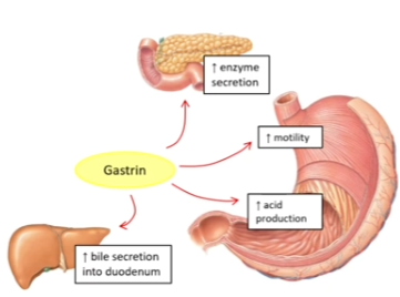
what is the function of gastrin?
Stomach: stimulates acid production by parietal cells; stimulates muscle contractions (increased gastric motility)
Duodenum: stimulates gallbladder to empty bile into the duodenum; stimulates the pancreas to release pancreatic enzymes into the duodenum
what is secretin?
endocrine hormone of the digestive tract
Source: enteroendocrine S cells in the duodenum
Released: when acidic chyme arrives in the duodenum (pH 2-4)
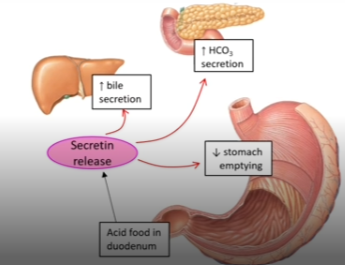
what is the function of secretin?
stimulates the pancreas to secrete bicarbonate/buffers (increases chyme pH/neutralise acidic pH
allows pancreatic enzymes to function optimally)
stimulates bile production by the liver
inhibits acid secretion from parietal cells in the stomach
inhibits/slows gastric emptying
what is gastric inhibitory peptide (GIP)?
endocrine hormone of the digestive tract
Source: enteroendocrine K cells in the duodenum
Released: when fats and carbohydrates (esp. glucose) enter the duodenum
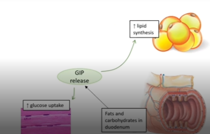
what are the functions of GIP?
Stimulates pancreas to release insulin
Stimulates lipid synthesis (fat accumulation)
Inhibits lipid breakdown
Stimulates glucose uptake by skeletal muscle
Regulates appetite
what is cholecystokinin (CCK)?
endocrine hormone of the digestive tract
Source: enteroendocrine I cells in the duodenum
Released: when fats and undigested proteins enter the duodenum
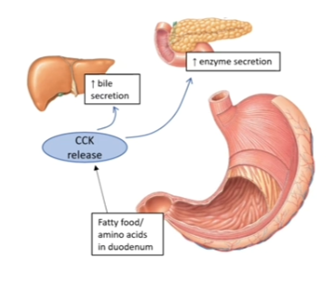
what are the functions of CCK?
Promotes digestion of fat and protein
Stimulates the pancreas to secrete pancreatic enzymes into the duodenum
Stimulates gallbladder to empty bile into the duodenum
Inhibits gastric activity
Suppresses hunger
what is peristalsis and segmentation?
The muscularis layer;
Made up of two layers of smooth muscle
Inner circular
Outer longitudinal
Coordinated contraction of these smooth muscle layers plays a vital role in peristalsis and segmentation of the bolus/chyme as it moves through tehb digestive tract
Peristalsis = moving material along the digestive tract (propulsion)
Segmentation = mechanical mixing
what is peristalsis?
Is what pushes bolus/chyme forwards through the GIT
Occurs throughout the GIT
Peristalsis is so powerful that if you swallow food while standing on your head it will enter your stomach
Bolus of food arrives
Circular muscles contract behind bolus -> narrows internal lumen diameter
Longitudinal muscles contract ahead of the bolus
Waves of circular muscle contractions behind bolus force it forwards

what is segmentation?
Occurs mainly in the small intestine (but also in the large intestine)
Not involved with pushing chyme forwards through the GIT - it occurs in a stationary way in localised regions of the intestine
Localised contractions of circular muscle isolate chyme in small sections of the intestine
Chyme is continually pushed back and forth between adjacent sections (like repeatedly squeezing a tube of toothpaste back and forth)
Thoroughly mixes chyme with digestive juices
Facilitates mechanical breakdown
Maximises contact between chyme and mucosa
When most of the chyme has been absorbed, segmentation stops

what is the large intestine?
Averages approx. 1.5m in length and 3 inches in diameter, and is divided into 3 regions
cecum
colon
rectum

what is teh cecum?
first region of teh large intestine
An expanded pouch (6cm) where material from the large intestine is stored and faecal compaction begins
The flow of chyme entering from the small intestine is controlled by the ileocecal valve
Has an attached appendix
what is the colon?
second region of the large intestine
Divided into four regions (1. ascending colon, 2. transverse colon, 3. descending colon, 4. sigmoid colon)
what is the rectum?
last region of the large intestine
The last 15cm of the digestive tract
Where faeces are stored prior to defecation
what is teh structure of the large intestine wall?
Mucosa
Simple columnar epithelium (cecum and colon)
Stratified squamous (rectum and anus)
No circular folds or villi
Mucosa drops down into intestinal glands
Contains lots of goblet cells (lubrication)
Muscularis - two layers of smooth muscle
Inner circular
Outer longitudinal (incomplete) - "teniae coli"

what are teniae coli of the large intestine?
The outer longitudinal layer is incomplete - it is present in three separate longitudinal ribbons running down one side
The teniae coli contract lengthwise to bunch up the colon into a series of pouches called haustra

what are the haustra of the large intestine?
Series of sac like pouches within the colon ; as the chyme passes through the large intestine, it progressively moves from one pouch to the next
The presence of chyme stimulates slow moving haustral contractions (->segmentation, primarily in the transverse and descending colon)
Contractions occur every 25min and last 1min
These contractions push the intestinal contents from one pouch to the next, facilitating mixing and H2O reabsorption

what are the functions of the large intestine?
Final absorption of nutrients and water
Only about 10% of nutrient absorption occurs in the large intestine
Chemical digestion
Facilitated by bacteria - no digestive enzymes secreted by the large intestine
Remaining indigestible carbohydrates serve as a nutrient source for bacteria in the colon
Bacterial activity; produces B vitamins, vitamin K, releases CO2 nad methane
Formation and elimination of faeces
Faeces = undigested food (cellulose), dead epithelial cells, mucus and bacteria
Frequency of defecation can be affected by diet, health, stress
The number of bowel movements varies greatly - ranges from 2-3 per day to 3-4 per week
what is absorption in the large intestine?
Less than 10% of nutrient absorption occurs in the large intestine - the chyme that enters the large intestine contains few nutrients except water
On average, 1.5L of material enters the large intestine every day, however only 200mL of faeces is eliminated
If faecal elimination (defecation) is delayed, additional water is absorbed -> constipation
If waste moves too quickly through the large intestine, not enough water is absorbed -> diarrhoea
what is the human microbiome?
Humans are colonised by an estimated 100 trillion microorganisms
These microorganisms account for approx. 1-2kg of a persons weight (roughly size of the brain)
Include bacteria, fungi, viruses
Distinct microbial populations reside al over our body
The bulk live in the GIT (esp. the cecum)
Most of the 1000s of species of bacteria in the GIT are symbiotic (benefits both host and microbiota)
The gut microbiota derives its nutrients from the hosts diet
Diet has a dramatic impact on the composition and metabolic activity of your microbiome
what is the gut microbiome?
The gut microbiome kicks in from the moment you are born, and is fully established by age 3
Microbiome development is affected by;
Gestational age (full term or prem)
Mode of delivery (vaginal or c section)
Type of feeding (breast milk or formula)
Maternal nutritional status (overweight or undernourished)
As you grow your gut microbiome begins to diversify (i.e. contain different types of microbial species)
A higher microbiome diversity = indicator of good health

what does the gut microbiome do?
Facilitates chemical digestion and absorption in the large intestine
Syntehsises specific vitamins (K and B), enzymes and amino acids
Ferments dietary fibre - produces a short chain of fatty acids (SCFAs)
Source of nutrients
Provides energy for epithelial cells; increased barrier integrity
Produces proteins to maintain desmosomes; increased barrier integrity
Used in cholesterol metabolism
Regulate appetite
Decreases pH of colon -> decreased growth of pathogenic bacteria
Affects drug metabolism - microbial metabolites can regulate liver function
Aids the immune system - activates intestinal immune cells to secrete immunoglobulins (IgA)
how does the gut microbiome impact overall health?
Mental health: hormones/neurotransmitters/immunological factors released from the gut send signals to the brain -> regulates mood
Immune health: signals derived from gut microbiota are critical for immune system development and resistance against infection (innate and adaptive immunity)
Metabolism: gut microbiota stimulate the release of hormones (e.g. CCK, PYY, serotonin) that regulate metabolic processes (e.g. glucose metabolism, fat storage, appetite)
Cardiovascular health: metabolites from gut microbiota affect cholesterol transportation and platelet aggregation
Inflammation: altered immune responses to gut microbiota can trigger inflammation -> disease

how does disease impact the gut microbiome?
Disturbance in the balance of gut microorganisms ("dysbiosis", due to infectious illness, prolonged use of antibiotics) -> decreased microbiome diversity -> increased susceptibility to disease
The challenge is to identify whether alterations to the gut microbiome are cause or effect
Changes in the gut microbiota have been associated with:
Inflammatory bowel disease - IBD patietns have decreased bacterial diversity (fecal transplants)
Diabetes - specific bacteria appear to be protective, toehrs are associated with diabetes
Obesity - gut micobiomes can influence appetite and metabolism
Eczema/allergies - lack of early life exposure to antigens (i.e. hygienic environments, C-section) alters the gut microbiome -> disrupts immune development
Manipulation of the gut microbiome (antibiotics/probiotics, microbiota transplant) may be useful to treat disease
what is the liver?
The second largest organ in the body (~1.5kg)
Sits just under the rib cage on the right side of the abdomen
The only organ that can regenerate - it can regenerate itself back to full size from as little as 51% of the original liver mass
Not just an accessory digestive organ - its the largest gland in the body - plays important roles in regulating metabolism
what is the anterior view liver anatomy?
The falciform ligament divides the liver into two primary lobes (left and right) and attaches the liver to the abdominal wall
The coronary ligament attaches the superior surface of the liver to the diaphragm
The round ligament is a thickening in poster end of the falciform ligament which connects the liver to the umbilicus

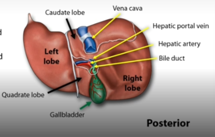
what is teh posterior view liver anatomy?
The caudate lobe lies between the left lobe and the inferior vena cava
The quadrate lobe is sandwiched between the left lobe and the gallbladder
Major vessels:
The hepatic artery brings oxygenated blood from the heart to the liver
The hepatic portal vein brings nutrient rich blood from the spleen and GIT to the liver
The inferior vena cava takes deoxygenated blood away from the liver, back to the heart
The bile duct carries bile away from the liver to the gallbladder and duodenum
Hepato = related to the liver
what are liver lobules?
the structural units of the liver
The liver is divided into approx. 100,000 lobules, each ~1mm in diameter
Each lobule is hexagonal in shape, are packed with liver cells (hepatocytes) and are separated by interlobular septa (connective tissue)
At each corner of these lobules is a portal triad consisting of:
An interlobular vein
An interlobular artery
An interlobular bile duct
Blood from the interlobular veins and arteries feed into the liver sinusoids (large, fenestrated capillaries)
At the centre of each lobule is a central vein which takes blood from the sinusoids to the inferior vena cava

what are hepatocytes?
part of the liver lobule
Make up 80% of the liver's mass
Have secretory, metabolic and endocrine functions
Within each lobule, hepatocytes are arranged in a series of irregular plates (like spokes of a wheel) -> plates are only one cell thick
Hepatocytes are tightly packed around fenestrated vessels (sinusoids) -> easy access to blood -> able to process the nutrients, toxins and wastes in the blood
what are sinusoids?
part of teh liver lobule
Liver sinusoids are open, fenestrated capillaries which lack a basement membrane
Also contain Kupffer cells (phagocytes) that remove dead blood cells, bacteria and foreign materials
how does blood flow in teh liver?
Oxygen rich blood arrives from the heart via the hepatic artery (1/3 blood supply)
Nutrient rich blood arrives from the spleen and GIT via the hepatic portal vein (2/3 blood supply)
Blood combines in liver capillaries (sinusoids)
Hepatocytes filter and process the blood
Regulate solute and nutrient levels
Absorb or secrete molecules such as proteins
Sinusoids all drain into a central vein
All central veins merge to form hepatic veins, which empty into the vena cava