Cell Phys
 Call Kai
 Learn
Learn Practice Test
 Spaced Repetition
 Match
Match Flashcards
 Knowt Play
1/137

Earn XP
Description and Tags
Basel Lectures - cytoskeleton, adhesions, and matrix
Name | Mastery | Learn | Test | Matching | Spaced | Call with Kai | Chat |
|---|
No analytics yet
Send a link to your students to track their progress
138 Terms
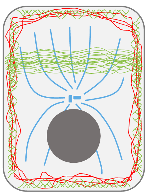
Cells are organized and held together by a set of structural proteins that are divided into three categories
Microtubules, Microfilaments, and Intermediate Filaments
Cell Shape • Cell Motility • Cell Strength • Cell Processes • Cell Polarization • Cell Signaling • Organelle Organization • Movement of Macromolecules • Membrane Organization are all important functions of the _____
Cytoskeleton
Hollow tubes that are often centered around nucleus radiating outward
Microtubule

- end = _____ exposed, and regress faster than Beta tubulin
Alpha

+ end = ____ exposed, and grow faster than Alpha tubulin
Beta
Micro tubules can be found in the
General cytoplasmic organization and cilia
During cell division (prophase, metaphase, etc.) where are microtubules present?
Mitotic spindle
T/F: Axons and dendrites have microtubules present within them.
True
Cell scaffolding, polarization, and Polarized movement of organelles, proteins, and DNA are primary functions of
microtubules
What other very important function do microtubes have?
Resists compression
Kinesin (toward ____ end) away from nucleus
positive
Dynein (toward ____ end) toward the nucleus
negative
Microtubule Transport Bind to cargo and ‘walk’ down the tubules, this is known as
polarized movement
T/F: Microtubules are the largest component of the microtubule complex
True
Two major nucleation sites of the Microtubule are
Centrosomes and Basal Bodies

Which component allow anchor of microtubules on the negative side?
Centrosomes
Nucleation _____ microtubule assembly
polarizes
Microtubules are hollow tubes ~___ nm in diameter made of tubulin dimers.
24
The microtubule network can be used for polarized transport of proteins and vesicles using motor proteins called
kinesins/dyneins
Microtubules are _____ structures regulated by nucleation, dynamic instability, and microtubule associated proteins.
dynamic

GDP - Tubulin Dimer =
Inactive/Shrinking State
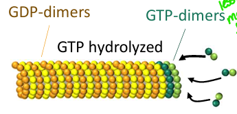
GTP-Tubulin Dimer =
Active/Growing State
Nucleotide required to be bound to tubulin dimers for addition to the positive (+) end of a microtubule
GTP - dimers
Tubulin subunit responsible for the slow hydrolysis of bound GTP to GDP
β-tubulin
Effect of high GTP-bound tubulin dimer concentrations on microtubule dynamics
Promotes microtubule growth and stabilization through the formation of a protective GTP cap (assembly occurs faster than GTP hydrolysis)
Effect of low GTP-bound tubulin dimer concentrations on microtubule dynamics
Promotes loss of the GTP cap, leading to dynamic instability, catastrophe, and rapid shrinking (GTP hydrolysis occurs faster than assembly)
Primary cellular advantage of dynamic instability despite its high energy cost
Allows rapid structural remodeling and fine-tuned spatial regulation of the microtubule network
Microtubule Associated Proteins (MAPs) have Many different proteins bind to MT that
regulate MT assembly and function
General functions of Microtubule Associated Proteins (MAPs) in a cell
Stabilizing, destabilizing, bundling, branching, mediating interactions with other cytoskeletal fibers or cell junctions, and transporting cargo
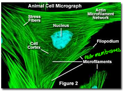
Double helical fibers and Found throughout the cell, but especially toward periphery
Microfilament
Monomeric protein subunit that polymerizes to form microfilaments
G-actin (globular actin)

Filamentous polymer formed by the assembly of actin subunits
F-actin (filamentous actin)
Key cellular structures and regions where microfilaments are found
Cell cortex, microvilli and stereocilia, lamellipodia and filopodia, and muscle fibers
Primary cellular functions of microfilaments
Cell membrane organization, ameboid movement, muscle contractions, cytokinesis, and resisting stretching
Type of mechanical stress that microfilaments primarily resist
Stretching
Microfilaments can be used for transport of proteins/vesicles or bulk cell movement using motor proteins called
myosins
Microfilaments are dynamic structures regulated by
nucleation, dynamic instability/treadmilling, and actin binding proteins
Functions of Microfilament Contraction and Transport
move vesicles, protrude membranes, and contract the cell
Microfilament Muscle fibers are specialized and densely organized fibers of
Actin and Myosin
Formation of G-Actin Monomers into tetramers is called ___ and occurs near the membrane
nucleation
After G-Actin binds to the (+) end of F-Actin, G-Actin binds ____ and becomes hydrolyzes to remove phosphate and becomes ____ as it moves towards the (-) end.
ATP ; ADP
Microfilament regulate length by
Rate of nucleation, rate of (+) end assembly, (-) end disassembly, and overall actin stability
Intermediate Filament functions
Mechanical strength (rigidity and flexibility), Non-motile scaffolding network, and resists shearing
T/F: All IF have the same functions for all tissue types and experience the same forces.
False
IF are made up of _____ proteins
fibrous (non globular)
The long fibrous proteins bundle and twist together into ____ like structures
rope
IF proteins are found in
Epithelium - cytokeratin - Connective tissue (fibroblasts) - Muscle - Glial cells/astrocytes - Nerves - Neuronal stem cells - Nucleus (all cells)
T/F: IF can deform and are able to spring back into position without breaking
True

Though relatively permanent, IF still need to change in response to changing conditions, a good example are ____ _____ because they support the nuclear membrane, are phosphorylated by lamin kinases during entry into prophase which destabilize the IF and are dephosphorylated on entry to telophase which allows reassembly.
A type of junction that connect two cells together very tightly (cells act in unison to compartmentalize) differentiate between apical and basolateral compartments
Tight Junctions
What are the two primary protein families of tight junctions?
Claudins and occludins
cells interact with each other is through ____ _____ ______ proteins expressed on the cell surface that bind the cell to other cells
cell adhesion molecules
Which tight junction protein has a much larger intracellular domains that regulatory domains
on them which can interact with different signaling pathways, changing how tightly the cells are in unison
occludin
There are 2 fluids that the tight junctions separate from the apical and basolateral
Extracellular fluid and Fat
Tight junctions forms 2 different types of regulations
Barrier (gate) and fence
Sodium Glucose Transport (SGLT) is a _____ active transport
secondary
Glucose Transporter (GLUT) is a ______ transport
passive
Tight Junctions form a permeability barrier, through action on claudins/occludins and interactions with the actin cytoskeleton can change permeability resulting in
leakiness
TJ leakiness and cytokine regulation may be associated with
Inflammatory Bowel Disease IBD
_____ are immune signals that activate pathways to make TJ more leaky.
Cytokines
A junction that causes cells to adhere to one another (cells act as one mechanical unit – ______network is joined across cells)
microfilament
Adhesion Belts are made of bundles of
Actin

What proteins are present on Adhesion Belts between intracellular space of both cells forming a zipper network
Cadherin Dimers
What is an example of small gTPAs in Adhesion regulation
Rho
When a stimulus is sent through Rho/ROCK what happens to the actin cytoskeleton that leasd to tension through the adherens junction?
contraction
Adherens junctions interact significantly with the ____ _______ and can be regulated through that interaction
actin cytoskeleton
Rho can also be important with
tissue modeling
What are the mechanical units in the intermediate filament network that join across the cell found in desmosomes
Keratin
What cadherin proteins are found in desmosomes that link the adjacent cell together
desmoglein and desmocolin
What adaptor protein is vital for preventing the separation of desmoglein and desmocolin from the membrane
desmoplankin
Desmosomes are abundant in tissues exposed to mechanical stress, including
stratified epithelia and cardiac muscle
Desmosomes are relatively _____ structures, but they still need to be remodeled at times during tissue growth and wound healing.
permanent
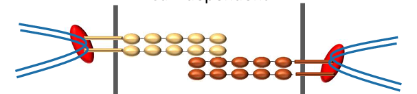
Protein kinase (PKCa) is a signal that acts on desmosomes, causing the desmoglein and desmocolin to form a weaker bond and become Ca dependent when
phosphorylated

_______ causes the desmoglein and desmocolin to form a stronger bond between desmosome complexes and become Ca independent, forming “hyperadhesive” state
dephosphorylation
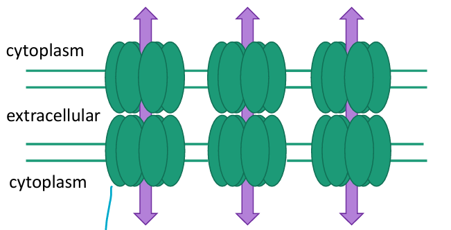
______ allow direct communication between the cytoplasm of two cells, allowing passage of ions and small molecules between neighboring cells
Gap junctions
Gap Junctions have six connexin subunits that form one connexon (hemichannel); connexons from adjacent cells dock to form a gap-junction channel. What is their function?
allow passage of different sizes and types of molecules
T/F: Most proteins can go through gap junctions.
False
T/F: If one cell becomes depolarized, the depolarization travels between the Gap junctions and the neighboring cells become depolarized as well.
true
Phosphorylation can change how permeable gap junctions are, but also ___, _____, and _____ can too.
pH, Ca, and Voltage
T/F: Cadherins usually bind to the same cadherin (homophilic)
True
Cadherins (“calcium-sensitive adhesion protein”) are important in
Anchoring junctions, initiation of anchoring junctions and tissue development and sorting
Usually Ca²⁺-independent; homophilic or heterophilic
Immunoglobulin-superfamily CAMs (IgCAMs)
I-CAM binds to LFA (Leukocyte Function Antigen) are important in
immune response
A family of cell adhesion molecules that bind carbohydrates
Selectins
T/F: Selectins are important when recognizing self vs non-self
True
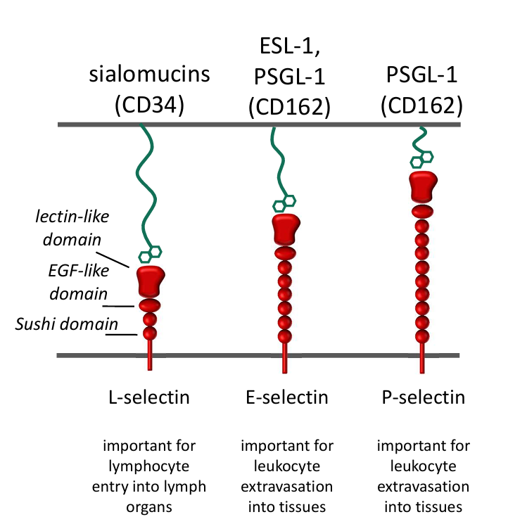
Selectins bind to specific carbohydrates (or glycoprotein) on the cell surface (heterophilic) which are important in
Inflammation/Immune Response and Uterine Implantation
primarily extracellular matrix binding proteins are called
Integrins
Integrins is a type of LFA, when inactive they are bent over, when stimulated, the Integrins errect and find to an I-CAM. This is important as adhesion molecule for
immune response
A family of fibrous proteins that give strength and rigidity to the extracellular matrix
Collagen
A family of fibrous proteins that give flexibility and elasticity to the extracellular matrix
Elastin
complex network of extracellular macromolecules that provides structural, mechanical, and biochemical support to cells
Extracellular Matrix
ECM is made up of generally 3 components:
Protein fibers, Complex carbohydrates, Adhesive glycoproteins
Triple helix Gives tensile strength and rigidity to extracellular matrix, is a Fibrous, helical protein
Collagen
Collagen Fibers can be oriented in _____ or ______ to create strength in different dimensions
parallel or obliquely
T/F: Triple helix are almost impervious to proteolytic degradation, making it difficult for proteases to it break down.
True
ECM strength and rigidity influenced by
Type, density, crosslinking and orientation of collagen.
Protein structures cannot make a tight bend on their own so they require the imino-acid ______.
Proline
To form a tighter kink in a triple helix structure proline is modified by a hydroxyl group to form
hydroxyproline