Module 6 Renal & Urologic systems and disorders
 Call Kai
 Learn
 Practice Test
 Spaced Repetition
 Match
 Flashcards
 Knowt Play
1/91

Earn XP
Description and Tags
patho
92 Terms
Starlings forces
hydrostatic
oncotic
The starling equation calculates
movement of edema fluid
depends on hydrostatic & oncotic pressure differences between capillaries & interstitium
Glomerular Filtration
depends on the same opposing forces that produce the exchange of fluids in every capillary in our body
hydrostatic pressure
force within a fluid compartment
major force that pushes water out of vascular system at capillary level

Oncotic Pressure
osmotic pressure exerted by colloids in solution
Albumin s a major colloid

Fluid movement in capillaries
amount & direction of movement determined by
capillary hydrostatic pressure
plasma oncotic pressure
interstitial hydrostatic pressure
interstitial oncotic pressure
capillary hydrostatic pressure
pushed H20 out of capillary
Plasma oncotic pressure
pulls H20 back into capillary
Interstitial oncotic pressure
pulls h20 out of capillary into interstitial spaces
filtration
controls the outward movement of fluid from capillary
reabsorption
pulls fluid back into capillary
fluid exchange between capillary & tissue

why does fluid exchange between capillary & tissue matter
low BP = drop in hydrostatic pressure > drop in filtration > acute kidney injury
low protein state = drop in oncotic pressure > fluid leaking out > edema
Fluid shifts
plasma to interstitial fluid shift results in edema
increase in hydrostatic pressure in capillaries
decrease in plasma oncotic pressure
increase in interstitial oncotic pressure
Interstital fluid to plasma - fluid shifts
fluid drawn into plasma space with increase in plasma osmotic or oncotic pressure
caused by: hypertonic solutions, osmotic diuretic (mannitol), colloids (albumin, dextran, or FFP)
compression stockings decrease peripheral edema
works by increasing interstitial hydrostatic pressure
A patient with low serum albumin is at risk for which complication related to Starling’s forces in the glomerulus?
edema
edema causes

Fluid distribution
intracellular (ICF) = 2/3 of body fluid
extracellular fluid (ECF) = 1/3 of body fluid
intravascular (plasma) - inside blood vessels
interstitial - between cells

What is the difference between interstitial fluid & extracellular fluid
edema - increase interstitial fluid volume
extracellular = all fluid outside the cells
interstitial = fluid in the spaces between cells (one part of extracellular fluid)
ICF
Increase potassium
moderate magnesium
decreased sodium, chloride, bicarbonate, phosphorus
very little calcium
ECF
increased sodium, chloride
moderate bicarbonate
decreased potassium, magnesium, calcium, phosphorus
Isotonic fluid
used to increase fluid in vascular space
cells stay the same
and fluids stay in the intravascular space

Hypotonic fluid - uses hypovolemia w/ hypernatremia and/or dehydration
cells swell as fluid goes into the cells
could cause cells to burst

Hypertonic fluid - used for severe hyponatremia or hypoglycemia

A nurse is administering Normal Saline (0.9% sodium chloride) and knows that this fluid type:
Has the same concentration of solutes as the fluid inside the body’s cells
Renin - Angiotensin - Aldosterone System (RAAS)

What sparks the RAAS to activate
dehydration
hyponatremia
hemorrhage
decreased cardiac output
decreased blood volume
decreased blood pressure
how does RAAS respond to a high salt diet

Antidiuretic hormone
ADH -also called vasopressin
helps kidneys control the amount of water your body loses through the urine
produced in the hypothalamus but released by the pituitary gland
if too much is produced - syndrome of inappropriate antidiuretic hormone (SIADH)
SIADH - syndrome of inappropriate antidiuretic hormone
kidneys hold onto too much water
hyponatremic because diluted by excess water
fluid overload
Hypervolemia
too much fluid volume typically intravascular but can become interstitial
Nurses call these patients WET
Common causes of hypervolemia
heart failure
kidney disease - chronic or acute renal failure or acute kidney injury
liver disease - cirrhosis & ascites
diabetes
Manifestations - hypervolemia
edema
bloating
increase weight
dyspnea
hypertension
crackles
Fluid volume deficit - hypovolemia
extracellular fluid volume deficit, can’t perfuse the body
nurses call these patients “dry”
think hypovolemic shock
Hypovolemia causes
diarrhea
hemorrhage (blood loss)
excessive sweating
hypovolemia manifestations
dizziness when standing
dry skin & mucous membranes
fatigue/weakness
muscle cramps
decreased urinary output, increase in urine specific gravity
hypervolemia treatment
remove fluid without changing electrolyte composition
give a diuretic
hypovolemia treatment
replace water and electrolytes with balanced IV solutions
replace whatever they are missing
Renal calculi: Nephrolithiasis
kidney stones
complex etiology
increase in stone components in blood and urine
anatomic changes
metabolic, endocrine & dietary influences
Renal calculi: Nephrolithiasis Risk Factors
family/personal Hx
dehydration
diet high in protein, NA & sugar
obesity
digestive diseases/surgery
Types of stones & causes - calcium
most common
high doses of vitamin D
intestinal bypass surgery (d/t metabolic changes)
metabolic disorders - renal tubular acidosis
Types of stones & causes - struvite
calcium, magnesium, ammonium & phosphate
usually caused by a UTI
types of stones and causes - Uric acid
chronic diarrhea or malabsorption
high protein diet
diabetes
metabolic syndrome
types of stones and causes - Cystine
amino acid
hereditary disorder - cystinuria
Nephrolithiasis signs and symptoms
hematuria
pain
renal colic: acute, intermittent, excruciating pain
may radiate down into groin for men
nausea/vomiting

Renal Calculi

Treatment for kidney stones
fluids - oral & IV
pain management
medications
tamsulosin (flomax)
Dutasteride & tamsulosin (Jalyn)
procedures/surgeries
extracorporeal shock wave lithotripsy (ESWL)
Percutaneus nephrolithotomy
ureteroscopy with or without stent placement
parathyroid gland surgery - if hyperparathyroidism caused
A 45-year-old male presents to the emergency department with sudden onset of severe flank pain radiating to the groin, accompanied by nausea and hematuria. A non-contrast CT scan reveals a 6 mm calculus in the left ureter.
Obstruction of urinary flow causing increased intrarenal pressure and ureteral spasm
Urinary Tract Infection risk factors
infection secondary to pathogens microorganisms
female anatomy (shorter urethra)
sexual activity
Birth control - specifically diaphragms & spermicidal agents
menopause
anatomical urinary tract problems
blockages-kidney stores or enlarged prostate
suppressed immune system
catheter use
recent urinary procedure
Lower UTI
Cystitis Bladder
Prostatitis Prostate
Urethritis Urethra
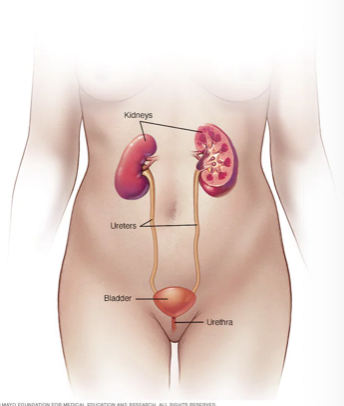
Upper UTI
Pyelonephritis Kidneys
Interstitial Nephritis Kidney
Renal Abscesses Tubules

UTI S/SX - Kidneys
back or side pain
high fever
shaking & chills
nausea
vomiting
UTI S/SX - Bladder
Pelvic Pressure
Lower Belly Discomfort
Frequent, painful urination
Blood in urine
UTI S/SX - Urethra
Burning with urination
dischange
lower urinary tract infection
#1 Cause: E.Coli
bacteria gain access to lower urinary tract → evade host defensive mechanism
due to
Immunosuppression
Decreased acidity of urine
Reflux of urine
decreased mucosal/IgA/prostatic secretions
damage to lining of system, etc
Urinalysis & urine culture - UTI
if bacteria found in UA → Pt will need a urine culture & sensitivity (C&S)
UTI Treatments
antibiotics to treat the cause
Commonly used for simple UTIs:
Trimethoprim and sulfamethoxazole (Bactrim, Bactrim DS)
Fosfomycin (Monurol)
Nitrofurantoin (Macrodantin, Macrobid, Furadantin)
Cephalexin
Ceftriaxone
+ Phenazopyridine (Pyridium)- for symptoms
To relieve pain, burning, urgency and frequency
Patient Education: Prevention of UTI
Drink plenty of liquids, especially water.
Try cranberry juice.
D mannose
Wipe from front to back.
Empty your bladder soon after having sex. Also drink a full glass of water to help flush bacteria.
Avoid potentially irritating feminine products: deodorant sprays, douches and powders
Change birth control method
Glomerulonephritis
Definition: inflammation of glomerular structures
What happens in the glomerulus? Filtration through the membrane
Etiology: exact cause unknown but immune reaction causes antibody-antigen complexes (hypersensitivity type III) to get trapped in the basement membrane.
Now things that shouldn’t get into Bowman’s Capsule do
Types
Acute, chronic
Primary – usually due to Group A Beta-hemolytic strep (GABHS)
Secondary – some other disease triggers – like SLE
Glomerulonephritis s/sx
Hematuria
Proteinuria
Azotemia – waste products stay in blood because nephron isn’t filtering them out
Fatigue
Oliguria – dramatic decrease in urine output
Edema – protein spilled into urine
Hypertension – kidney isn’t regulating NA-K like normal
Decreased GFR
Increased glomerular membrane permeability
Glomerulonephritis treatment acute
Symptomatic support
Resolution
Glomerulonephritis treatment Chronic
Permanent scarring
Can lead to renal failure
Glomerulonephritis treatment
ACE inhibitors protect blood flow into the kidneys
Corticosteroids decrease inflammation less scar tissue
Diuretics remove excess fluid
Diet changes eating less protein, Na, & K
If renal failure: Dialysis
pyelonephritis
inflammation of the kidneys (tubules, interstitium, pelvis)
pyelonephritis etiology
Acute: ascending infection (from GU)
Gram-negative bacteria (E.Coli) most common
Chronic
Reflux and infection
pyelonephritis s/sx
Fever, chills, malaise
Back pain (CVA tenderness = costovertebral angle)
Cloudy urine, foul odor
Dysuria, é frequency & urgency
WBCs & protein in urine
Elevated WBC in CBC
Chronic = change in ability to concentrate urine
What will specific gravity and urine osmolality look like?
pyelonephritis treatment
Antibiotics:
Trimethoprim-sulfamethoxazole (TMP-SMX)
Amoxicillin or Augmentin
Ceftriaxone or cephalexin
Ciprofloxacin or levofloxacin
Hydration:
IVF
Symptom management:
Pain
Nausea
Kidney Labs - Creatinine
Normal range: 0.6-1.2 mg/dL
Waste product from digestion of protein and normal breakdown of muscle tissue
Healthy kidneys filter creatinine from blood
Creatinine and BUN ↑ when kidneys are stressed
Kidney Labs - Blood urea nitrogen (BUN)
Normal range: 8-20 mg/dL
Liver produces ammonia (contains nitrogen)
Nitrogen combines with carbon, hydrogen and oxygen = urea
Healthy kidneys filter urea from blood
If elevated but creatinine isn’t- usually dehydration
Creatinine and BUN ↑ when kidneys are stressed
Kidney Labs - Glomerular Filtration Rate
measures kidney function
decreases with kidney disease & age

Chronic renal failure
Chronic renal failure is the deterioration & destruction of nephrons with progressive loss of renal function
Renal function lost cannot be regained
Function well below our anatomic reserve
Most common causes of CRF = long term uncontrolled DM & HTN
Chronic Renal Failure - stages
Renal insufficiency (stages 3-4)
Renal failure (stage 5)
End stage renal disease (ESRD)

Stage 1 Chronic Renal Failure
possible kidney damage (e.g. protein in the urine) with normal kidney function
eGFR 90 or above
kidney function 90-100%
Stage 2 Chronic renal failure
kidney damage with mild loss of kidney function
eGFR 60-89
kidney function 60-89%
Stage 3a Chronic renal failure
mild to moderate loss of kidney function
eGFR 45-59
kidney function 45-59%
stage 3b chronic renal failure
moderate to severe loss of kidney function
eGFR 30-44
kidney function 30-44%
stage 4 chronic renal failure
severe loss of kidney function
eGFR 15-29
kidney function: 15-29%
stage 5 chronic renal failure
kidney failure
eGFR less than 15
less than 15%
what is uremic frost
a rare sign of severe kidney failure in which white, powdery crystals form on the skin because waste products build up in the blood.

chronic renal failure: altered homeostasis
Problems associated with CRF reflect inability of the kidneys to perform necessary functions
Waste excretion
Fluid & electrolyte balance
Acid-base regulation
Vit D synthesis
Erythropoietin synthesis
Regulation of BP
Chronic renal failure complication
1) Accumulation of nitrogenous wastes (urea, creatinine, uric acid)
Azotemia
2) Declining ability to produce urine
Oliguria, anuria, uremia
3) Electrolyte imbalances
Hyperkalemia
4) Fluid imbalance
HTN, Increase risk of HF
6) Decreased erythropoietin production
Anemia
7) Acid-base imbalance
Acidosis
8) Decreased activation of vitamin D
Hypocalcemia
Increased phosphorous
Renal Diet
limit proteins to high biologic value proteins
may need a fluid restriction as well
Renal Diet Avoided
foods higher in potassium
oranges, bananas, potatoes, tomatoes, brown rice
foods higher in phosphorus
processed meat, dairy, soft drinks
deep fried foods
foods higher in saturated & trans fat
large amounts of meat & poultry
prepared or packaged foods
typically higher in sodium
renal diet recommendation
fresh fruits & veggies
apples, peaches, carrots, green beans
low potassium & phosphorus
white bread, pasta, rice, wheat cereals, rice milk
moderate amounts of protein
plant based options
ex tofu nut butter, soy yogurt
renal diet moderation
foods high in potassium
bran cereals, oatmeal, beans, lentils & nuts
alcohol
no more than 1-2 drinks/day
when does a patient get put on dialysis
usually when GFR is 15% or less + s/s
renal dosing across the lifespan - why does it matter
kidneys remove many drugs from the Body
impaired kidney function → drug build up → toxicity
dosing must be adjusted to patients age, renal function, clinical condition
renal dosing across the lifespan - how do we keep patients safe
size dose to kidney function + age (pharmacy and provider will do this, but you need to report the information in front of you)
age extremes (neonates/elderly) = most risk
follow levels + labs
evaluate + be ready to adjust often
neonates & infants
kidneys immature at birth → decreased clearance
drug half-life prolonged
require smaller doses or longer intervals (eg antibiotics)
by 1 year renal function approaches adult levels
children
after infancy renal clearance may be faster than adults
often require weight based or body surface area dosing
careful monitoring essential
adults
typically stable renal function if health
adjust dosing if kidney disease or acute illness (eg. dehydration, sepsis)
older adults
normal aging → decrease GFR & renal blood flow
BEERS criteria
“normal” creatinine can be misleading (low muscle mass)
higher risk of accumulation (digoxin, aminoglycosides, lithium)
start low, go slow