NROB60 Neuroanatomy lab 4-6
 Call Kai
 Learn
 Practice Test
 Spaced Repetition
 Match
 Flashcards
 Knowt Play
1/156

Earn XP
Description and Tags
Human brain
157 Terms

Early development embryo germ layer (18 days)
Ectoderm
Mesoderm
Endoderm

Notochord during 18 days
forms at the midline of the mesoderm, develop ventral to the neural tube. It is transient determining position of the nervous system and required for early neural differentiation

Germ layer
one of the three primary layer of cells in an early embryo that give rise to all tissues and organs of the body

Neural plate in 18 days
Ectoderm above the notochord becomes the neural plate

Neurulation (20 days)
Process of developing the neural tube from the ectoderm of the embryo, which then lead to the developing of the brain and spinal cord. Neural plate (midline start) fold → Neural groove → Neural tube

Floorplate during neurulation
Arises from the neural plate immediately above the notochord

Neural crest during neurulation
Emerges at the lateral margins of the neural plate (furthest from the notochord)

Neural tube completion (22 days)
complete once the edges of the neural plate meet in the midline

Somites during completion of neural tube (22 days)
begin to form in mesoderm around the neural tube, that develop into axial musculature and skeleton

Neural tube (22 days)
Will differentiate into forebrain, midbrain, hindbrain, and spinal cord, by 4 weeks will fully close

After neural tube completion (24 days)
Neural tube close to somites will become rudimentary spinal cord, neural crest gives rise to sensory and autonomic ganglia (major elements for PNS). Anterior neural folds continue expand eventually give rise to brain

3 Primary swelling of neural tube (4 weeks)
Prosencephalon (forebrain), Mesencephalon (midbrain), Rhombencephalon (hindbrain). In CNS, spinal cord differentiate from more posterior of neural tube

5 distinct secondary swellings (5 weeks)
Prosencephalon (forebrain): Telencephalon (cerebrum) , Diencephalon (thalamus, hypothalamus)
Mesencephalon (midbrain): Mesencephalon (midbrain)
Rhombencephalon (hindbrain): Metencephalon (pons, cerebellum), Myelencephalon (medulla)

Fetal brain at (24 weeks)
fetal brain and spinal cord clearly differentiated by the end of the second trimester. Thalamic nuclei and basal ganglia are differentiated, sulci and gyri of the cerebral cortex begins emerge and development.
28-40 weeks
Brain surface continue fill with gyri and sulci, myelins complete untill adulthood with last region of frontal cortical structure about decision making

Prosencephalon (Forebrain)
Divides into
Telencephalon: give rise cerebral hemispheres
Diencephalon: thalamus , hypothalamus, and retina
Associate with lateral and third ventricle

Mesencephalon (Midbrain)
does not divide just rise to midbrain. Associate with cerebral aqueduct

Rhombencephalon (hindbrain)
Divides into
Metencephalon: give rise cerebellum pons
Myelencephalon: medulla
Associate with fourth ventricle and central canal
Neural tube deflects
congenital conditions that occur when portions of the neural tube do not develop properly
spinal bifida
Anencephaly
~1/1000 pregnancies in Ca
Cause and risk of neural tube deflects
Complex combination of genetic nutritional and environmental factors, low levels of folic acid is a risk

Spinal bifida
occurs when the caudal portion of the neural tube does not close completely somewhere along spine. Results in missing backbone that normally protects the spinal cord and nerves. Types:
Spinal bifida occulta
Menningocele
Myelomeningocle

1.Spinal bifida occulta
mildest and most common type of spinal bifida, small gap in spine but no opening or fluid-filled sac and spinal cord and nerves are usually unaffected. Discover until late childhood or adulthood

2.meningocele
Light and least common type of spinal bifida, a sac of fluid comes though an opening in the back, but spinal cord and nerves are not contained within it. Usually little to no damage

Myelomeningocele
Most sever type of spinal bifida, parts of the spinal cord and nerves are contained within the sac of fluid and damaged, resulting in moderate to severe damage. Sometime surgery prior to birth or within 72-hrs can be performed to correct

Anencephaly
“Without a brain” fatal neural tube defect in which the baby is missing significant portions of the brain and skull, usually lack of telecephalon (rostral end of neural tube fails to close)

The neurons of the cerebral cortex (grey mater) are arranged in six distinct layers
White matter → IV → V → cell body → VI → III → II → I
Cerebral cortex
One of the component of grey mater, outer covering of the cerebral hemispheres that consisted largely of neuronal cell bodies
Nissl stain
Counts cell and measure density in brain regions
cells with larger cell bodies or more dealt packed will be darker
White material religions have fewer cells course mostly are myelinated axons
The outermost layer of cortex looks clear cuz have few cell bodies and mostly comprised with dentrites and axons projections into cortex

6 stages of neuronal development
1.Neurogenesis → 2.Cell migration → 3.Differentiation → 4.Synaptogenesis → 5.Neuronal cell death → 6.Synapse rearrangement

1.Neurogenesis
mitotic division of no neuronal cells to produce neurons, some cells leave the ventricular zone begin transforming into either neurons of glia

Ventricular zone
Tight packed precursor cells mitosis into, where the cells mitosis, all neurons and glia cells derived from cells originating in the ventricular zone

Marginal zone
Some precursor cells would migrate our from ventricular zone to here, while some are sent back down to ventricular zone to divide again

Early neural tube layer
First with Ventricular + Marginal layer → the wall thickens and form an intermediate layer
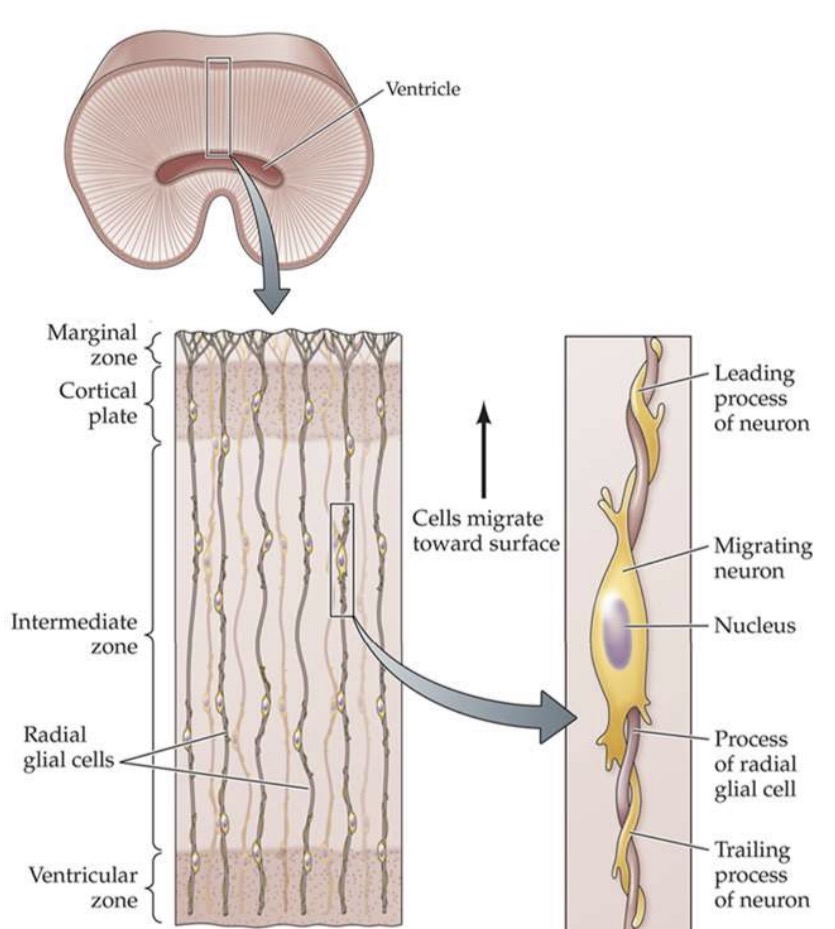
2.Cell migration
cell migrate away from the ventricular zone by moving along the surface of radial glia cells, establish distinct nerve cell populations (ex. Nuclei, layers)

Radial glial cells
generated before neurogenesis and extend from the inner to outer surfaces of the brain (like spokes on a wheel). After migration they become astrocytes.

The movement of cell during cell migration
cells goes along radial glial cells → cells of cerebral cortex arrive in “wave” → new cells become the outer layer of the established cells → form 6 layer

3.Differentiation
The transformation of precursor cells into distinctive types of neuron and glial cell, neural stem cells have the capacity to give rise o 3 major cell classes

Neural stem cells give rise to 3 classes:
1.Neurons 2.Astrocytes 3.Oligodentrocytes

Factor contribute to differentiation
Local environment: neurons look and act like their neighbor around them
young neural cells can turn into variety of neurons depending on where they end up
Notochord: can release proteins that induces some spinal cord cells to differentiate into motor neurons
Lately research indicates some fate restriction set early in development

Extent of neural connection and differentiation increase
increase with age during development of human cortex

4.Synaptogenesis
The axons and dendrites of young neurons grow extensively and form new synapses

Growth cone
growing tip of an axon or dendrites, have filopodia on it. Seeking out signal that attracts them or repel them (CAMs) so they can know which type of cells they should be connecting with

Filopodia
very fin ,tubular outgrowths form the growth cone

Call adhesion molecules (CAMs)
Protein found on the surface of a target cells that guides cell migration and/or axonal pathfinding. Some attract certain growth cones (chemoattractant) and some repel them (chemorepellent )

Myelination during 4.synaptogenesis
Once axons reach their final destination, the process of myelination (exWrapping repeated of a Schwann cell ) takes places to greatly speed conduction,

5.Cell death
Also a crucial phase of Brian development, especially during embryonic stages. Important sculpting process, that we will have more neuron to sort out then apoptosis the ones we don’t want

Apoptosis
naturally occurring cell death. In mice can be interrupt by knocking out pro-apoptoic genes, or overexpression of anti-apoptotic gene
6.Synapse rearrangement
The loss of some synapse (synaptic pruning) and development of others, to refine synapse rearrangement

Disrupting the balance between proliferation and differentiation produces a disorganized, nonfunctional brain
This shows bigger doesn’t necessarily mean better, the mice have more cells and cortical folding, but cells have divided excessively instead differentiate

Lissencephaly
rare gene linked brain malformation (DCX gene mutation), resulting the absence of convolutions in cortex. Children with lissencephaly have very small heads (microcephaly)

Mutation in DCX gene
the ventricles are dramatically enlarged, subcortical white matter is nearly absent, and sulci and gyri are completely absent

Symptoms of lissencephaly
facial difference, difficulty swallowing, failure to thrive, muscle spasms, seizures, severe psychomotor impairment
Histology
scientific study of the composition of tissues at a microscopic level. Aim to understand the structure and how this structure relates to the function with technique like staining procedure
Staining procedure for histology
Nissl: count cells and measure density in brain regions
Golgi: examine the morphology of individual neurons
IHC: map expression of cellular products
Tract tracers: trace interconnections between neurons
Transcardiac perfusion
the process of removing blood from the body/brain and replacing it with fixative solution (ex. Formaldehyde). During sacrifice, pump saline into the left ventricle and into the aorta.
Microtome
a specialized precision cutting instrument that accurately and repeatedly slices extremely thin sections from a block of embedded frozen tissue at a consistent thickness

Nissl stain
outlines all cell bodies in the tissue by staining RNA, and is useful for defining cytoarchitecture
Advantage:determining cell size and density, detecting lamination, and distinguishing white vs grey matter
Disadvantage:limit on detailed morphology of the neuron is not stained (ex. Dendrite and axons don’t have RNA so they don’t get stained)

Cytoarchitecture
The study of the structural organization and arrangement of cells within tissues, and they vary from brain region to brain region.

Function of Cytoarchitecture
show us the cell size, density, and distribution of the total population of brain cells are not uniform.
In several regions of the brain, such as cerebral cortex, neurons are arranged in layers (pr lamina) that can be clearly defined by differences in cell density

Golgi staining
useful for examining the precise shape and structure of individual neurons, classifying cell types based on morphology and revealing fine details of axons and dendrites, only die 5-10% of cells in the sample at random, but deep and complete (advantage and limitation)

More modern technique of golgi staining
inject fluorescent dyes into individual neurons, often after physiological recording to relate structure to function

Immunohistochemistry (IHC) and Immunofluorenscence (IF)
Map expression of cellular products. Both use antibodies to target cells with a specific protein, antibody seek out and attach themselves to target protein and reveals a distribution of only those neurons that make the target protein

Target protein
commonly referred to as the antigen

Process of Immunohistochemistry (IHC) and Immunofluorenscence (IF)
Primary antibody attach to the target cell, then the secondary antibody attach to the primary antibody which is coupled with an enzyme, like horseradish peroxidase (HRP), it is brown under light microscopy

Immunofluorence (IF)
commonly use with fluorescent microscopes, also allows for multiple antigens using different-colored fluorophones to be used on a single slide

Example of using immunohistochemistry to target c-fos, revealing brain regions with recently activated cells
Activation of receptors by neurotransmitters can lead to c-Fos expression, as well as increases in intracellular calcium from synaptic activity. Shows researchers all of the cells in the brain that were highly active during the specified time period of interest

Example of immunofluorescence used to indicate which neurotransmitter is contained within cells of interest
Antibody against the inhibitory neurotransmitter GABA. Found localized in the cells surrounding the special glomeruli structures in the olfactory bulb

Neuron processes information
electrical signals are the vocabulary of the nervous system
Synapses transmit information from one neuron to another
Action potential cause the release of the neurotransmitter into the cleft to signal neighboring cells

Electric charge inside and outside the cell
All living cells have electric charge. Intracellular (K+ and large anions) < extracellular (Na+ and Cl-)

Membrane potentials generation
by differential distribution of ions across the cell membrane

Cell membrane
a lipid bilayer, act as barrier of cell, ion channels of transport pumps located. Have different ion concentration each side of membrane→help active sodium-potassium pumps

Ion channels
Tube-like pore that selective for certain ions mainly:
Cation: Sodium (Na+), Potassium (K+), Calcium (Ca2+)
Anion: nonselective to small anions, but mostly referred to Cl-
Can be in open or close state, can have different types for same ion

Activation of ion channels
Voltage-gated channels: changes in membrane potential
Binding of neurotransmitter
Mechanical force

What happen when ion channel open
ion move across the membrane according to their electrochemical gradients

Electrochemical gradient
Electrical driving forces/Electrostatic forces: K+ wants to move out to Cl-
Chemical driving forces/Diffusion: Higher conc to lower conc, intracellular have more K+ than extracellular

Reached equilibrium potential
This is when the electrochemical gradient is balance and no net flow of ions.

Nernst equation
a mathematical function that can predict the Eq potential for a given ion based on the conc of the ion on both sides of the membrane

Electrochemical diving forces
From the electrochemical gradient, acting on an ion determines the direction and extent f ion flow. The goal is to reach Eq

K+ when depolarization
efflux, leave the cell

K+ when hyperpolarized
Influx, enters the cell

Intracellular microelectrode
To record membrane potential, or V, across the membrane.
recording electrode (red): placed intracellular fluid (cytoplasm)
reference electrode (black): placed extracellular solution
Electrode connected to amplifier and view the electrical difference on oscilloscope

Resting membrane potential
The difference in electrical potential potential across the membrane when at rest (-65mV)

Why RMP is -65mV?
1. Membrane permeability to K+: many more K+ channels are open at rest (potassium leak channels)
2. Ion concentration gradients: electrochemical forces are in balance for K+ close to -65mV
3. The Na+-K+ pump: acts to maintain the differential distribution of K+ and Na+

Three types of neuronal electrical signals
Receptor potential
Synaptic potential
Action potential
Results from transient changes in the resting potential

1.Receptor potential
elicited in receptor neuron during sensory transduction, grade (proportion to the magnitude of the sensory stimulus), passive

2.Synaptic potential
subthreshold, graded(amplitude vary according to the number of synapses activated, strength of each synapse, previous amount of synaptic activity), EPSPs and IPSPs, passive

Action potential
actively propagated, long distance, all or none

Hyperpolarization
occurs if the current delivered makes the membrane potential more negative than its resting potential (e.g., if resting was -65mV, the membrane potential is now -70 mV)
Depolarization
occurs if the current delivered makes the membrane potential more positive than its resting potential (e.g., if resting was -65mV, the membrane potential is now -60 mV)
Subthreshold graded responses
these are a passive response that can be
depolarizing or hyperpolarizing. The greater the stimulus, the greater the response. → All graded potentials (EPSPs + IPSPs) are summed together within a postsynaptic neuron and an action potential is generated if it meets threshold for triggering spike
Threshold
The level of membrane potential at which an action potential is generated (usually around -40mV, but varies among different types of neurons) • In the example above the threshold happens to be at -50mV
• Larger depolarizations above threshold produce more action potentials, not larger action potentials; the amplitude of APs is independent of the strength of the stimulus

Action potential process: 1. At rest
K+ leak channels opens/ Na+ leak closed, Eq for K+

Action potential process: 2. Rising phase (rapid depolarization)
membrane potential depolarize above threshold and closer to Eq potential for Na+ (~+60mV) → voltage-gated Na+ channels opens → Na+ rush in cell → further depolarization

Action potential process: 3. Overshoot
the brief period when the interior of the cell is positive (above 0mV)

Action potential process: 4. Falling phase (repolarization)
membrane potential falls back down towards the Eq potential for K+ to restore the resting potential
Voltage-gated Na+ channels quickly inactivate and close
Voltage-gated K+ channel (delayed rectifier K+channel) open → K+ leave (there was too much Na+)

Action potential process: 5.Afterpotential
immediately following an action potential, there may be a positive or negative change in membrane potential before it eventually returns to baseline
Ex. Undershoot phase

Action potential process: 6.Refractory periods
refers to transiently inactivated or exhausted axonal membranes, that make another action potential less likely to occur
Absolute refractory phase: a brief period of complete insensitivity to input stimuli (due to the inactivated Na+ channels); it is impossible to trigger a second action potential during this phase
Relative refractory phase: a period of reduced sensitivity, during which only strong stimulation can produce another action potential, due to the higher K+ efflux that is still occurring due to voltage-gated K+ channels still open.

Action potential transfer within axon
There is delay for the next, however they don’t have to go to threshold

Rapid saltatory conduction along myelinated axons
15x faster than in unmyelinated axons, myelin insulates the axons allowing the depolarization to passively spread further before requiring regeneration

Exocytosis
the process by which a synaptic vesicle fuses with. The presynaptic terminal membrane to release neurotransmitter into the synaptic cleft