3-2 Pulmonary Circulation
 Call Kai
 Learn
 Practice Test
 Spaced Repetition
 Match
 Flashcards
 Knowt Play
1/42

There's no tags or description
Looks like no tags are added yet.
43 Terms
Blood supply to the lungs comes from what 2 sources
bronchial artery system
pulmonary artery system
Unoxygenated blood leaves the… by..
right ventricle by way of the pulmonary artery trunk, which branches into the right and left pulmonary arteries
where does gas exchange occur
alveolar capillary membrane
the pulmonary arteries further divide into smaller arteries and arterioles that feed into the capillary network
pulmonary artery blood vs blood from pulmonary veins
arteries- unoxygenated
veins- oxygenated
pulmonary and bronchial circulation fxns
facilitates gas exchange
delivers nutrients to lung tissues
acts as blood reservoir for the left ventricle
pulm circ has lower pressure (18) than systemic circ (18)
only 1/3 of vessels are filled with blood at any given time
Pulm circulation pressure and volume
it’s a low pressure, high volume system
what 2 systems do pulm circ use to lower pulmonary pressure
in the setting of increased blood flow
1. recruitment - opening of these closed vessels
2. vessel distention, which allows more blood to circulate through. This dynamic system allows the lungs to efficiently match blood flow with ventilation and oxygen needs, ensuring optimal gas exchange under varying conditions.
bronchial circulation
part of the systemic circulation
received 1% of the cardiac output
does not participate in gas exchange
Under normal resting conditions:
During rest:
When the body requires more oxygen (e.g., during exercise):
some pulmonary capillaries are closed and not perfused.
only a fraction of the pulmonary capillaries are needed to oxygenate the blood, so many remain closed.
increased cardiac output raises pulmonary blood pressure, which recruits (opens) these previously closed capillaries to allow for greater gas exchange
Alveolar capillary membrane is
normal pulmonary arterial pressure in healthy adult vs arterial pressure of systemic circulation
very thin (the width of a single cell), crucial for efficient gas exchange
pulmonary: 22/8 systemic: 120/80
Ventilation:
movement of air into and out of the lungs
Distribute air within lungs to alveoli for maintenance of oxygenated and removal of CO2
carried by pulm system
Diffusion:
movement of gases between air spaced in the lungs and the bloodstream
carried by pulm system
Perfusion
movement of blood into and out of the capillary beds of the lungs to body organs and tissues
carried by cardiovascular system
Dead space
3 dimensions:
Some air stays within the airways and does not participate in gas exchange
anatomic dead space: includes gas in the conducting airways
Alveolar dead space: composed of ventilated but unperfused areas of the lung (portion of the air…)
Physiologic dead space: sum of the anatomic and alveolar dead space
Minute Ventilation
the total volume of air that is inhaled or exhaled from the lungs in one minute
product of tidal volume xRR per minute
can help determine whether a pt can or not come off the vent
Alveolar ventilation
the difference between tidal volume and anatomic dead space volume multiplied by RR per minute, useful when looking at integrity of alveolar capillary membrane on PFTs
4 steps of gas transport for oxygen delivery
1. ventilation of lung
2. diffusion of oxygen from alveoli into capillary bed
3. perfusion of systemic capillaries w oxygenated blood
4. diffusion of oxygen from systemic capillaries into the cell
4 steps in reverse order for removal of CO2
1. Diffusion of CO2 from cells into systemic capillaries
2. Perfusion of pulm capillary bed by venous blood
3. Diffusion of CO2 into the alveoli
4. Removal of CO2 from the lungs by ventilation
Diffusion
movement of gases btw air spaced in lungs and bloodstream
Effective gas exchange:
needs ~ even distribution of ventilation(o2) and blood (perfusion) in all portions of lungs
HOWEVER distribution is uneven
why is distribution of ventilation and perfusion uneven
- Gravity and alveolar pressure affect distribution of blood and diffusion (depend on body position)
- Ventilation perfusion ratio (perfusion > ventilations in base of lung) (vent > perfusion in apices of lungs)
Relationship btw ventilation and perfusion:
•is expressed as a ratio
•the normal V/Q ratio is 0.8 - which is the amount perfusion exceeds ventilation under normal circumstances (this means that, overall, the lungs receive slightly more blood flow than air, but this is close to optimal for gas exchange)
Zone 1
reflects blood flow in apices of lung
blood flow is minimal bc enlarged alveolar sacs in apices create alveolar pressure higher than capillary pressure ( V/Q ratio >1)
Zone 2
reflects the middle region of lung
pulm arterial pressure varies due to right ventricle systole, so there’s intermittent perfusion
Zone 3
continuously perfused throughout the cardiac cycle
pulm arterial pressure exceeds alveolar pressure (V/Q <1)
Diffusion across alveolocapillary membrane
idea for oxygen diffusion
large total surface area
very thin= one cell thick under normal circumstance
Diffusion of gas from high to low concentration
Partial pressure of oxygen molecules (Po2) is much greater in alveolar gas (PAO2) than it is in capillary blood (PaO2): Promotes rapid diffusion from the alveolus into the capillary.
Determinants of arterial oxygenation
Hemoglobin: oxygen-carrying oxygen
Oxygen content: total amount of O2 carried in blood
Cardiac fxn: ability to deliver oxygenated blood to tissues
as oxygen diffuses across alveolar capillary membrane, what does it do FIRST
what other steps does it take
dissolved in plasma, where it exerts pressure - PaO2
PaO2 increases= oxygen moves O2 from plasma into RBC and bind with hemoglobin molecules
•Oxygen continues to bind with hemoglobin molecules until hemoglobin binding site are filled (or saturated)
•Oxygen continues to diffuse across the alveolar-capillary membrane until PAO2 and PaO2 equilibrate, eliminating the pressure gradient
Components of Hemoglobin
what does it contain?
Each can bind up to
heme- oxygen binding pigment containing iron
globin- protein! envelop that provides a pocket for a heme molecule
4 heme groups, 4 globin chains (beta&alpha)
bind up to 4 molecules of oxygen
Cooperative
phenomenon where binding of oxygen to one subunit of a hemoglobin molecule increases the affinity of oxygen at the other binding sites
Oxyhemoglobin:
•Hemoglobin molecules bind with oxygen: Oxyhemoglobin
•the majority of oxygen in the blood (97%) is bound to hemoglobin
•the oxygen saturation (SaO2) is the percentage of hemoglobin that is bound to oxygen. This can be measured with an oximeter (pulse oximeter-SpO2)
what happens at the tissues where partial pressure of Oxygen is low
o2 is released from the hemoglobin molecule
•When hemoglobin saturation and desaturation are plotted on a graph, the result is
a distinctive S-shaped curve known as the oxyhemoglobin dissociation curve.
Oxyhemoglobin association and dissociation
•Hemoglobin molecules bind with oxygen: Oxyhemoglobin
•the majority of oxygen in the blood (97%) is bound to hemoglobin
•the oxygen saturation (SaO2) is the percentage of hemoglobin that is bound to oxygen. This can be measured with an oximeter (pulse oximeter)
•The PaO2 measures only thr small amount of oxygen dissolved in plasma and is measured by obtaining an arterial blood gas (ABG)
•A po2 of 60 mm Hg corresponds to an spo2 of about 90%
•At the tissues where partial pressure of Oxygen is low, O2 is released from the hemoglobin molecule
•When hemoglobin saturation and desaturation are plotted on a graph, the result is a distinctive S-shaped curve known as the oxyhemoglobin dissociation curve.
For PaO2 lvls <60, oxygen is
For PaO2 lvls >60:
readily unloaded to peripheral tissues and hemoglobin’s affinity for oxygen diminishes
curve flattens reflecting maximum saturation of hemoglobin w oxygen
Oxygen-Hemoglobin Dissociation Curve
Shift to the left
Shift to the right
left- depicts the hemoglobin’s increased affinity for oxygen, which promotes association in the lungs and inhibits dissociation in the tissues.
•Alkalosis (high pH) and hypocapnia and hypothermia
right- depicts the hemoglobin’s decreased affinity for oxygen or an increase in the ease with which oxyhemoglobin dissociates and oxygen moves into the cells.
•Acidosis (low pH) and hypercapnia and hyperthermia
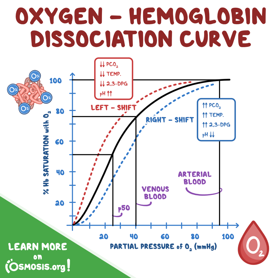
large changes in PaO2 causes
only small changes in hemoglobin saturation (SaO2)
CO2 Transport
what 3 ways is it transported in blood:
~ 200 ml of CO2 produced by tissues per min= byproduct of cellular metabolism
1. Dissolved in plasma
2. As Bicarbonate (60-70%)
3. Attached to hemoglobin
Solubility of CO2 vs O2
Reduced hemoglobin is able to..
CO2 is 20x more soluble than O2= it wants to get out quickly! (from tissue cell into blood)
carry move CO2: •therefore, the drop in SaO2 at the tissue level increases the ability of hemoglobin to carry CO2 back to the lungs
Presence of RBS enzyme carbonic anhydrase, what happens
CO2 combines w water to form carbonic acid —>
Carbonic acid almost instantaneously breaks down into H+ and Bicarbonate ions —>
•Hemoglobin attaches to H+ (RBC buffer) and HCO3- diffuses into the plasma
•in exchange for chloride - a process known as “chloride shift”
In CO2 transport, when venous blood reaches the lungs,
the chemical process is reversed!
oxygen diffuses into blood and attached to HG → HG releases H to bind with O2 → HCO3- reenters RBC in xchange for Chloride → HCO3 combines w H ( bc of enzyme carbonic anhydrase) to form carbonic acid →

CO2 transport to the lungs
diffusion gradient for CO2 from blood to the lung is only 6mm Hg (46-40)
BUT, because CO2 is so soluble in the alveolar capillary membrane, it quickly diffuses into alveoli (Co2 wants to get out!)
•Diffusion of CO2 in the lung is so efficient that diffusion defects that cause hypoxemia do not cause hypercapnia