Chapter 4 - Sesay slides
 Call Kai
 Learn
Learn Practice Test
 Spaced Repetition
 Match
Match Flashcards
 Knowt Play
1/95

Earn XP
Description and Tags
genes and cellular function/ DNA and RNA
Name | Mastery | Learn | Test | Matching | Spaced | Call with Kai | Chat |
|---|
No analytics yet
Send a link to your students to track their progress
96 Terms
Deoxyribonucleic acid (DNA)
-Long, thread-like molecule with uniform diameter, but
varied length
-46 DNA molecules (chromosomes) in nucleus of most
human cells
-Average human DNA molecule about 2 inches long
3 components:
sugar- dioxyribose
nitrogenous base- changes between DNA
phosphate-
DNA doesnt have uracil it has thymine.
Nucleotides
DNA and other nucleic acids are polymers of nucleotides
Each nucleotide consists of three components:
1-A sugar—deoxyribose
2- A phosphate group
3- A nitrogenous base
• DNA bases: A, T, C, G
• Purines—adenine (A), guanine (G)
• Have double-ringed structure
• Pyrimidines—cytosine (C), thymine (T), uracil (U)
• Have single-ringed structure
The Five
Nitrogenous
Bases found in
DNA and RNA
Purines: Adenine (A) and Guanine (G) (double ring)
Pyrimidines: Cytosine (C), Thymine (T), Uracil (U) (you cut the pie

Draw DNA structure

What is the essential function of DNA?
The essential function of DNA is to carry instructions (genes)
for the synthesis of proteins. genes only contain information for production of proteins
which nitrogen base is not in DNA
Uracil is only in RNA, but Thymine is in DNA
What is a Gene?
segment of DNA coding for the synthesis of a specific
protein/aka polypeptide
• Humans have about 20,000 genes
• Only about 2% of total DNA code for protein
• Other 98% is noncoding DNA- they regulate gene activity
• Plays role in chromosome structure
• Regulates gene activity
Chromatin—
•
fine filamentous DNA material complexed
with proteins called histones
not dividing- loose chromatin
Occurs as 46 chromosomes in most cells
• 6 feet long thread packed in cell nucleus of 5 μm diameter
• In nondividing cells, chromatin is so slender it cannot be seen with
light microscope
• Granular appearance under electron microscope
DNA is chromatin and chromosome
about to divide- chromosome
non dividing- chromatin
Chromatin sctructure

Histones—
proteins around which DNA winds
• Histones cluster in groups of eight molecules
transition from chromatin to chromosome
when chromatin transitions to chromsome it loses histones and coils tighter.
how does chromatin ‘behave”?
In a nondividing cell, the chromatin is not static
• Structure and location changes moment to moment
according to genetic activity of cell
• Genes get turned off and on:
Amylase
breaks down carbohydrates
after cell division
each chromosome becomes to parallel chromosomes called sister chromatids.
in early stages of prophase/ cell division these sister chromatids coil even more, getting thicker- enables them to fit into nucelous and to split apart!
Ribonucleic acids (RNAs)
General structure:
• Contain the sugar ribose
• nitrogenous Bases A, U, G, C; uracil (U) replaces thymine found in DNA
• Single nucleotide chain (not a double helix) except in short regions
• Smaller than DNA, can have less than 100 or just over 10,000
bases per molecule
• Functions mainly in cytoplasm
RNA function
Produce proteins with three main rna’s; messengers rna, ribosomal rna, and transfer rna.
Three RNAs are important for protein synthesis
Messenger RNA (mRNA)
• Ribosomal RNA (rRNA)
• Transfer RNA (tRNA)
What Is a Gene?
Previous (classical) definition: a gene is a segment of
DNA that carries the code for a particular protein
• But body has millions of proteins and only about 20,000 genes
• Current definition: a gene is an information-containing
segment of DNA that codes for the production of a
molecule of RNA that most often plays a role in
synthesizing one or more proteins
• Amino acid sequence of a protein is determined by the nucleotide
sequence in the DNA
how many chromosomes in a human?
The 46 human chromosomes come in two sets of 23
chromosomes, one set from each parent
Genome—
all the DNA in one 23-chromosome set
• 3.1 billion nucleotide pairs in human genome
• Individual variation comes from single-nucleotide polymorphisms
Genomics—
study of the whole genome
• How genes and noncoding DNA interact to affect structure and
function of the organism
Genomic medicine—
application of knowledge of the
genome to the prediction, diagnosis, and treatment of
disease
• Relevant to many disorders (examples: cancer, Alzheimer disease,
schizophrenia, obesity, AIDS, tuberculosis)
4.2b The Genetic Code
code—system that enables these 4 nucleotides to
code for the 20 amino acid sequences of all proteins
Body can make millions of different proteins (the proteome),
from just 20 amino acids, and encoded by genes made of
just 4 nucleotides (A, T, C, G)
Base triplet—
sequence of three DNA nucleotides that stands
for one amino acid
codon- must know start and stop codons
3-base sequence in mRNA
in nucleus- speaks codon language
cytoplasm- speaks amino acid/protein language
• 64 possible codons available to represent 20 amino acids; 61
code for amino acids, 3 are stop codons
• Start codon—AUG codes for methionine, and begins the amino
acid sequence of the protein
• Stop codons—UAG, UGA, and UAA: signal “end of message,” like a period at the end of a sentence
4.2c Protein Synthesis
When a gene is activated, messenger RNA (mRNA) (by RNA polymerase) is
made; serves as a code for a particular protein
• DNA → RNA → protein
• DNA → mRNA is called transcription; occurs in nucleus
• mRNA → protein is called translation; occurs in cytoplasm
Transcription—
copying genetic instructions from DNA to
mRNA in the nucleus.
RNA polymerase rewinds the DNA helix behind it; allows gene to be
transcribed by several polymerase molecules.
MRNA is small enough to leave nucleus.
• Terminator: stop sequence at the end of a gene
Pre-mRNA
is “immature” RNA produced by transcription;
undergoes processing
has introns
• Enzymes within the nucleus remove introns from the preRNA and
splice exons together to make mRNA (only has exons)
Alternative splicing—
pre-mRNA is spliced- introns are removed
variations in the way exons are
spliced allow for a variety of proteins to be produced from
one gene
• One gene can code for more than one protein
• Exons can be spliced together into a variety of different mRNAs
alternative splicing of mRNA

Translation—
nucleotide language converted into amino acid
language
A ribosome “reads” the mRNA codons (group of three nucleotides) and forms a protein in the cytoplasm
Three main participants in translation:
• Messenger RNA (mRNA)
• Carries code from nucleus to cytoplasm
• Has protein cap that is recognition site for ribosome
• Transfer RNA (tRNA)
• Finds free amino acid in cytosol, Delivers a single amino acid to the ribosome
• Contains an anticodon—series of 3 nucleotides that are
complementary to the mRNA codon
Initiator tRNA always carries AUC methione.
• Ribosomes
• Organelles that read the message and build a peptide chain
• Contain a large subunit and a small subunit
• Free in cytosol, on rough ER, and on nuclear envelope
• E site, P site, and A site involved in translation process
• Messenger RNA (mRNA)
• Carries code from nucleus to cytoplasm
• Has protein cap that is recognition site for ribosome
Transfer RNA (tRNA)
Finds free amino acid in cytosol, Delivers a single amino acid to the ribosome
• Contains an anticodon—series of 3 nucleotides that are
complementary to the mRNA codon
Initiator tRNA always carries AUC methionine.
• Ribosomes
• Organelles that read the message and build a peptide chain
• Contain a large subunit and a small subunit
• Free in cytosol, on rough ER, and on nuclear envelope
• Has three pockets that span both small and large subunits: A(accepts) site, P(growing protein) site, and E (exits)site involved in translation process
tRNA lands inside the Ribosome
Three main steps in translation: (not on test)
• Initiation
1- Ribosome assembles with mRNA in cytosol
•2- Small subunit binds leader sequence of mRNA, slides along mRNA until AUG is recognized.
3- Initiator tRNA tRNA comes along, with anticodon UAC, pairs with the AUG start codon and settles into P (middle site) of the ribosome with MET methionine as cargo. (subsequent tRNA enter A site)
4-Large subunit ribosome then joins complex- whole complex then slides along mRNA
• Elongation
1- Next tRNA with amino acid arrives, binds to A site
2- tRNA anticodon paris with second Codon of mRNA. For example mRNA codon GCU recieves anticodon CGA.
3- an Enzyme in the Ribosome creates peptide bond between first and second amino acids.
4- Ribosome moves down mRNA by one codon, so growing peptide is now attached in P site
5- Initiator tRNA shifts to E site and exits.
• Process continues and peptide grows
Termination
• Ribosome reaches a stop codon
• A site binds a protein called a release factor
• Ribosome disassembles and dissociates from mRNA
often the subunits reassemble on same mRNA, because they are so close to the leader sequence, and make more protein!
Initiation
1- Ribosome assembles with mRNA in cytosol
•2- Small subunit binds leader sequence of mRNA, slides along mRNA until AUG is recognized.
3- Initiator tRNA tRNA comes along, with anticodon UAC, pairs with the AUG start codon and settles into P (middle site) of the ribosome with MET methione as cargo.
4-Large subunit ribosome then joins complex- whole complex then slides along mRNA
• Elongation
•
1- Next tRNA with amino acid arrives, binds to A site
2- tRNA anticodon paris with second Codon of mRNA. For example mRNA codon GCU recieves anticodon CGA.
3- an Enzyme in the Ribosome creates peptide bond between first and second amino acids.
4- Ribosome moves down mRNA by one codon, so growing peptide is now attached in P site
5- Initiator tRNA shifts to E site and exits.
• Process continues and peptide grows
Termination
•
• Ribosome reaches a stop codon
• A site binds a protein called a release factor
• Ribosome disassembles and dissociates from mRNA
often the subunits reassemble on same mRNA, because they are so close to the leader sequence, and make more protein!
Making proteins for packaging or export
•
Proteins destined for lysosomes or secretion are modified by ER
• Ribosome docks on ER and synthesizes protein into ER cistern
• ER modifies and packages protein into transport vesicles
• Multiple ribosomes, in clusters called polyribosomes, can
translate the same mRNA molecule simultaneously
Relationship of a DNA Base Sequence to Peptide
Structure

Chaperone proteins—4.2d
Protein synthesis is not finished when the amino acid
sequence (primary structure) has been assembled
To function, protein must fold into precise secondary and
tertiary structures
• Chaperone proteins (older proteins)—guide the folding of newly
synthesized proteins
• Help prevent improper association between different proteins
• Some also called stress proteins or heat shock proteins- Chaperones produced in response to heat or stress and then come to cytosol to help heat damaged proteins.
• Help damaged proteins fold back into correct functional shapes
image of protein processing and secretion- very important image

4.2e Gene Regulation
Genes can be turned on and off- depending on need
Cells can turn some genes permanently off
Example: liver cells turn off hemoglobin genes
adult cells live longer so we dont need the extra hemoglobin from the liver.
Cells can turn genes on only when needed
The level of gene expression can vary from day to day or hour to
hour
• This can be controlled by chemical messengers such as hormones
• Example: In response to prolactin, mammary gland cells turn on
gene for casein protein only when breast milk is produced
Gene Regulation 2
Example: Casein synthesis and secretion
1. Hormone prolactin binds to receptors on membrane of mammary cell- signal that we need the protein
2. Receptors trigger activation of a regulatory protein (transcription
activator) in cytoplasm
3. Regulatory protein moves into the nucleus and binds to the DNA near
the casein gene
4. The binding enables RNA polymerase to bind to the gene and
transcribe it, producing the mRNA for casein
5. The casein mRNA moves to the cytoplasm, and ribosomes on rough
ER translate it
6. The Golgi complex packages casein into secretory vesicles
7. The secretory vesicles release the casein by exocytosis, and it
becomes part of the milk

4.2f Synthesizing Compounds Other Than Proteins
Cells synthesize glycogen, fat, steroids, phospholipids, pigments,
and other compounds
• No genes for these products, but their synthesis is under
indirect genetic control
• They are produced by enzymatic reactions, and enzymes are
proteins encoded by genes
example of 4.2f Synthesizing Compounds Other Than Proteins
• A cell of the testes takes in cholesterol
• Enzymatically converts it to testosterone
• Only occurs when genes for enzyme are active
Genes may greatly affect such complex outcomes as behavior,
aggression, and sex drive
image of
Testosterone
Synthesis by DNA
step 5 requires an enzyme.

4.3 DNA Replication and the Cell Cycle 2
Before a cell divides, it must duplicate its DNA so it can
give a complete copy of all its genes to each daughter cell
• Since DNA controls all cellular function, this replication
process must be very exact
• Law of complementary base pairing enables us to predict
the sequence of one DNA strand if we know the other
DNA replication- def on exam
dna is double stranded.
chromatin- very thin and wrapped around histones
first we must unwind from the histones.
now its in chromosome state
now chromsome unzips
creates a complementary pair (by adding complementary base pair)
4.3a DNA Replication1
Four steps of DNA replication: Definitely on exam
1. Unwinding the helix from the histones
2. Unzipping small portion of helix by DNA helicase enzyme;
separates the strands, forming a replication fork
3. Enzyme called DNA polymerase moves along each strand,
reads the exposed bases, and synthesizes complementary
strands. towards replication fork on one strand, and away on the other strand- On the discontinuous strand, segments(okazaki fragments) are connected by DNA ligase enzyme.
• Semiconservative replication—each new DNA molecule
contains old (parental) DNA and newly synthesized DNA
4. New histones synthesized in cytoplasm, transported into the nucleus- used to organize new DNA strands into nucleosomes.
image of semi-conservative DNA replication- this picture will be on exam

4.3b Errors and Mutations (not on test)
DNA Damage Response (DDR)
Mutations
DNA polymerase occasionally makes mistakes
a-DNA Damage Response (DDR)
• b- Mutations (not on test)
•a-
• Mechanisms in place to correct replication errors
• DNA polymerase double checks the new base pair and tends to
replace incorrect, biochemically unstable pairs with more stable,
correct pairs
• Result is only one error per 1 billion bases replicated
b-
• Changes in DNA structure due to replication errors or environmental
factors (radiation, viruses, chemicals)
• Some mutations cause no ill effects, others kill the cell, turn it
cancerous, or cause genetic defects in future generations
• Genetic variation within one’s body, due to mutations and
replication errors, is called genetic mosaicism
4.3c The Cell Cycle 1
The cell cycle includes interphase and the mitotic phase
Interphase includes three subphases:
• First gap phase (G1)
• Synthesis phase (S)
• Second gap phase (G2")
Mitotic phase includes multiple subphases:
• Prophase
• Metaphase
• Anaphase
• Telophase
• Cytokinesis

The Cell Cycle
Interphase
• G1
: first gap phase
• Interval between cell division and DNA replication
• Cell carries out normal tasks and synthesizes proteins and
materials for next phase, accumulates enough material to replicate DNA in next phase. (carries out semiconservative replication described earlier).
• S: synthesis phase
• Cell replicates all nuclear DNA and duplicates centrioles
identical sets of DNA molecules are ready to be put into daughter cells.
• G2
: second gap phase
• Interval between DNA replication and cell division
• Cell repairs DNA replication errors, grows and synthesizes
enzymes that control cell division, finishes replicating centrioles,
Mitotic phase
• Cell replicates its nucleus
• Pinches in two to form new daughter cells
G0
(G zero) phase- on test
•
describes cells that have left the cycle and cease dividing
for a long time (or permanently)
Mitotic phase stages:
Prophase:
1-At outset of mitosis, chromosomes shorten and thicken into compact rods. there are 46 chromosomes each made of two chromatids (one molecule of DNA per chromatid).
2-Nuclear envelope disintegrates- releasing chromosomes into cytosol
3-Centrioles sprout microtubules called spindle fibers, which push centrioles apart as they grow- arriving at opposite poles.
4-spindle fibers grow toward the chromosomes, attach at kinetochores on the centromere sides, and then the spindle fibers tug the chromosomes to align in the center…
Metaphase
1- Chromosomes are aligned on cell equator, awaiting to be split apart
2- spindle fibers form a lemon like shape called mitotic spindle.
3- shorter microtubules called aster, attach centriole to plasma membrane.
Anaphase
1- special enzyme activates the cleavage of the sister chromatids at the centromere
2- each chromatid is now considered a single stranded daughter chromosome that migrates to each end of the cell- centromere leading and arms flailing behind.
-motor proteins in the kinetochore crawl up/”chew” up the spindle fiber bringing it closer to the centriole.
Telophase:
1- daughter chromosomes cluster at each pole.
2- rough ER produces a new nuclear envelope around each cluster.
3- chromosomes begin to uncoil into loose chromatin in their new nuclear “home”.
4- mitotic spindle breaks up
5- nucleus forms nucleoli, meaning it has formed RNA
6- overlaps with cytokinesis…
Cytokinesis
Division of cytoplasm into two cells:
1-motor protein myosin pulls on microfilaments of actin in terminal web of cytoskeleton. creates a cleavage furrow along equator of cell, eventually pinching the cell in two.
…and now interphase begin!…G1…S….G2
—-beware in some cells cytokinesis does not happen!
4.3d Mitosis 1
Mitosis is cell division resulting in two genetically identical
daughter cells
• In contrast to meiosis, which produces eggs and sperm
Functions of mitosis
• Development of individual from fertilized egg to about 50 trillion
cells
• Growth of all tissues and organs after birth, replacement of cells
that die, and repair of damaged tissues
Four phases of mitosis
• Prophase
• Metaphase
• Anaphase
• Telophase
Mitosis image stage 1 and 2
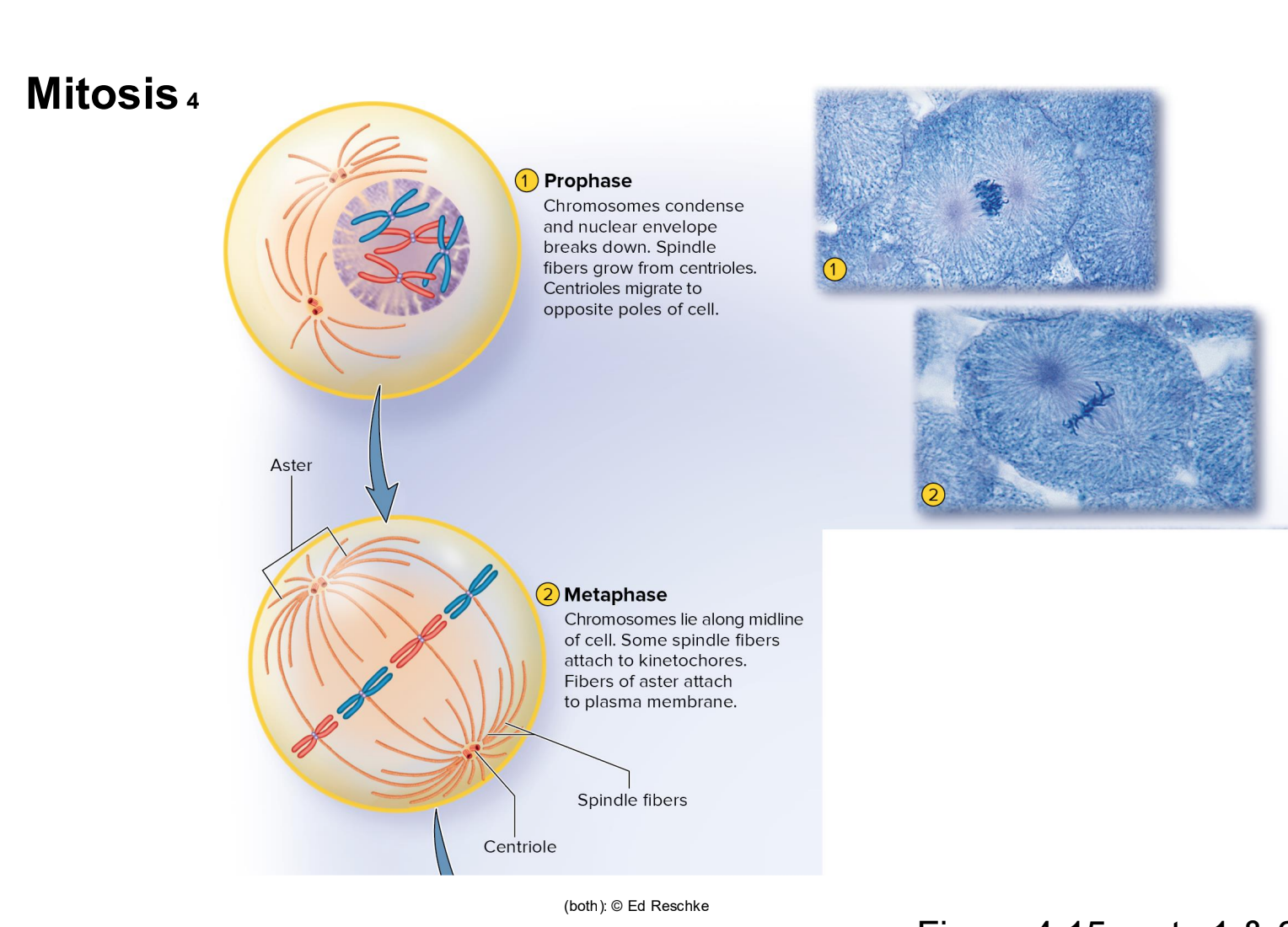
Mitosis image stage 3 and 4

genetic mosaicism
body’s cells is a patchwork of genetic variation.
contact inhibition
when cells proximity/touching inhibits cell division
Regulation of Cell Division 3
Checkpoints
within the cell cycle
• Controlled by protein called Cyclin–Cdk complexes- adds protein to other proteins thus inhibiting or activitating it. cyclin binds to CDK and triggers cascade of reactions that prepare cell for next steps.
• Start or G1 checkpoint
• Either allows the cell to proceed toward the S phase or, if it doesn’t,
the cell goes into the noncycling G0 phase
• G2
∕ M checkpoint
• Late in the G2 phase determines whether the cell is able to proceed
to mitosis
• A third checkpoint at the transition from metaphase to
anaphase determines whether the cell can proceed to
anaphase, leading to separation of its sister chromatids
4.4a The Karyotype
• Chart of all 46 chromosomes laid out in order by size
• 23 pairs of homologous chromosomes
Heredity—
transmission of genetic characteristics from
parent to offspring
Homologous chromosomes
Two members of each pair of the 46 chromosomes•
1 chromosome from each pair inherited from each parent
• 22 pairs of autosomes
• Look alike and carry the same genes
• 1 pair of sex chromosomes
• Female has homologous pair of X chromosomes
• Male has one X and one much smaller Y chromosome
autosomes
the 22 pairs of chromosomes that are similar
sex chromosomes
XX for female
XY for male
Simply labeled x and y because they perplexed scientists initially.
Locus—
a gene’s position on a chromosome
Alleles—
different forms of a particular gene
• Alleles are found at same locus on homologous chromosomes
Dominant allele
(represented by capital letter)
• If present, corresponding trait is usually seen in the individual
• Masks effect of recessive allele
• Often produces protein responsible for visible trait
Recessive allele
(represented by lowercase letter)
• Corresponding trait only seen when recessive allele present on
both homologous chromosomes
• Often codes for a nonfunctional variant of the protein
Genotype—
the alleles an individual possesses for a particular
gene
Homozygous
• Heterozygous
individuals have two identical alleles for the gene
individuals have different alleles for the gene
Phenotype—
an observable trait
• An allele is expressed if it shows in the phenotype of an
individual
• Usually determined by multiple genes, but some straits arise
from a single gene
• Sickle-cell disease caused by homozygous recessive alleles
• Carriers have a single allele for the disease; have sickle-cell trait (SCT)
Punnett square—
diagram showing possible genotype and
phenotype outcomes from parents of known genotype
Sickle-Cell Disease image
determined by just one gene.

The Roles of Environment and Heredity in Producing a
Phenotype

4.4h Epigenetics
Epigenetic effects—
gene expression is altered without
changes in base sequence;
• Can be passed to offspring, called epigenetic inheritance
• Examples:
• DNA methylation: mechanism of epigenetic change in which methyl
groups (—CH3) are added to DNA
• Variations in packaging of chromatin
• Gene silencing by noncoding RNA (ncRNA)
• Many epigenetic changes are normal, but some may
trigger diseases such as cancer
Cancer 1
Malignant tumors cause cancer
• Grow rapidly
• Tend to metastasize—give off cells that spread and seed
the growth of tumors elsewhere
• May stimulate tumor angiogenesis—growth of blood
vessels by energy-hungry tumors
• Distinguished from slow growing, encapsulated benign
tumors

Oncology—
medical specialty dealing with tumors
Cancers are named for tissue of origin
• Carcinomas: in epithelial tissue
• Lymphomas: in lymph nodes
• Melanomas: in pigment cells of epidermis (melanocytes)
• Leukemias: in blood-forming tissues
• Sarcomas: in bone, other connective tissue, or muscle
cancer causes
Cancers are always genetic, but only 5 to 10% are hereditary
Most cases caused by mutations
• During replication or exposure to carcinogens—
environmental cancer-causing agents
• Radiation: ultraviolet rays, X-rays
• Chemicals: cigarette tar, food preservatives, industrial chemicals
• Viruses: human papillomavirus, hepatitis C, type 2 herpes simplex
Oncogenes
cause cell division to accelerate out of control
• Excessive production of growth factors or growth-factor
receptors that stimulate mitosis
Tumor-suppressor (TS) genes
block development of
cancer, but can be mutated
• Mutated TS genes or silenced TS genes leave oncogene
action unopposed
lethal effects of cancer
Lethal effects of cancer
• Replace functional tissue in vital organs
• Often invade blood vessels, lung tissue, or brain tissue
• Tumors can put pressure on organs, blocking their normal
function
• Weaken one’s immunity
• Open the door for opportunistic infections
• Steal nutrients from the rest of the body
• Cachexia: severe wasting away of depleted tissues