Systems Review and focused history taking
 Call Kai
 Learn
 Practice Test
 Spaced Repetition
 Match
 Flashcards
 Knowt Play
1/13

There's no tags or description
Looks like no tags are added yet.
14 Terms
Anatomical position
Standing erect with palms and feet facing forward
Palms face anteriorly with thumbs pointed away from the body
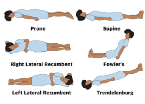
Positioning diagram
Anatomical Reference points diagram

Lateral definitions
Unilateral - pertaining to 1 side
Ipsilateral - on the same side as the reference point
Bilateral - pertaining to both sides
Contralateral - on the opposite side of the reference point
Terms of motion diagram

What are the anatomical plans?
Sagittal plane
Transverse plane
Coronal plane

Explicit and implied/implicit consent definitions
Explicit consent:
Can be spoken or written when a patient gives you permission to do something
Implied/implicit consent:
When consent is indicated indirectly e.g. by bringing a prescription for dispensing. It’s only valid if the patient understands what they are consenting to
General survey (inspection) - observing the patient
Level of consciousness
Mental awareness – agitation/ drowsiness/ confusion
Cachexia/obesity
Position/posture
Oxygen/therapeutic adjuncts
Walking aids
How comfortable?
Able to talk in sentences
Cyanosis/pallor/mottling
Feverish?
Effort in breathing – accessory muscles
Fever
Audible wheeze/stridor/grunting
Intercostal recession
Sweating
Tremor/Fasciculations/Unintended movements
Vital signs
Blood pressure - lying, standing, inter arm difference
Pulse - rate, rhythm, volume, character
Temperature
Respiratory rate
Height/weight
Capillary Refill Time
Pulse Oximetry
Familiarise yourself with normal ranges
Peripheral Examination
Skin: i.e. turgor/colour/lesions/temperature/integrity
Finger nails: i.e. clubbing (check Schamroth’s window sign)/ koilonychia (spooning)/splinter haemorrhages/ leukonychia (white)
Hands: i.e. palmer erythema/Dupuytren's contractures/ wasting /Herberdens Nodes/ Oslers nodes/ xanthomas/ Janeway lesions/ tremor / asterixis
Arms: i.e. radial/ brachial pulses, blood pressures, Collapsing Pulse
Eyes: i.e. conjunctival pallor/ xanthalasma/ corneal arcus/ sclera/ petechial haemorrhages/ Kayser Fleischer rings
Face: i.e. Malar Flush
Mouth: i.e. dentition/cyanosis/angular chelitis (stomatitis)/ glossitis/ulceration/hydration
Neck: lymph nodes, central venous pressure, carotid pulse
Legs: pulses, skin, oedema
Focused Systems Review - IPPA
Inspection - use of vision, hearing and smell to detect characteristics and physical signs
Palpitation - using the hands to touch and make measurements of physical signs
Percussion - striking the body’s surface with a finger to produce a vibration
Ausculation - listening to sounds created in body organs to detect variations from the normal
Jugular Venous Pressure
JVP provides an indirect measure of central venous pressure (right atrium)
The jugular venous pulse is:
Not palpable
Obliterated by pressure
Characterised by a double waveform
Variable with respiration - it decreases with inspiration
Enhanced by the hepatojugular reflux
JVP can be difficult to detect and has been superseded by other diagnostic methods
Measurement of JVP
Stand at patients RHS – patient supine, reclined at 45 degrees
Look across the neck from the patients RHS – good light source
Locate the surface markings of the IJV - this runs from the medial end of the clavicle to the ear lobe, under the medial aspect of the sternocleidomastoid muscle
Identify the jugular vein pulsation ( 2 beats per heartbeat) in the suprasternal notch or behind the sternocleidomastoid muscle
Use LUQ pressure to confirm it is the JVP
Measure the JVP as the vertical height in cm between the upper limit of the venous pulsation and the sternal angle (junction of the manubrium and sternum at the level of the second costal cartridges)
JVP in health = ≤ 4cm above the sternal angle

Lymph node distribution palpitation diagram
