GI physiology
 Call Kai
 Learn
 Practice Test
 Spaced Repetition
 Match
 Flashcards
 Knowt Play
1/46

There's no tags or description
Looks like no tags are added yet.
47 Terms
Monogastric
Single chambered stomach
Ruminants
Rumen, reticulum, omasum, then “stomach” = abomasum
Hind-gut fermentation
Monogastrics but specialized for fermentation in large intestine
Ex: Horses and rabbits
Animals that have a forestomach
Ruminants and camelids
Which species is an obligate carnivore?
Cat
Which species is a facultative carnivore?
Dogs
Flow of feed
Mouth
Esophagus
Stomach (± forestomach)
Small intestine
Large intestine
Rectum/anus
Order from stomach to anus
Stomach
Duodenum
Jejunum
Ileum
Cecum
Ascending colon
Transverse colon
Descending colon
Rectum
Anus
Major functions of the stomach
Mechanical digestion
Acidic digestion
Some enzymatic digestion
Major functions of the small intestine
Most similar among species
Enzymatic digestion
Absorption
Immune functions
Major functions of the large intestine
Fermentation
Absorption
Major function of the cecum
Between the ileum and colon
Fermentation
Function of Descending Colon/Rectum/Anus
Absorption
Transport
Storage

What is in red?
Luminal

What is in blue?
Mural

Mucosa
The Epithelium that lines the lumen of the GI tract

Submucosa
Underlying connective tissue- Collagen with blood vessels and nerves (provides structural integrity)
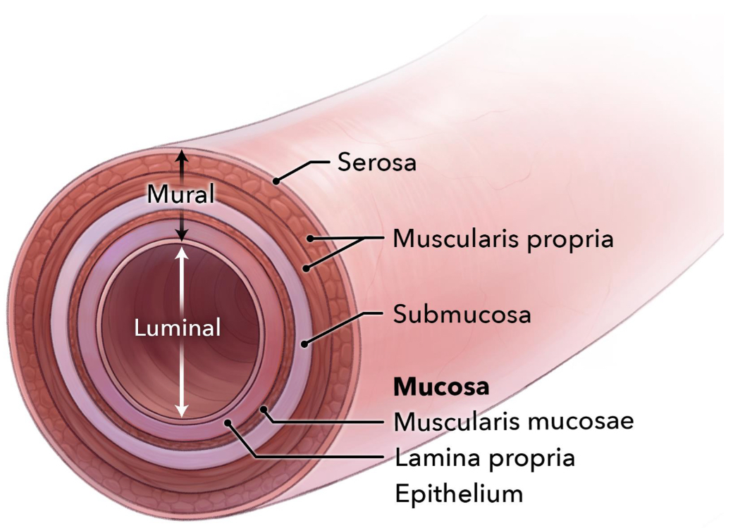
Muscularis
Smooth muscle that produces wave-like contractions (peristalsis)
Peristalsis- progression of coordinated contractions and relaxations of the smooth muscles of the GI tract to propel food

Serosa
Connective tissue; outermost covering (visceral peritoneum is on top of the serosa)
Where does the blood supply and innervation from the GI system come from?
It comes through the mesentery (the sheet of fat that organs hang by)

The GI tubular system is covered by two layers, what are they?
Serosa
Muscularis
Outer longitudinal
Inner circular
Enteric Nervous System
Independent and intrinsic to the GI tract
Controls nearly all aspects of GI function
Motility
Secretions
Blood flow
Second Brain
Can operate independently of CNS, but normally does receive input from autonomic nervous system
Complex- the number of neurons = number of neurons in spinal cord
The enteric nervous system two interconnected systems of ganglia
Myenteric (Auerbach)
Between the muscle layers (longitudinal and circular)
Regulates motility
Submucosal (Meissner)
Within the submucosa
Regulates secretions from mucosa
Sensory neurons too
Interneurons
Connect myenteric and submucosal ganglia
Parasympathetic input on ENS
Vagus nerve
Pelvic splanchnic nerve
Gives input for rest and digest
Promotes motility and secretions
ACH is the neurotransmitter
Sympathetic input on ENS
Thoracic/lumbar splanchnic nerves
Stops motility, decreases blood flow, stops secretions
Fight or Flight response
Postganglionic fibers
Norepinephrine is the neurotransmitter
Gut Brain Endocrine Axis
Stress can impact the GI system
The GI tract makes serotonin
Sphincters
A ring of muscle surrounding and serving to guard or close an opening or tube
The 6 sphincters of the GI tract
Upper esophageal sphincter
Lower esophageal sphincter
Pyloric sphincter
Ileocecal valve/sphincter (dogs and cats ileocolic junction)
Internal anal sphincter
External anal sphincter (voluntary control through the pudendal nerve)
Intestinal barrier importance
Poop is bad if it is in the blood
Luminal contents contains
Bacteria and bacterial products (LPS- massive inflammatory response)
Digestive enzymes capable of tissue injury (acids, biles and reactive metabolites)
Food antigens and toxins
The mucosal barrier prevents these from entering the patient’s system
Even small breaches (ulceration) → massive inflammation
Translocation of bacteria or endotoxin → systemic inflammatory response, sepsis, shock, death
Intestinal mucosa layers (from luminal to mural)
Epithelium
1 later of cells
Lamina propria
Loose collagen
Immune cell rich
Muscularis mucosa
Thin layer of smooth muscle
Intestinal epithelial cells
Simple columnar
Absorptive epithelial cells (absorb nutrients or water/electrolytes)
Enterocytes
Colonocytes
Other types of epithelial cells
Goblet cells- Mucin for protection and lubrication
Paneth cells- immune cell that secretes antimicrobial peptides and growth factors
Stem cells- progenitor cells
Enteroendocrine and Neuroendocrine- releases hormones regulating motility, secretion, and appetite
Many others
Crypts vs villi
Both small and large intestine have crypts
The bottom of crypts contain critical cells (stem cells)
Stem cells at bottom of the crypt migrate towards the lumen, differentiate into other cell types, migrate, die, and slough (3-5 days)
Only small intestine has villi
Villi provide an increased surface area
Mucosal blood flow
Tremendous metabolic demand
Tremendous blood flow
around 25% of cardiac output at rest to GI tract
75% of that to mucosa
Homeostasis
Mucosal surface is hypoxic (tip of villi have low oxygen to accommodate for anaerobic microbes)
Counter current blood flow
Needed for microbes in lumen
Anaerobic environment
Intestinal barrier, mucosal layer cell specifics
Epithelial cells
Tight junctions between cells
Barrier
Controlled transport for absorption and enzymes
Mucus layers
Produced by goblet cells (mucin)
Physical barrier
Food for beneficial bacteria
In the crypt
Antimicrobial peptides made by paneth cells
Lamina propria
Immune cell rich
Last line of defense
Layers of mucosal barrier from lumen to tissue
Microbiota
Mucus
Antimicrobial peptides and immunoglobulins
Epithelial cells
Lamina propria (immune cells)
Visceral pain
Abdominal organs
Pathway- visceral afferent neurons
Character- dull and vague (poorly localized/referred)
Stimuli/cause
Stretch of mesentery, distension of bowel
Mechanoreceptors
Inflammation
Chemoreceptors
Somatic pain
Parietal peritoneum/body wall
Pathway- somatic spinal nerves
Character- sharp, localized, intense
Causes/stimuli
Mechanical, thermo, stretch, chemical, etc.
GI organs are relatively insensitive to:
Cutting
Crushing
Burning
Touching the mucosa
GI organs are relatively sensitive to:
Distension and excessive stretch
Traction of the mesentery
inflammation/ischemia
Forceful smooth-muscle contraction
Chemical (bile peritonitis)
Infarction
Tissue death due to loss of blood supply
Peritoneal fluid
On ultrasound fluid is black
Normal amount is small (<3mm depth)
Functions for lubrication
Normal parameters
Clear (yellow/straw colored)
Can read through it
On cytology low cell and protein count
Gut microbiota
Collection of micro-organisms
bacteria, fungi, viruses, and archaea
Colon has the greatest number of bacteria
Ruminants have the greatest number of microorganisms
Horses have the greatest number of species
These organisms are anaerobic and have specific requirements so they are difficult to culture
20-60% not cultivable
pH order of organs from most acidic to most basic
Stomach
Large intestine
Small intestine
Gut microbiota, what do they do?
Part of the intestinal barrier
Provide competition with pathogens
GI motility
Serotonin and other neurotransmitters are made by some bacteria
Healthy vs. Unhealthy microbiome
Healthy- is diverse and prevents infection from enteric pathogens
Unhealthy- Has reduced diversity of microbiota
GALT
Responsible for differentiating between nutrients on the one hand and potential harmful substances
Largest immune organ in the body
Where are these cells:
Individual cells throughout
Lymphoid follicle throughout (cluster of immune cells usually B cells in mucosa of intestine
Peyer’s patches (ileum) (large aggregation of lymphoid tissue in the ileum and sometimes jejunum)
Lymphatics to mesenteric lymph nodes (lymph fluid then goes to thoracic duct)
Immune cells get out of GALT and into circulation through the thoracic duct
Specialized cell types
Paneth cells - AMPs
M- cells- Antigen sampling
The importance of GALT
Mucosa constantly exposed to antigens
Food allergies
IBD
Enteric pathogens
Regulated response (Regulatory T cells)