Pathologial conditions of the gallbladder
 Call Kai
 Learn
Learn Practice Test
 Spaced Repetition
 Match
Match Flashcards
 Knowt Play
1/122

There's no tags or description
Looks like no tags are added yet.
Name | Mastery | Learn | Test | Matching | Spaced | Call with Kai | Chat |
|---|
No analytics yet
Send a link to your students to track their progress
123 Terms
what are some indications for a GB exam
Rt shoulder pain
positive murphy sign
nausea
vomiting
jaundice
loss of appetitie
abnormal LFTs
with GB imaging you want the ____ frequency when possible
highest
what are some exam limitations for the GB
bowel gas
artifact (reverberation)
pt cant hold breath
pt ate
obesity
what is the normal GB length measurement
8-12 cm
what is the normal GB wall thickness
3 mm
what is the normal CBD AP measurement
0.1-0.7 cm
the GB neck is ____ and the fundus is _____
superior, inferior
what are 3 common normal variants of the GB
phrygian cap, junctional fold, and hartmanns pouch
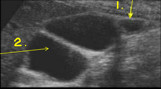
phrygian cap
junctional fold
phrygian cap occurs at what part of the GB
fundus
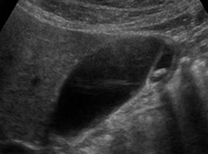
what GB variant is this
phrygian cap
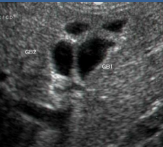
what GB variant is this
double gallbladder
GB lumen should have what echogenicity
anechoic
the GB wall should be measured on the ____ border when the wall is _____ to the transducer
anterior, perpendicular
what are rokitansky-aschoff sinuses
rugae or pocket like invaginations of the epithelium into the muscularis
what should you associate rokitansky-aschoff sinuses with
adenomyomatosis
Rugae or pocketlike invaginations of the epithelium into the muscularis
rokitansky-aschoff sinuses
what is viewed bewteen the RPV and the GB
main lobar fissure
rupture is most common at what part of the GB
fundus (it has the least amount of blood supply)
what is the most common dz of the GB
cholelithiasis (GB stones)
_____ can be single large stones or multiple tiny stones
cholelithiasis
are large singular stones or tiny multiple stones more dangerous in the GB
tiny stones (can enter bile ducts and obstruct bile flow)
_____ can lead to stone formation
bile stasis
what are the five F’s associated with cholelithiasis risk factors
fat, female, forty, fertile, and fair
what are 4 clinical symptoms for cholelithiasis
RUQ pain
Epigastric pain
Nausea and vomiting
elevated serum bilirubin
does gallstones cause shadowing
yes (gallstones attenuate and reflect causing shadowing)
when you identify gallstones on an exam and then decube the pt the gallstones are expected to _____
move (they are mobile and gravity dependent)
what positions can you move the patient in to evaluate the mobility of gallstones
erect, decubed
gallstones may not shadow if they are ____
small (<3mm)
when using color, gallstones will ____
have no blood flow to them

cholelithiasis
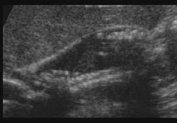
cholelithiasis

what sonographic sign is this
WES sign (wall echo shadowing)
what does the WES sign indicate
GB packed with stones
the WES sign is associated with what kind of cholecystitis
chronic cholecystitis
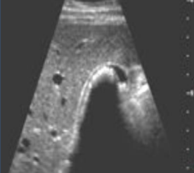
what sonographic sign is this
WES sign
What is sludge
thick viscous bile
what are the clinical symptoms associated of sludge
asymptomatic (could have classic biliary symptoms)
what causes sludge
bile stasis, obstruction of flow, cholecystitis, sickle cell anemia
sludge is a normal finding in patients who are ______
fasting for a prolonged period of time
does sludge cause shadowing
no
when you identify sludge on an exam and then decube the pt the sludge is expected to _____
move
if the GB is completely filled with sludge the GB can be isoechoic to liver called ______
hepatization of GB
sludge can form solid masses called _____
sludge balls

sludge

sludge

sludge (hepatization of gb)
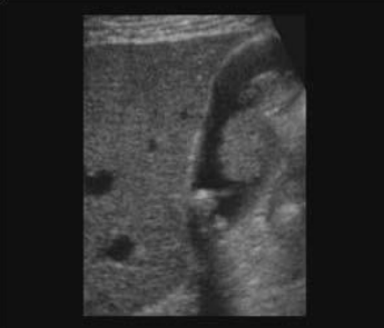
sludge (sludge balls)
diffuse GB wall thickening is usually not caused by an issue with the GB itself except for _______
acute cholecystitis
what are 5 causes of diffuse GB wall thickening
renal failure
pancreatitis
acute cholecystitis
hepatitis
CHF
what are 5 causes of focal GB wall thickening
adenomyomatosis
carcinoma
adenoma
gangrenous cholecystitis
mets

GB wall thickening
rare condition where GB wall becomes calcified
porcelain gb
porcelain gallbladder is associated with ___ in the majority of patients
gallstones
porcelain GB is common in older ____
women
there is a ___% association between porcelain GB and GB carcinoma
25%
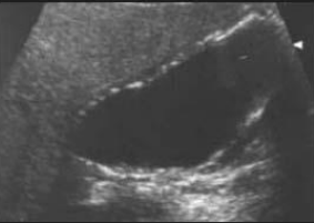
porcelain GB

is this a porcelain GB or a WES sign
WES sign
____ are masses projecting into the GB lumen
polyps
what is the most common thing polyps are made of
cholesterol polyps
what are the clinical symptoms for polyps
asymptomatic
do polyps cause shadowing
no
when you identify polyps on an exam and then decube the pt the polyps are expected to _____
not move
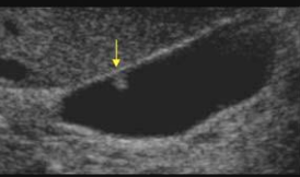
polyp
over growth of the GB wall is called ____
hyperplastic GB wall
Hyperconcentration, Hyperexcitability, Hyperexcretion are all dengenerative and proliferative changes of the GB related to ______
hyperplastic GB wall
what are 2 hyperplastic conditions of the gallbladder
cholesterolosis and adenomyomatosis
a non-inflammatory hyperplastic condition of in GB wall causing thickening
adenomyomatosis
what is the clinical symptoms of adenomyomatosis
biliary colic
Hyperechoic, non movable hyperplastic diverticuli within the GB wall cause elevation appearance in wall
adenomyomatosis
adenomyomatosis can appear as crystal depostis which result in comet tail artifiact but it could also appear as _____
multiple cystic spaces (usually near the fundus)

adenomyomatosis
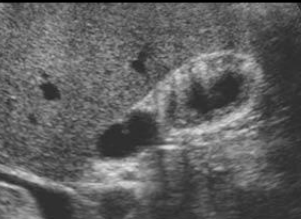
adenomyomatosis

adenomyomatosis (comet tail artifact)
abnormal build up and deposition of lipids within the lamina propria of the GB which form tiny nodules on the surface of the mucosal lining
strawberry GB
GB stones present ____% of the time with strawberry GB
50-70%
strawberry GB is common in ____
males (over 40)
what are strawberry GB clinical symptoms
asymptomatic (or biliary symptoms)

strawberry GB

strawberry GB
when you identify cholesterolosis/strawberry GB on an exam and then decube the pt the echogenic masses are expected to _____
not move
hydropic GB (mucocele) is associated with obstruction of the _____
cystic duct or GB neck
globular appearance of GB with abnormal distention
hydropic GB (mucocele)
what causes hydropic GB (mucocele)
prolonged total obstruction of cystic duct (bile gets reabsorbed)
hydropic GB is obstruction of the _____ while courvoisier’s GB is obstruction of the ______
neck or bile duct, distal CBD
An enlarged, usually painless GB, indicates an extrahepatic mass compressing the CBD
courvoisier’s sign
inflammation of the GB
cholecystitis
what are the 5 forms that cholecystitis can take
acute
chronic
acalculous
emphysematous
gangrenous
what is acalculus cholecystitis
inflammation of the GB without presence of stones

cholecystitis

cholecystitis

describe this sonographic image
edematous GB in pt with acute cholecystitis, thickened edematous walls

acute cholecystitis (acalculus cholecystitis)

what is the arrow pointing to
pericholecystic fluid

inflamed fat (secondary to inflammation)

inflamed fat (secondary to inflammation)
what are 2 complications of acute cholecystitis
gangrenous cholecystitis and emphysematous cholecystitis
serious, painful necrosis develop in GB wall, sloughed mucosa of wall. Wall may bleed causing an abscess. Pericholecystic fluid and air. May rupture causing pericholecystic collection and peritonitis. caused by infection or absence of blood supply
gangrenous cholecystitis
Gb becomes infected with GAS-FORMING organisms. Gas has ring down or comet tail artifact or dirty shadowing. Highly reflective air may also be seen
emphysematous cholecystitis

gangrenous cholecystitis