5011- Cardiac Theory 1 (overall)
 Call Kai
 Learn
 Practice Test
 Spaced Repetition
 Match
 Flashcards
 Knowt Play
1/117

There's no tags or description
Looks like no tags are added yet.
118 Terms
Describe long axis imaging planes
POM @ 11 o’clock
Divides: left and right

Describe short axis imaging planes
POM @ 1 o’clock
Divides: superior and inferior

Describe 4CH imaging planes
POM @ 3 o’clock
Divides: anterior and posterior

Describe 2CH and 3CH imaging planes
2CH: POM @ 1 o’clock
divides left and right
3CH: POM @ 12 o’clock
divides anterolateral and posteriomedial

Describe subcostal imaging planes
POM @ 3 o’clock

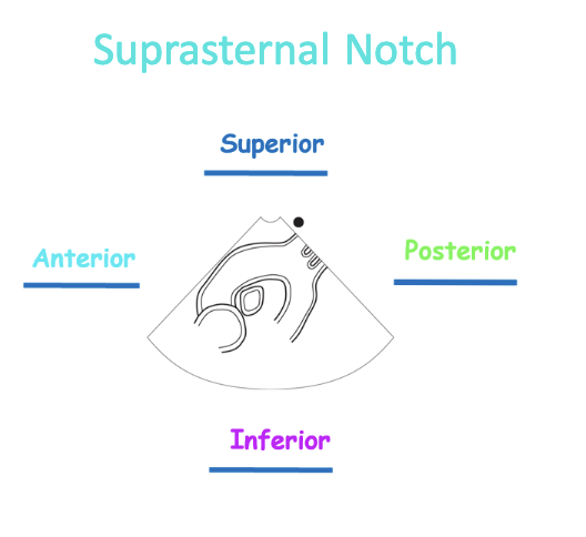
Describe suprasternal imaging planes
POM @ 1 o’clock
Difference between arteries and veins?
veins: blood returning to the heart
arteries: blood moving away from the heart
Which 3 structures drain into the RA?
SVC, IVC, and CS
Describe characteristics of RA (2)
smooth walled except anterior surface
anterior surface contains pectinate muscles, prominent ridges of muscle
right atrial appendage (RAA) on anterior surface
contains pectinate muscle and increases RA volume
Describe characteristics of RV (5)
walls lined with trabeculae carneae
contains moderator band
thick walls, but not as much as LV
chordae tendineae that connects TV leaflets to pap muscles
3 pap muscles: anterior, posterior, and septal
What are trabeculae carneaes?
ridges of muscle lined with endocardium
help push and rings out the blood
What is moderator band?
thick band of the cardiac muscle that extends from the IVS to anterior papillary muscle
helps prevent over-distention of the RV, similarly to rubberband
Describe characteristics of LA (3)
receive oxygenated blood from 4 pulmonary veins
smooth walled
left atrial appendage (LAA) contains pectinate muscles
Describe characteristics of LV (4)
thick walls
contain trabeculae carneae
chordae tendineae connect MV to pap muscles
2 pap muscles: anterolateral and posteromedial (Prime Minister At Lunch)
What are the main roles of chordae tendineae and pap muscles?
prevent eversion of MV into LA and TV into RA
What are the main roles of semilunar valves
the crescent shapes valves help prevent backflow
Rank the valves from most superior to inferior
Pulmonary
Aortic
Mitral
Tricuspid
What are the valves relation to the aortic valve?
Most posterior: MV
LIMP
Most anterior: PV
PALS
Most inferior: TV
TRIP
Furtherest to the right: TV
Difference between pericardium and heart wall
pericardium: sac/membrane that surrounds the heart
heart wall: layers of the heart
What is the role of the pericardium? (4)
protects the heart from infection
protects the heart from over-distention
acts as lubricant to reduce friction between the heart and surrounding structures
made up of two layers: serious pericardium (inner) and fibrous pericardium (outer)
Describe fibrous pericardium
outer most layer heart sac
tough, dense, connective tissue
What are the main roles of fibrous pericardium (3)
protects the heart from infection
anchors the heart in place during beating and bouncing
prevents overfilling of the heart
What are the 2 parts of the serous pericardium
Visceral or epicardium layer - inner most layer, part of the heart wall
Parietal layer - fused to fibrous pericardium
Note: pericardial fluid found between the two serous layers
What are the 2 main roles of serous pericardium
cushions the heart
lubricates the heart
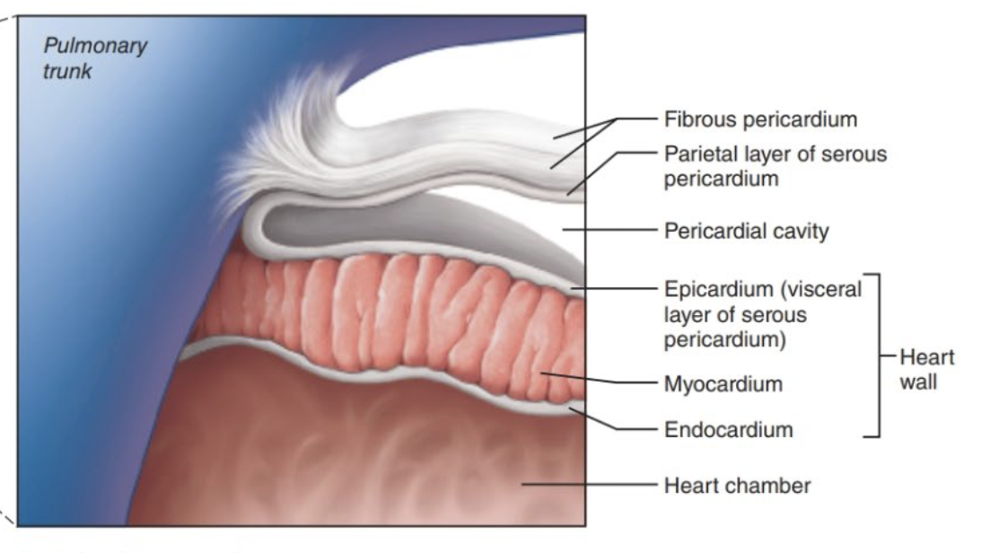
What makes up the heart wall (3)
epicardium or visceral serous pericardium: outer most layer, but inner most layer of serous pericardium
myocardium: middle muscular layer
endocardium: inner most layer of the heart lining the chambers and valves
What are the main roles of epicardium, myocardium, and endocardium?
Epicardium: protects heart layers, produces pericardial fluid, houses coronary blood vessels
myocardium: pumps blood through the heart
endocardium: protects the myocardium and the heart valves
Why are the LV walls thicker than RV ?
pumps same amount of blood but LV needs to generate greater forces to overcome high resistance of the systemic circuit
RV needs only small amount of force to overcome the low resistance of pulmonary circuit

Label the correct structures of the wall
Difference between coronary arteries and veins
coronary arteries deliver oxygen and nutrients to myocardium
coronary veins remove waste material
What are the 3 types of sulci/grooves, and the artery and vein pairing to each?
anterior interventricular sulcus
artery: LAD
vein: GCV
posterior interventricular sulcus
artery: PDA
vein: MCV
coronary sulcus (left)
artery: CS
vein: GCV
coronary sulcus (right)
artery: RCA and marginal artery
vein: SCV
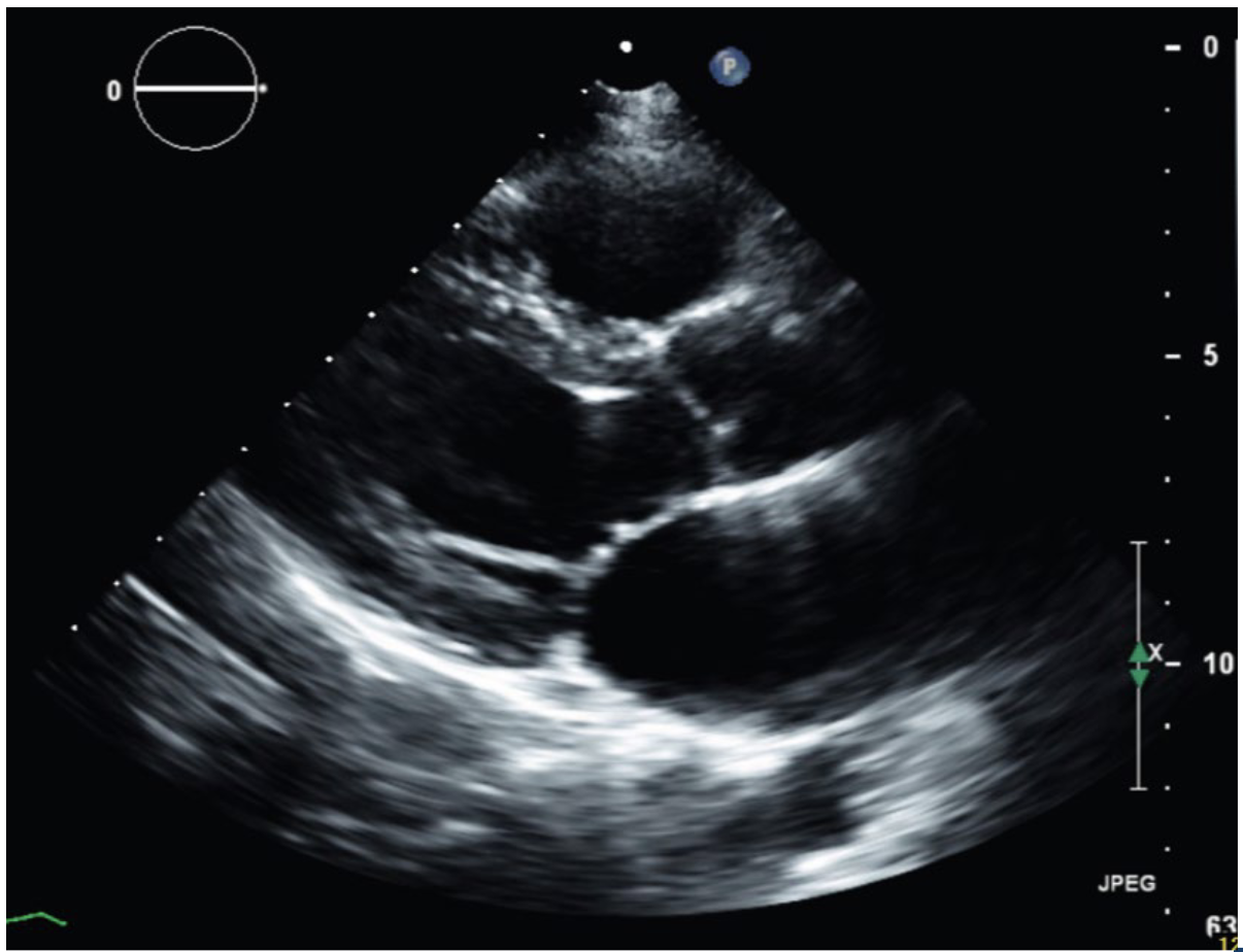
Label PLAX structures


Label RVIT structures

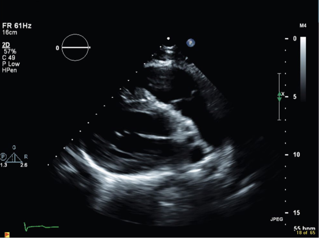
Label RVOT structures


Label PSAX overview structures


Label PSAX pulmonary level


Label PSAX tricuspid level
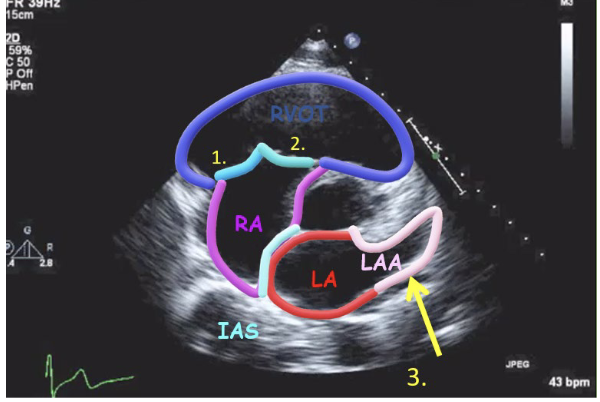
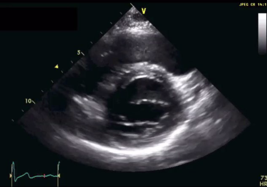
Label PSAX mitral level

How are the scallops of the mitral valves labeled

What are the two types of cardiomyocytes
ventricular and atrial muscle cells
pacemaker cells (SA & AV node)
Describe ventricular and atrial muscle cells
cells shorten and lengthen to contract and relax
contract when they receive an electrical signal (action potential) from a neighboring cell
Describe pacemaker cells (SA & AV node)
modified myocytes, found in specialized cells
autorhythmic: spontaneously generate and send out electrical signals without external stimulus
respond to stimulation from the brain, and continue to contract even when pulled out
Describe a cardiomyocyte, what are the important structures?
muscle cell of the heart
myofibril and sarcomere structures
Describe myofibril
contractile portion of the cell, many per cardiomyocyte
made of repeating contractile units called sarcomere
Describe sacromere
contain actin/myosin myofilaments
thick filaments: myosin
thin filaments: actin
How does the contraction and relaxation work in regards to cardiomyocytes
binding and unbinding of actin and myosin results in contraction and relation of muscle fibre
What are intercalated discs? what are they made up of (3)?
found in cardiac tissues ONLY
communication network between cells that allows heart to work as single functioning unit
gap junctions
desmosomes
fascia adherens
Describe gap junctions
protein lined tunnels that directly connect the cytoplasm of the two adjoining cells
allow action potential to spread quickly from one cell to the next
lots gap junctions = faster impulse transmissions = contractions occur in unison
Describe desmosomes
provide cell to cell adhesion
maintain mechanical integrity and resist shearing forces
bridge the space between cells
Describe fascia adherens
anchors the actin filaments to the cell membrane
Describe the conduction system in the heart
SA Node (chief pacemaker, where specialized cells are located at RA)
Bachmann’s Bundle
AV node
Bundle of His (Atrioventricular bundle)
Right/Left bundle branches
Purkinje Fibers
Note: higher up = faster to generate a pulse, at a higher rate and it is lower as it goes down
Describe SA node, and main purpose
located in RA wall
automatically produces a consistent and constant electrical signal, 60-100 bpm
sends the signals to the atria, causing the atria to contract
natural pacemaker of the heart, it determines heart rate because reaches threshold for depolarization the fastest
Describe Bachmann’s Bundle, and main purpose
travels from RA to LA
initiates contraction of the LA
Describe AV node, and main purpose
located at the crux
delays electrical impulses slightly to allow atria to finish emptying before the ventricles can contract
sends signal down to Bundle of His
Describe Bundles/Purkinje signals
Bundle of His: sends signal from AV to bundle branches
Bundle branches: signal travels down the IVS down the left and right bundle branches
Purkinje fibres: travel deep into the heart to supply the muscle of ventricles
start at apex and work their way up
Briefly describe the conduction of action potential (AP) in the heart, starting with SA node
SA node is primary pacemaker of the heart, fires spontaneously generating an AP that spreads thru the atria
myocytes are connected via gap junctions, ions flow from one cell to another, trigger AP in neighboring cells and propagate rapidly
impulse gets to AV node and slows down to allow atria to contract then goes down the conduction pathway to ventricular myocytes
How do the action potential differ from pacemaker cells and cardiac myocytes?
pacemaker cells: creates own signal, unstable resting potential (slope on graph), autorhythmic
atrial and ventricular cells: needs to be stimulated to contract, stable resting potential (flat on graph), non-autorhythmic
What is the unique plateau seen in atrial and ventricular cells?
phase that allows cells to contract and relax fully before release
cannot be restimulated until the end
Describe action potential
cardiac cells are filled and surrounded by solutions that contain positive and negative ions
More Na+, Ca2+ outside of the cell, and more K+ inside the cell
electrical stimulation is created by sequence of ion fluxes through specialized channels in the membrane of cardiomyocytes that lead to contractions
AP: reversal of cell membrane electrical polarity due to voltage gated ion channels
Describe depolarization and repolarization in relation to cell action potential
Depolarization: negative to positive (up the slope), cell is contracting
Repolarization: positive to negative (down the slope), cell is relaxing
Briefly explain all 3 phases of the action potential contractile cell
Phase 0: AP triggered in neighboring cardiomyocyte or pacemaker cells reaches next cell via gap junction, ions leaking in raising RMP to threshold → rapid depolarization: Na and Ca in, K out
Phase 1: brief, early repolarization phase with some K leaving and lower transmembrane potential
Phase 2: plateau phase where Ca coming into cell triggers more Ca from storage → sets off muscle contractions
plateau caused by balance between inward movement of Ca and the outward movement of K via leaking down [gradient] and VGC
allows cardiac muscles cells to pump blood effectively before firing a second time
Phase 3: rapid repolarization phase, K leaking out and VGC slowly close, while Ca is actively leaving cell and pumps are restoring ion balance
Phase 4: cell goes back to rest, K leaks out following chemical gradient, Na and Ca pumps closed
What is calcium doing at the muscle fiber level?
Calcium influx
calcium binds to troponin → causes troponin to change shape, which exposes the actin-myosin binding sites in the myofibril
actin and myosin bind and shortens the muscle fiber → contraction!
Calcium effux
calcium levels drop and remainder gets used up → calcium unbinds from troponin complex → shape changes
shape no longer allows actin and myosin to bind together because the site is no longer exposed → cell at resting length/ relaxation
What leads do sonographers use?
Lead II, strongest deflection bc it is parallel to wave of depolarization: negative to positive
What does the different segments in the ECG represent?
P wave: atrial depolarization
PR segment: delay at the AV node → no electrical activity = flat line → atria contraction while ventricles filling
QRS complex: ventricular depolarization and atrial repolarization
Q: septal depolarization from left to right
R: ventricular depolarization (not contracting)
S: Purkinje fiber depolarization
ST segment: isoelectric period, refractory period of ventricles → contracting
T wave: ventricular repolarization
PR interval: onset of atrial depolarization to ventricular depolarization
QT interval: ventricular systole
Based on a box grid for ECG, how to measure HR from R to R?
thick solid line to next until next QRS
300 → 150 → 100 → 75 → 60 → 50
Compare and contrast of sinus bradycardia, tachycardia and arrhythmia
brady: <60 bpm
tachy: >100 bpm
arrhythmia: rhythm varies with respiration
What are premature atrial contractions? (PAC)
premature beat originates somewhere in the atria and interrupts the underlying rhythm
p wave may look different since its not from the SA node, but are still followed by QRS
What are atrial fibrillations?
most common cardiac arrhythmia
atria receive multiple pacemaker signals, thus “quivers” instead of contracting
no P waves, and ventricles depolarized at irregular intervals

What are atrial flutters?
abnormal rhythm either in RA or LA, the atria are depolarizing at an extremely rapid rate
“saw-like”, several P waves for each QRS, varying R-R intervals because not all electrical signals make thru the AV

What are premature ventricular contractions (PVCs)
initiated by Purkinje Fibers in the ventricle instead of the SA node, thus ventricles contract first before the atria can fill with blood
“skipped beat”, usually random
every second beat: bigeminy
every third beat: trigeminy

What are ventricular tachycardia (VT)
extremely fast heart rhythm that originates in the ventricles, either LV or RV
more than 3 PVCs in a row
since ventricles do not have enough time to fill, the blood isnt pumped to the body → can lead to emergency
non-sustained VT: < 30s
sustained VT: > 30s
What are ventricular fibrillations?
uncoordinated contraction of the cardiac muscle → ventricles quiver
can also quickly turn into asystole within mins
do not want to >5min or else brain damage

What are the 3 types of AV heart blocks?
First degree heart blocks: AV conduction slowed
Second degree heart blocks: AV conduction incompletely blocked
Third degree heart blocks: AV conduction completely blocked
Characteristic of first degree AV block
conduction at AV delayed beyond normal amount, where PR interval is longer than 0.2s followed by QRS
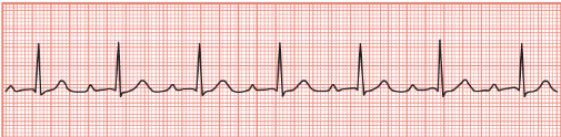
Characteristic of second degree blocks, and what are the two types?
P wave is blocked from initiating a QRS complex → more p waves than QRS
Mobitz I/ Wenckebach and Mobitz II
Explain the 2 types of second degree AV block
Mobitz I/ Wenckebach: PR interval gets longer with each beat until a P wave occurs, but not followed by QRS (QRS eventually dropped)
Mobitz II: No PR prolongation and QRS complex just fails to occur after a P wave (QRS drops occasionally)

Characteristic of third degree AV block
complete heart blocks → atria and ventricles do not communicate
atria controlled by one pacemaker, the ventricles by another
p waves occur at regular intervals with QRS, but no apparent patterns, so p waves interrupt
What are bundle branch blocks
loss of ventricular synchrony
ventricular depolarization is prolonged
impulse has to move through muscle fibers instead of bundle
QRS wider than 3 squares
Differentiate right vs left bundle branch blocks
RBBB: damage to the right:
RV after the LV
LBBB: damage to the left
LV contracts after the RV
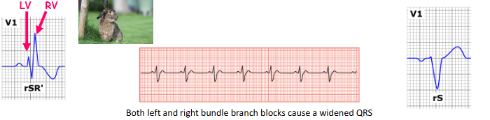
What is asystole
cardiac arrest rhythm → no electrical signal on the ECG monitor
flat line → code blue!
What is pulseless electrical activity (PEA)
cardiac arrest
dying heart → weak electrical activity, but too dead to respond mechanically
no ventricular activity → no pulse
Characteristic of pacemaker on ECG
release electrical impulses to regulate beating of the heart
spike on trace every time the electrode fires

Compare STEMI vs NSTEMI
STEMI: ST segment elevation
coronary artery completely blocked off by clot
NSTEMI: depression ST segment or T wave inversion
partial occlusion to the coronary artery
What are holter monitors
used to monitor ECG rhythms over 24 hrs
patient wear ECG leads and record activities in logbook
r/o structural reasons in heart causing arrhythmias and/or assess hemodynamic impacts
What are stress ECH or cardiac stress tests
12 lead stress test evaluates coronary circulation at rest vs maximum physical exertion
stress echo looks at decreased heart function during exercise
What is pulse oximetry
measured oxygen saturation of hemoglobin relative to total hemoglobin
hypoxemia = insufficient oxygen in blood
What are the 4 (7) phases of a cardiac cycle
isovolumetric relaxation phase (part of diastole)
ventricular filling:
rapid filling
diastasis
atrial systole
isovolumetric contraction phase (part of systole)
ventricular ejection
Elaborate the 4 stages of diastole briefly
IVRT: all valves closed as ventricles just finished contracting, dicrotic notch
Rapid filling stage: MV opens, blood leaves LA and pressure drops rapidly
diastasis: mid diastole, pressure in LA and LV almost equal, MV leaflets start to close
atrial kick: end of diastole, second push of blood, a wave= atrial contraction
What are the 2 phases in systole?
IVCT: all valves closed, ventricles preparing to contract, c wave is the MV buldging back
Ventricular ejection: blood is being ejected, eventually v wave to signify LA pressure slowly rising as blood enters LA
Whats the lub and dub sounds
Lub: 1st heart sound for closure of AV valves
Dub: 2nd heart sound for closure of semilunar valves

What does the DEFAC signify in m-mode of mitral leaflets?
D:valve opens, onset of diastole
E: rapid filling, maximal opening
F: end rapid filling, diastasis
A: atrial kick/atrial systole, MV reopens after atrial contraction to eject more blood into LV
B: seldom seen, elevated LV end diastole pressure shown as 3rd bump, usually absent
C: valve closes, final leaflet position before LV systole
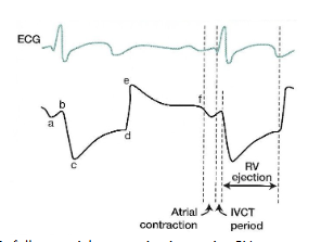
What does the ABCDEF signify in m-mode of pulmonary level?
a: follows atrial contraction, increasing RV pressure with extra blood
b: denotes small anterior deflection occurring at onset of ventricular systole
c: large posterior deflection immediately following ventricular ejection, abrupt opening
d: gradual anterior motion of leaflets during ejection period as the RV pressure decreases, valve moves to center of the root
e: denotes closed position of leaflet upon completion of ventricular ejection
f: slight posterior movement of leaflet during diastole immediately prior to atrial contraction
Why do we wanna assess pulmonary valve with m-mode?
pulmonary hypertension: early systolic closure and absent A wave
severe PHT, PA diastolic pressure is higher than RV, so RA contraction will have no effect on PV → no A
Flying “W” = mid systolic closure and late systolic opening
can help confirm PHT in absence of TR
pulmonary stenosis:
increased A wave
look for valve thickening, doming, veg


Label Apical 4CH structures


Label Apical 2CH

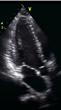
Label Apical 3CH


Label Apical 5Ch


Label subcostal 4CH


Label suprasternal notch

What are the 3 types of effusions
pericardial effusion: fluid in the pericardial sac above normal levels
pleural effusion: fluid around lungs
ascites: fluid in abdominal cavity

Name all 17 wall segments
