Anatomy TBL 3
 Call Kai
 Learn
 Practice Test
 Spaced Repetition
 Match
 Flashcards
 Knowt Play
1/68

There's no tags or description
Looks like no tags are added yet.
69 Terms
3 types of muscle tissue
skeletal
cardiac
smooth
what is the germ layer origin of skeletal muscle?
paraxial mesoderm (myotomes)
what is the germ layer origin of cardiac muscle?
viceral/splanchnic lateral plate mesoderm
what is the germ layer origin of smooth muscle?
multiple sources
visceral/splanchnic lateral plate mesoderm
NCC: large arteries and coronary arteries of heart
what is the exception of the germ layer origins of smooth muscle?
pupillary, mammary gland and sweat glands come from ECTODERM
do not come from mesoderm like most other smooth muscle
glands come from invagination in surface ectoderm
pupillary muscles come from neural ectoderm of the optic cup
skeletal muscle formation in week 5
myoblast cells fuse to form myotubes
skeletal muscle formation by end of 3rd month
myofibrils and striations form
tendon formation
tendons = NOT muscle
from sclerotome
¾ of tissue types are formed from what embryologically?
MESODERM
all except for nervous
mesoderm divides into separate areas on each side of the embryo
axial
paraxial
intermediate
lateral plate
axial mesoderm
not bilateral- at midline
becomes notochord

paraxial mesoderm
paired
form somitomeres first → commit to cellular pathway
compacted somitomeres → somites

intermediate mesoderm

lateral plate mesoderm
somatic (parietal)
visceral (splanchnic)
somatic (parietal) lateral plate mesoderm

visceral (splanchnic) lateral plate mesoderm

embryonic folding will do what?
bring ectoderm and amniotic cavity around entire embryo
endoderm folds into tube
humans are coelomates
have body cavities
somitomeres
formed from paraxial mesoderm
commit to cellular pathway

somites
week 3-4
42-44 pairs
used to stage development
7 cranial unnumbered and do not become somites, remain somitomeres

how does a somite form
paraxial mesoderm → somitomere → somite
3 regions of somite
week 4-5
sclerotome
myotome
dermatome
sclerotome

myotome

dermatome

eye muscle somitomere source

where is the somitomere source of pharyngeal arch muscles?
CN I-IV
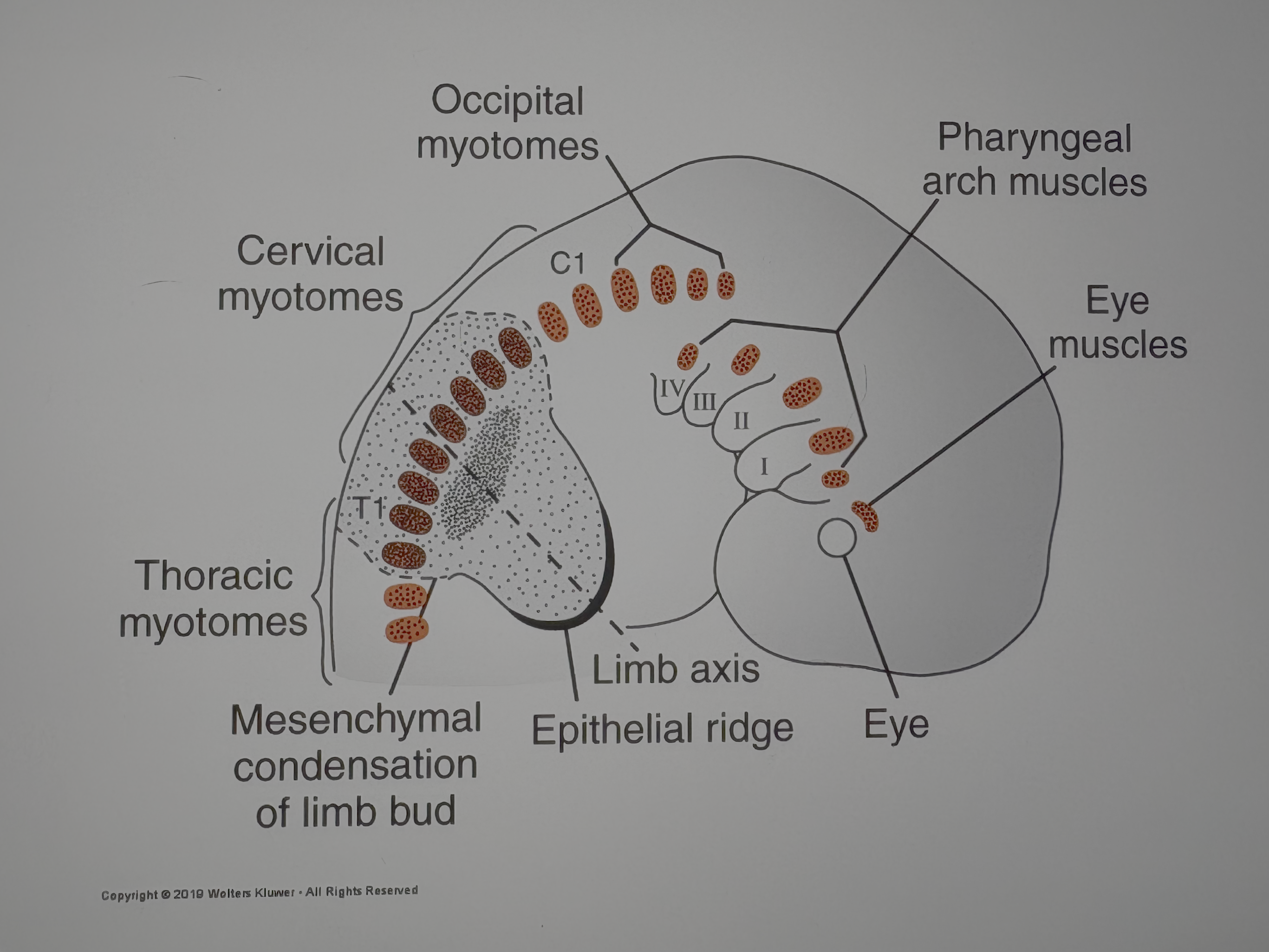
what is the somitomere source of occipital myotomes

where is the somitomere source for cervical myotomes?
C1-T1
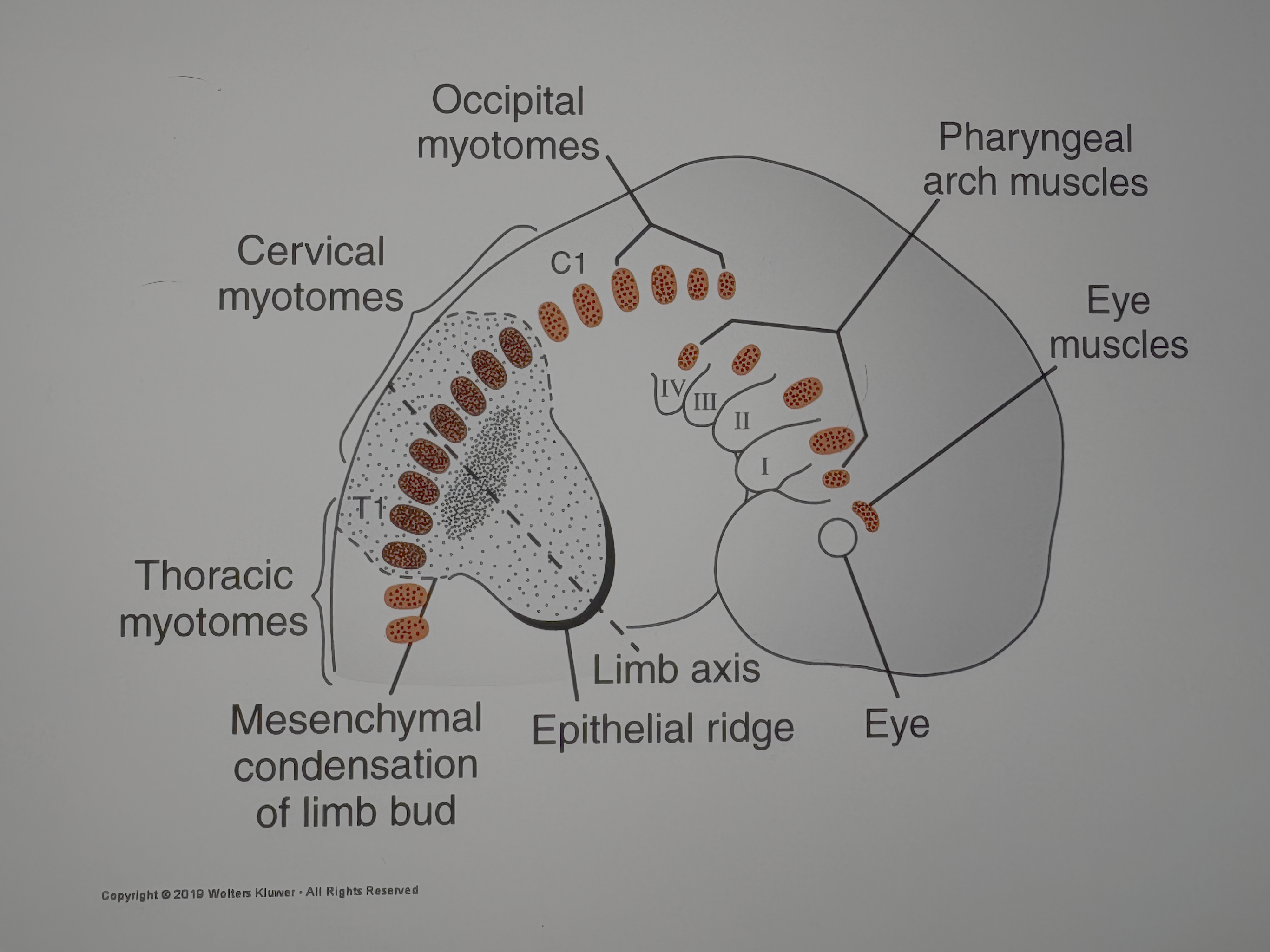
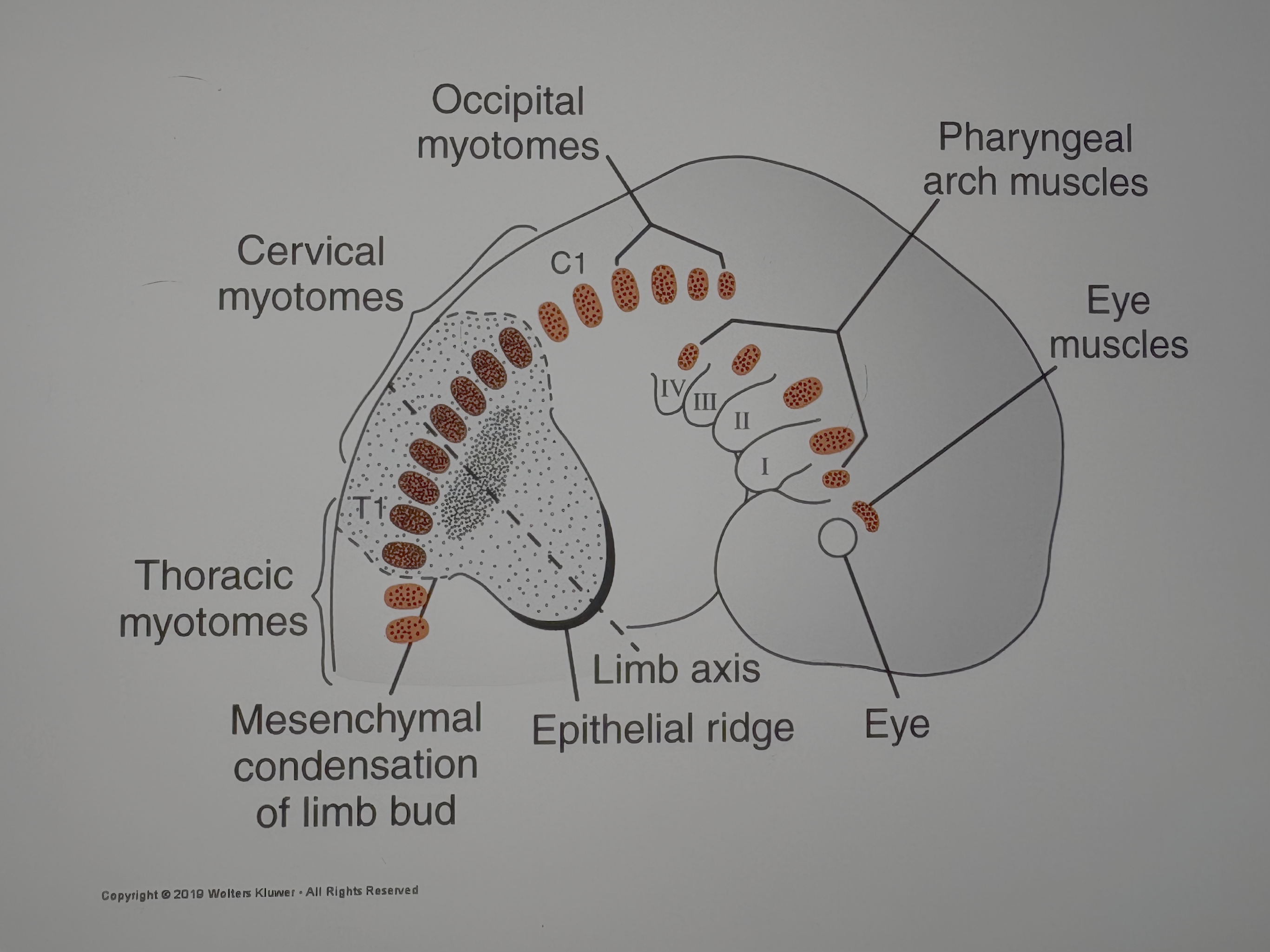
where is the somitomere source of thoracic myotomes?
T1 and below

mesenchymal condensation of limb bud

limb axis

epithelial ridge
role of somites
formation of a myotome and the somitic frontier
which cells migrate ventrally?
ventrolateral and dorsomedial cells of the somite migrate ventrally
which cells migrate to the lateral somitic frontier
some cells of the ventrolateral group migrate to the lateral somitic frontier into the parietal lateral plate mesoderm
lateral somitic frontier
separates the somite from the parietal lateral plate mesoderm
primaxial domain [separated by lateral somatic fronteir] abaxial domain

primaxial domain
myotome cells from somite
around the neural tube
neural tube and notochord give developmental signals for muscle formation
primaxial domain: dermis from where?
dermatome
primaxial domain produces what?
scalenes
intercostals
rhomboids
levator scapulae
latissimus dorsi
abaxial domain
migrated somitic ventrolateral cells
lateral plate mesoderm gives developmental signals for muscle formation
abaxial domain: dermis from where?
lateral plate mesoderm
abaxial domain produces what?
pectoralis major and minor
abdominal muscles (all 4)
distal upper limb muscles
all lower limb muscles
epaxial
above the axis
hypaxial
below the axis
epaxial muscles
innervated by dorsal rami of spinal nerves
intrinsic back muscles

hypaxial muscles
innervated by ventral rami of spinal nerves
body wall and limb muscles

myotomes receive innervation from where?
spinal nerves of the same segment of their somitic origin
upper limb is associated with which myotomes?
C5-T1 myotomes
lower limb is associated with which myotomes?
L4-S3 myotomes
dorsal and ventral muscle masses form in each limb
dorsal = extensors
ventral = flexors
for limb and girdle muscles, what happens when original location changes?
limb rotation
myogenic cells
myogenic cells that form muscles migrate to pharyngeal arches first
from cranial somites and somitomeres
use neural crest cells to guide cells to arches
head and neck muscle innervation
cranial nerves form connection with muscle in arches, prior to migration out of arches
arch 1 is innervated by:
CN V
arch 2 is innervated by:
CN VII
arch 3 is innervated by:
CN IX
arch 4/6 is innervated by:
CN X
pharyngeal arch muscles
1-2: muscles to open jaw and middle ear muscles
1-4: tongue
arch 1
muscle of mastication
arch 2
muscle of facial expression
arch 3
stylopharyngeus muscle
arch 4/6
muscle of soft palate, pharynx, larynx
germ layer origin of cardiac muscle
splanchnic lateral plate mesoderm
myoblasts do not fuse…
instead form special junctions called intercalated discs
what is the germ layer origin of smooth muscle in large arteries
lateral plate mesoderm and NCC
what is the germ layer origin of smooth muscle in coronary arteries
proepicardial cells and NCC
what is the germ layer origin of smooth muscle in gut wall and derivatives
splanchnic lateral plate mesoderm
what is the germ layer origin of smooth muscle in pupil/mammary gland/sweat glands
ectoderm