Foundations 2 Exam 1 Histology
 Call Kai
 Learn
 Practice Test
 Spaced Repetition
 Match
 Flashcards
 Knowt Play
1/71

There's no tags or description
Looks like no tags are added yet.
72 Terms
Lips
Vermillion Zone: transition from keratinized stratified squamous of skin on outer lip to wet keratinized stratified squamous of the mucosa of the inner lip

Oral Cavity
keratinized/wet keratinized stratified squamous(depends on diet)

GALT
found in lamina propria. Tonsils are an example of nodules of this

Tounge
Keratinized/WK stratified squamous. Lingual papillae
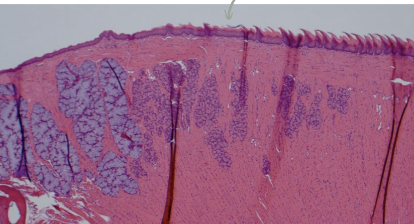
Filiform Papillae
Most numerous in most species. No taste buds


Fungiform papillae
fewer in number. Thin, Keratinized/WK stratified squamous. Have taste buds

Foliate Papillae
Large animals, pigs, several taste buds

Vallate Papillae
Few in number, largest, have a crypt, taste buds on sides, salivary glands ope in crypts

Taste Buds
Made up of: taste receptor cells, supporting cells, and basal cells

Outer Enamel Epithelium

Dental Papilla

Predentin
Pink


Ameleoblasts
Pink


Odontoblasts
Orange-Produces predentin which mineralizes into dentin


Developing Brachydont tooth

Dentin
Light Blue-70% mineral

Dential Tubules
Turquoise-odontoblastic processes travel through dentin via this

Enamel
Orange-96% mineral

Alveolar Bone
Pink

Periodontal Ligament
Turquoise

Cementum
Orange

Gingiva
Light Blue- Keratinized/WK stratified squamous

Pulp Cavity
Houses blood vessels, lymphatic vessels, nerves

Mucosa
-epithelium, lamina propria, and muscularis mucosa

submucosa
fibrous CT, submucosal glands, larger blood vessels, nerves, lymphatics, submycosal ganglia

muscularis externa
smooth muscle, inner circular layer, outer longitudinal, myenteric ganglia between inner and outer layer

Serosa/Adventitia
Adventitia: CT of variable thickness. Serosa: simple squamous mesothelium

Esophagus

Myenteric Ganglia

Duct
Stratified cuboidal. Exocrine cells have these, endocrine don’t
Secretory
Simple cuboidal
Serous Glands
Nuclei near basal side-parotid gland

Mucous glands
heterochromatic nuclei-sublingual gland

separate mixed glands
mandibular gland

serous demilunes
serous cells look like they form a cap

myoepithelial cells

Striated duct
simple columnar epithelium

intercalated duct
low cuboidal, simple- green arrows

Interlobular ducts


Fundus and Body of Stomach
Contain surface mucous cells, mucous neck cells, parietal cells, chief cells, and enteroendocrine cells

Gastric Pits
Blue

Neck/Isthmus
Pink

Gastric Glands
Orange

Surface Mucous Cells

Mucous neck cells

parietal cells
eosinophilic

chief cells
basophilic

cardia and pyloric part
surface mucous cells and primarily mucous secreting cells in glands. Rare parietal cells, no chief cells
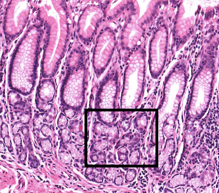
surface mucous cells

enteroendocrine cells
communicate with nervous system. Only seen with special stain

margo plicatus
separates non-glandular and glandular portions of equine stomach

Gastric Wall

gastric muscularis externa
mainly for churning and mixing

oblique smooth muscle
internal to circular and longitudinal muscle layers

circular smooth muscle

longitudinal smooth muscle

Gastric Serosa

Villi
Blue

Crypts
Orange

Enterocytes
Simple columnar epithelium with microvilli


Enteroendocrine cells
regulate pancreatic secretions, energy homeostasis, digestion, and absorption

Stem Cells
Located in crypts. continuous replacement of intestinal epithelial cells

Plicae circulares
amplified SA. Permanent folds of mucosa and submucosa in small intestines. Don’t flatten out like rugae of simple stomach

lacteals
enterocytes transport absorbed lipids into these

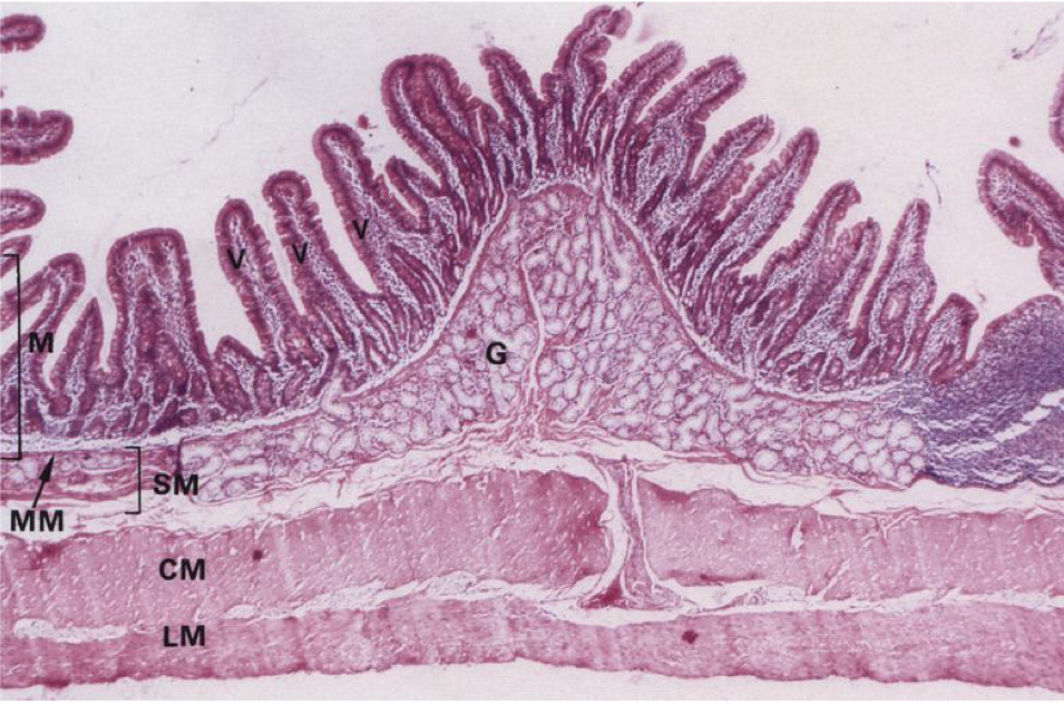
duodenum
has brunners glands(G) which secrete alkaline mucus to protect against acidic secretions from the stomach
Cecum/Colon
Have no villi, only crypts. epithelium is primarily goblet cells and stem cells for regeneration

Ano-Rectal Junction
Rectum mainly has goblet cells and Anus has stratified-squamous wet keratinized epithelium

Rumen
stratified-squamous wet keratinized epithelium. Papillae increase SA for absoprtion. No muscularis mucosa, submucosa is very thin


Reticulum
Smaller papillae found on floor and base of walls. stratified-squamous wet keratinized epithelium. squeezes out fluid/small particles into omasum. contracts larger particles back into rumen for further fermentation. muscularis mucosa only found in walls


Omasum
squeeze out fluid/electrolytes. May absorb residual VFAs and bicarbonate. stratified squamous epithelium(K/WK). Both muscularis mucosa and externa in core of laminae. Papillae found on laminae and floor


Abomasum
12 abomasal folds. break down microoraganisms, products of fermentation. Have surface mucus cells, mucus neck cells, parietal, chief and enteroendocrine cells


primarily mucous secreting cells in glands
