Microbiology Microscopy and the Six I's
 Call Kai
 Learn
 Practice Test
 Spaced Repetition
 Match
 Flashcards
 Knowt Play
1/35

Earn XP
Description and Tags
Flashcards covering microscopy techniques, the Six I's of microbiological culture, types of light and electron microscopes, resolving power, and microscope anatomy based on lecture notes.
36 Terms
Inoculation
The process of introducing a sample onto a sterile culture medium to begin the culture of microorganisms.
Incubation
Maintaining a culture at suitable temperature, time, and atmospheric conditions to allow microorganisms to grow and multiply.
Isolation
The technique of separating cells by streaking on agar to obtain a pure culture containing one species.
Inspection
Observing colony appearance and examining cells microscopically after staining to describe growth, cell shape, arrangement, and other characteristics.
Information gathering
Performing biochemical, genetic, or immunological tests and recording results to collect evidence about an organism's characteristics.
Identification
Comparing observations and test results with known organisms or databases to determine an organism's identity.
Refraction
The phenomenon occurring when a beam or ray of light transmitted through the air strikes and passes through a material.
Resolution
The ability of a microscope to distinguish two points as separate, allowing better resolving power to reveal finer details.
Resolving power formula
rp=2×numerical aperture of objective lenswavelength of light (nm)
Oil immersion
A technique using an oil that has the same optical qualities as glass, preventing refractive loss as light passes from the slide into air, thereby increasing numerical aperture (NA) and resolving detail.
Bright-field microscope
A microscope where light transmitted through the specimen absorbs some light before passing to the eyepiece, causing the specimen to appear darker than the brightly illuminated field; used for live, unstained and preserved, stained specimens.
Dark-field microscope
A microscope featuring a special stop disc in the condenser that blocks all light except peripheral rays from entering the objective lens, producing a brightly illuminated specimen on a dark/black field; used for living cells distorted by drying, heat fixation, or that cannot be standardly stained.
Phase contrast microscope
A microscope containing devices that convert subtle changes in light waves passing through a specimen into differences in light intensity, used to observe intracellular structures and locomotor structures in living eukaryotic cells.
Confocal microscope
A microscope utilizing a laser beam to scan various depths of a specimen and deliver sharp images focused on a single plane, producing CT scan-like slices using fluorescent tags and computer-aided imagery.
Fluorescence microscope
A modified compound microscope equipped with an ultraviolet (UV) radiation source and protective eye filters, used with fluorescent dyes or minerals that emit visible light when bombarded by shorter UV rays.
Transmission electron microscope (TEM)
An electron microscope that transmits electrons through extremely thin slices of metal-coated dead specimens to visualize detailed internal structures of cells and viruses in black and white.
Scanning electron microscope (SEM)
An electron microscope that scans a beam of electrons back and forth over the metal-coated surface of a whole specimen to collect deflected electrons, producing an extremely detailed three-dimensional view.
Scanning tunneling microscope (STM)
A microscope that uses a thin metal probe to scan the surface of an unprepared specimen to reveal atomic bumps and depressions, offering greater resolving power than an electron microscope and the ability to visualize DNA.
Atomic force microscope (AFM)
A microscope that moves a metal and diamond probe gently along the surface of an unprepared specimen to record surface movements and generate three-dimensional images of biological molecules.
Eyepiece
The microscope component labeled A that contains the ocular lens.
Nosepiece
The rotating microscope component labeled B that holds the high- and low-power objective lenses to change magnification.
Objective lenses
The microscope lenses labeled C whose magnification ranges from 10× to 40×.
Stage clips
The mechanical clips labeled D on a microscope that hold the slide in place.
Stage
The platform labeled E that supports the slide being viewed under the microscope.
Diaphragm
The microscope component labeled H that regulates the amount of light directed onto the specimen.
Coarse adjustment knob
The control knob labeled J that moves the microscope stage up and down for focusing.
Fine adjustment knob
The control knob labeled I that moves the stage slightly to sharpen the image focus.
Arm
The structural portion of the microscope labeled K used to support the instrument when carried.
Field of view (FOV)
The visible circle of light and area seen through the microscope, which decreases in diameter as magnification increases.
brightfield microscope image

darkfield microscope image

phase contrast microscope image
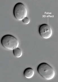
confocal microscope image

fluorescence microscope image

transmission electron microscope image
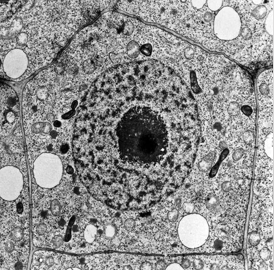
scanning electron microscope image
